
In this article we will discus about:- 1. Nature of Venom 2. Effects of Poison 3. Anti-Snake Venom Serum 4. Poison Apparatus and Biting Mechanism 5. Ligaments and Muscles of Snakes 6. Transference of Venom.
Nature of Venom:
It is a kind of highly evolved saliva which the snake uses to both kill as well as digest its prey. There are two kinds of snake venom. One affects the nerves (the venom of the Cobra and Krait), the other affects blood (that of the Vipers).
1. Snake venom is a clear, transparent, pale yellow or straw-coloured fluid having a specific gravity of 1.03-1.07.
2. The pH of the venom varies in different species, e.g. in Russell’s viper pH is 5.8 while that of cobra’s venom is 6.6.
ADVERTISEMENTS:
3. It is protein in nature containing many enzymes, viz. Proteinase, Hyaluronidase, L-Arginine hydrolases, Transaminase, L-Amino acid oxidase, Phospholipase-A, B and C, Phosphodiesterase, Cholinesterase, Ribonuclease, Deoxyribonuclease, Alkaline phosphatase, Acid phosphatase, Exopeptidases etc. The composition of venom varies from species to species.
4. Most snake venoms are tasteless but cobra venom’s taste is slightly bitter.
5. It is acidic in reaction and soluble in water and glycerine.
6. It is destroyed by coagulating agents like KMnO4, AgNO3, as well as alcohol and strong alkalies like NaOH and KOH etc.
ADVERTISEMENTS:
7. The venom also contains metals like zinc (Zn), sulphur (S), copper (Cu) etc.
8. Venom contains several active non- enzymatic proteins having at least two or three peptides.
9. Venom is thermolabile.
10. It can be dried as crystals or lyophilized. Dried venom is thermo-stable.
ADVERTISEMENTS:
11. It is a good digestive enzyme and can be easily swallowed if there is no wound in the buccal cavity and alimentary canal.
Effects of Poison:
Venom is generally introduced into the subcutaneous tissue. It then reaches the general circulation. When introduced directly into the vein the effect is instantaneous. The venom of different snakes acts differently.
(a) Cobra poison:
The effects are observed within half an hour. Symptoms are giddiness, high pulse rate, extreme salivation, partial paralysis of tongue and larynx, vomiting and contraction of pupil. The eyes remain sensitive to light and consciousness remains undisturbed. Respiration is shallow, which ultimately ceases and death follows. Cobra venom is a neurotoxin.
(b) Viper poison:
The effects are observed within a quarter of an hour. Symptoms are swelling of the wounded region, discolouration due to extravasation, acute burning pain, pupil dilatation, high pulse rate, profuse vomiting and watery discharge from rectum. Eyes lose sensitivity to light and consciousness is affected. Extravasation becomes extensive, swelling spreads and death occurs. Viper venom is haemo-toxic.
(c) Krait poison:
Kraits are highly poisonous but the absence of local pain, swelling, oozing or bleeding raise doubt of krait bite in the first hour. Even fang marks do not reveal any visible spot. Symptoms are very much similar with the signs of neurotoxicity of cobras.
The first sign of neurotoxicity is ptosis. The other signs are severe abdominal pain, drooping, dribbling of saliva, cyanosis and respiratory failure. The krait venom acts both as a neurotoxin and haemo-toxin. The cause of death is asphyxia through paralysis of the respiratory centre.
Anti-Snake Venom Serum:
ADVERTISEMENTS:
In India anti-snake venom serum is prepared at the Hoffkine Institute, Mumbai, and Central Research Institute, Kasauli, Himachal Pradesh. At Hoffkine, anti-venom is prepared by immunizing horses. A mixture of venom of four common poisonous snakes (Cobra, Naja naja; Common Krait, Bungarus caeruleus; Russell’s viper, Vipera russelli and Saw scaled viper, Echis carinatus) is injected into horse’s body and the dose is increased gradually. When the horse acquires a certain level of immunity, the plasma is collected by drawing blood from the body.
After collection, it is packed in 10 ml doses. Anti-venom of Banded krait, King cobra and Sea snake is not produced in India. Nearest source of anti-venom of Banded Krait and King cobra is the Queen Saovabha Institute, Bangkok, Thailand.
Poison Apparatus and Biting Mechanism:
The poison apparatus consists of a pair of poison glands, their ducts, fangs and muscles.

(i) Poison glands (Fig. 2.46A):
The glands are situated one on either side of the upper jaw. The poison glands are possibly the modified superior labial or parotid glands. In Naia naja, it is in the shape and size of an almond kernel, thickly encapsulated with fibrous tissue.
In vipers it is large and tubular shaped, and the shape may vary in different genera. The capsule supports vascular fibrous septa, separating the gland into secretory pockets, the Poison lakes of Bobeau. The section of the poison gland reveals the true structure of an exocrine gland.
(ii) Poison ducts:
The gland is provided with a narrow duct at its anterior end. The duct passes forward along the side of the upper jaw and loops over itself just in front of the fang and opens either at the base of the fang or at the base of the tunnel on the fang.
The duct actually opens in a pocket of mucous sheath that covers the basal part of the fang. In spitting cobra (Naja nigricollis), the poison duct is modified into an “L” shaped bend, just prior to exiting the fang, with the discharge orifice being located on the front of the fang.
(iii) Fangs (Fig. 2.46):
In poisonous snakes a few maxillary teeth are modified to act as poisonous teeth or fangs. The fangs are conical, curved, sharply pointed and are enlarged maxillary teeth which regenerate when lost. An intake aperture is at the basal part of the fangs and the discharge aperture is sub-terminal.
In spitting cobra (Naja nigricollis) and South African ringhals (Hemachatus haemachatus), the discharge orifice is much smaller, presumably to increase the force of venom. The fangs are one on each side of the maxillary bone of the mouth. In the Viperidae, there is a single, large, poison fang on the maxilla with small reserved fangs at its base.
Fangs are of the following types, depending on the structure and position of the venom canals (Fig. 2.46):
(a) Proteroglypha (Proto = first, glyph = hollowed):
Fangs are small, relatively non-movable and situated at the front of the maxillary bone. An open groove runs on the anterior surface of the fang. Fangs of cobras, kraits, mambas (Fam. Elapidae) and sea snakes (Fam. Hydrophidae) are included in this category.
(b) Solenoglypha (Solen = Pipe):
Fangs are long, hollowed and situated at the rear end of the maxillary bone. They are capable of vertical movement and are movable in which the fangs are folded against the roof of the mouth when the jaws are closed. In this type, fangs are pierced by venom canals, thus acting as hypodermic syringe. The fangs of pit vipers and true vipers (Family Viperidae) fall in this category. The long movable fangs of true vipers can penetrate deep into the tissues of the victims.
(c) Opisthoglypha (Opistho = behind):
Fangs with open groove on the posterior surface, situated at the posterior extremity of the maxillary bone. The fangs are either one or two in number with few smaller teeth in front. The fangs of vine snake (Ahaetulaa nasutus), common cat snake (Boiga trigonata), flying snake (Chrysopelea ornata), South African boomslang (Dispholidus typus), egg-eating snakes (Dasypeltis) are in this category. The bite of opisthoglyph snakes is not usually lethal to human beings except South African boomslang. Generally, their bite may be lethal to lizards, and sometimes birds, mice and rats.
Ligaments and Muscles of Snakes:
(i) The poison gland is held in position by ligaments, which extend from maxilla-lacrimal junction to pterygoquadrate.
(ii) Fan-shaped ligaments present along the sides of the poison gland.
(iii) Associated muscles are anterior and posterior temporalis (masseter), digastric muscles and protractor-pterygoid muscle that help in biting mechanism.
(iv) The fan-shaped temporalis muscle originates from the post-frontal and parietal ridges. It embraces most part of the poison gland. Its sudden contraction helps to eject out the venom from the gland, and also controls the biting mechanism.
(v) Digastric muscle: It arises from the squamosal and quadrate junction and is attached to the articular of the lower jaw (Fig. 2.47B). Contraction of these muscles help in opening the mouth by depressing the lower jaw.
(vi) Sphenopterygoid or protractor-pterygoid muscle: It arises from the anterior margin of the basal orbitosphenoid region and inserts to the dorsal side of the pterygoid (Fig. 2.47B). It assists in pulling the pterygoid forward, resulting in pushing the ectopterygoid which rotates the maxilla and erects the fang.
(vii) Anterior temporalis muscle: It is the closing muscle of the mouth. It arises from the pterygoid bones of the upper jaw and is inserted to the articular region of the mandible (Fig. 2.47B).
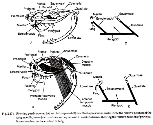
Structural elements of the skull (Fig. 2.47):
The peculiarity of the skull is that all the bones are loosely attached i.e. connected by elastic ligaments so that each bone is capable of movement independently. The structural elements of the skull are arranged in such manner that it acts like a lever system to erect the fangs during strike.
The following bones of the skull are associated in activating the poison apparatus:
(i) Maxilla:
These are paired irregular bones, forming an anterior larger part of upper jaw (Fig. 2.47A). These maxillae bear fangs.
(ii) Pterygoid:
It is paired irregular bones. At the posterior end each bone joins loosely with the quadrate and anteriorly it joins with the palatine that meets the maxilla (Fig. 2.47A). It is a part of alisphenoid.
(iii) Ectopterygoid or Trans-palatine:
It is a small bone that passes from the pterygoid and joins with the maxilla (Fig. 2.47A).
(iv) Quadrate:
This bone is large rod-like and highly movable (streptostylic). It remains at the posterior end of the upper jaw. The anterior part joins the squamosal while the posterior part articulates with the lower jaw. Mandible articulates with skull by ligaments and the rami are united by ligaments.
Biting mechanism:
The mechanism of biting is a complicated process and the sequences of biting may be discussed in three observable steps.
These are:
(a) Opening of the mouth:
The digastric muscle contracts, as a result the mouth opens (Fig. 2.47).
(b) Rotation of maxilla:
As the mouth opens, the lower jaw moves forward and a rotation of the squamosal, quadrate and mandible in relation to each other occurs. Now the sphenopterygoid muscles contract. This contraction results in the forward movement of pterygoid and up-pushing of the ectopterygoid.
The upward movement of the ectopterygoid brings about a rotation of the maxilla on its own axis round the lacrimal and as the end result the fang is raised and becomes directed forward (Fig. 2.47C, D). The fang is nearly horizontal in position when the mouth remains closed. But during opening of the mouth to bite, the fang assumes almost a vertical position.
(c) Closing of mouth:
This is brought about by the contraction of the temporalis muscles and sphenopterygoid muscles. The point of the fang is directed backward while the mouth is closed. It takes a longer time to open the mouth than to close it.
Transference of Venom:
During the contraction of the digastric muscle the posterior ligament is relaxed and during the rotation of the squamosal bone the fan-shaped ligaments are stretched to squeeze the walls of the gland. This makes the poison to come out of the gland through the duct and fang.