
Tissue is a group of similar cells having a common origin that act together in the performance of a particular function. Early during development, the cells of the growing embryo differentiate into three fundamental embryonic tissues called germ layers namely ectoderm, mesoderm and endoderm. These germ layers, in turn, differentiate into different cell types and tissues that are characteristic of the vertebrate body.
Most animal cells are surrounded by a narrow space filled with extracellular or intercellular fluid. This forms the immediate environment of the cell. From this fluid, the cell receives all the material needed by it and also transfers the waste materials produced by it to this fluid.
In adult vertebrates and also in most invertebrates, there are four principal kinds of tissues. They are epithelial, muscular, connective and nervous tissues.
The epithelial tissues originate from ectoderm, mesoderm and endoderm layers. They are mainly concerned with protection, secretion, absorption, excretion and reproduction functions.
ADVERTISEMENTS:
The muscular and connective tissues are of mesodermal origin. If muscular tissue helps in the movement of parts and locomotion, the connective tissue assists in attachment, support, protection, storage and transport.
The ectoderm derived nervous tissue controls and coordinates the conduction of nerve impulses.
1. Epithelial Tissues or Epithelia (sing., epithelium):
The tissue that covers the outside of the body and lines organs and cavities within the body is called epithelial tissue or epithelium. The epithelia are derived from all three germ layers. The epidermis derived from ectoderm constitutes the outer portion of the skin. The inner surface of the digestive tract is lined by an epithelium derived from endoderm. The inner surfaces of the body cavities are lined by epithelia derived from mesoderm.
Structure:
ADVERTISEMENTS:
i. The epithelial tissues consist of variously shaped cells.
ii. The epithelial cells are closely arranged forming a single layer or multiple layers.
iii. The intercellular spaces between the cells are practically absent.
iv. The epithelial cells are held together by very little amount of intercellular matrix containing a carbohydrate derivative called hyaluronic acid.
ADVERTISEMENTS:
v. The epithelial cells always rest on a thin non-cellular basement membrane consisting of glycoproteins and collagen fibres. The basement membrane is formed of two layers – outer thinner basal lamina secreted by the epithelial cells, and inner thicker fibrous reticular lamina secreted by the underlying connective tissue.
vi. The neighbouring epithelial cells are held together by 3 types of intercellular junctions as shown in the figure such as tight junctions, adhering junctions and gap junctions. The tight junctions prevent the leakage of substances, the adhering junctions cement the adjoining cells with desmosomes, interdigitation, etc., and gap junctions allow the movement of ions and biomolecules between adjoining cells.
vii. The epithelial tissues are non-vascular. They get nutrition from the blood capillaries present in the underlying connective tissue and also transfer the waste matter to the blood.
viii. Although the blood vessels are absent in the epithelial tissues, the nerve ending may penetrate the epithelium.
ix. The free surface of the epithelial cells may be smooth or may have fine hair like cilia, stereocilia (though their name is similar to cilia, they are actually more closely related to microvilli, and some sources consider them to be a variant of microvilli, they are characterized by their length – distinguishing them from microvilli and their lack of motility – distinguishing them from cilia) and microvilli.
Epithelium possesses remarkable regenerative powers, constantly replacing its cells throughout the life of the animal. For example, the liver, a gland formed from epithelial tissue, can readily regenerate after substantial portion of it has been surgically removed. The epithelium inside the stomach is replaced every two or three days. The epidermis is renewed every two weeks because of the continuous wear and tear.
Based on the cell shape and cell layers, the epithelial tissues are classified as simple epithelia and compound epithelia.
The simple epithelia are one cell layer thick. They rest on the basement membrane. They cover moist surface where there is minimum wear and tear. They never occur in the surface which is exposed to mechanical or chemical abrasions because they cannot protect the underlying tissues. Simple epithelia occur mainly on secretory and absorptive surfaces.
Simple epithelium is further classified into five types based on the form and structure of the cells into squamous, cuboidal, columnar, ciliated, glandular and pseudostratified types. The compound epithelia are into two types: stratified and transitional.
2. Connective Tissues:
ADVERTISEMENTS:
The tissue that either supports other tissues or joins one type of tissue to the other, muscle to bone or bone to bone is called a connective tissue. It includes areolar tissue, adipose tissue, reticular tissue, cartilage, bone, tendons, ligaments, blood and lymph. The connective tissue is mesodermal in origin.
Location:
Connective tissue is found throughout the body. It occurs in between different tissues and organs. It also forms sheaths around various organs of the body.
Structure:
The connective tissue comprises of three basic components, viz., matrix, cells and fibers.
The connective tissue is characterized by the presence of a large amount of nonliving intercellular or extra cellular, amorphous, transparent ground substance called matrix. The matrix may be fluid (E.g. – blood) or jelly-like (E.g. – cartilage) or solid (E.g. – bone). It is mainly formed of mucopolysaccharides and glycoproteins. Embedded in the matrix, there are several types of protein fibres and various kinds of cells. The matrix permits the diffusion of nutrients, gases and wastes to and from the cells.
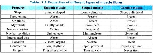
3. Muscle Tissue:
Muscle tissue is composed of long cells called muscle fibres (myocytes) that are capable of contracting, usually when stimulated by nerve signals. The muscle tissue is mesodermal in origin. In mammals they make up about 40 % of the body weight.
Types of Muscular Tissue:
Based on the location, structure and function, the muscle tissue is classified into following three types (Table 7.1):
General Functions:
The muscle tissues perform the following functions:
i. The muscles support the bones and other structures of the body. They give shape and beauty to the body.
ii. The muscle tissue enables the body parts like hands, arms, legs, etc., to move and helps the organism in locomotion.
iii. The muscle tissues are responsible for the food to pass through the alimentary canal, for the blood and lymph to flow through the vessels, for the respiratory gases to flow through the respiratory tracts, for the heart, to pump the blood and for the propulsion of secretions and waste products through the ducts.
iv. The muscle tissues also help in ingestion, egestion, urination, mating, egg-laying, parturition, feeding the young ones, maintenance of equilibrium and in gathering information about the external environment by moving the sense organs like eyes, nose and pinnae.
v. The muscles also play an important role in gesture and facial expression.
4. Nervous Tissue:
Nervous tissue is a specialized tissue with the property of excitability and conductivity i.e., the cells of this tissue are specialized for receiving the stimuli and to react to stimuli by conducting impulses to various organs in the body which bring about responses to the stimuli. The nervous tissue is of ectodermal origin.
Structure:
The nervous tissue is formed of four types of cells:
1. Neurons (Nerve Cells):
A neuron is a structural and functional unit of nervous system.
2. Neuroglia (Glial Cells):
The neuroglial cells are usually referred to as glial cells or glia. They are quite different from nerve cells. The major difference is that glia cells do not participate directly in synaptic transmission, although their supportive functions help in synaptic contacts and to maintain the signaling abilities of neurons. Glia is more numerous than nerve cells in the brain, outnumbering them by a ratio of approximately 2:1.
3. Ependymal Cells:
Ependymal cells are the cells which line the ventricles of the brain and the central canal of spinal cord. They are typically cuboidal and often have cilia which are best seen in younger brains. They secrete cerebrospinal fluid.
4. Neurosecretory Cells:
Neurosecretory cells are the special type of nerve cells found in the hypothalamus of the brain. They release chemical substances which influences the activities of another structure. For example, the neurohormones secreted by the hypothalamus influences the anterior lobe of pituitary gland and stimulate it to secrete hormones like TSH, STH, FSH, LH, ACTH, etc. The neurosecretory cells release the chemicals from their axon endings directly into the blood stream instead of into the synaptic cleft.