
The following points highlight the top six types of tissues found in animals. The types are: 1. Epithelial Tissue 2. Connective Tissues 3. Muscular Tissue 4. Blood and Lymph Tissue 5. Nerve Tissue 6. Reproductive Tissue.
Type # 1. Epithelial Tissue:
It forms a continuous layer covering the body externally or internal surfaces. It is protective in function. Epithelial tissue covering the external body surface is external epithelium and that covering the internal surface, viz. body cavity and other organs are internal epithelium. Epithelial cells are usually supported by a basement membrane.
The outer epithelium, to some extent, protects the body from mechanical injury, from chemicals and from the invasion of microorganisms. The epithelium in the intestine absorbs water and nutrients; in the kidney glucose, amino acids, salts and, in some cases, water.

Epithelial tissues (Fig. 5.1) are classified according to their shape:
ADVERTISEMENTS:
a. Simple or non-stratified epithelium:
The tissue is made of a single layer of cells. Present in the intestine.
b. Stratified epithelium:
ADVERTISEMENTS:
The tissue is made of many layers. Present in the palate of rabbit.
c. Pavement or Squamous epithelium:
The cells of the tissue are thin and flat. Present in outer skin of frog.
d. Cuboidal epithelium:
ADVERTISEMENTS:
The cells are cuboid in outline. Present in liver, kidney tubules etc.
e. Columnar epithelium:
The cells are elongated, and supported by a basement membrane. The length is more than the width. Present in intestinal mucous membrane.
f. Ciliated epithelium:
It is modified columnar epithelium. The cells are provided with cilia at the free end. Present in oral cavity and genital ducts. Some epithelial cells, both external and internal, respond to stimuli. These are sensory cells. Some cells are capable of secreting certain substances. These are glandular cells.
Type # 2. Connective Tissues:
These tissues form supporting structures of the body and connect various parts of the body. Large amount of nonliving intercellular substance, the matrix, is present in these tissues holding the cells together. The nature and function of the connective tissue is determined by its intercellular matrix.
Six major types of connective tissues are found:
a. Fibrous connective tissue:
The cellular part of the tissue consists of small, scattered cells. The intercellular structure is constituted by un-branched, wavy fibres. The white fibres or collagen fibres are arranged in bundles and embedded in a clear homogeneous matrix. The yellow elastic fibres or elastin are straight and arranged closer (Fig. 5.2). Other types of fibrous tissues are present in muscular sheath, tendon and ligament.

b. Tendon:
Thick, closely packed bundles of collagen fibres constitute a tendon. Tendons are flexible and join a muscle to a bone or to another muscle.
c. Ligament:
Ligament is similar to tendon, it connects one bone to another.
d. Cartilage:
The cartilaginous tissue is semitransparent. Oval cells—singly or in groups, are embedded in the semitransparent intercellular matrix which forms the main bulk of the cartilage. Cartilage can grow from within by divisions of cartilage cells, die modified fibroblasts. They secrete more matrix around themselves (Fig. 5.3).
Hyaline or semitransparent cartilage is present in skull, limb girdles, between the vertebrae and other parts of the body. The cartilage is a firm but elastic structure and most bones develop from cartilage by calcification.

e. Bone:
The bones are of two types—the long bone and membrane bone. The bone is almost similar to cartilage but the matrix is rendered hard by a rich deposition of lime salts. Membrane bone is formed directly from mesenchyme. A long bone is surrounded by a thin, fibrous periosteum (Gr peri = around + osteum = bone), except articular surfaces.
A bone consists of an outer layer of compact bone, and a medullary cavity at the centre (Fig. 5.4A). The compact bony layer is made of many Haversian systems, closely packed. A system has a narrow, central Haversian canal, around which are concentric bony rings or lamellae, having narrow lacunae in between them. Osteoblasts, osteocytes and osteoclasts are present in the lacunae.
Narrow canaliculi connect the lacunae, with each other and also with the Haversian canal. Interstitial lamellae separate the Haversian systems. The Haversian systems are enclosed in a secondary lamella (Figs. 5.4B, C). The medullary cavity is occupied by bone marrow, which is the site for production of blood cells. The bones support and add rigidity to the body.

f. Adipose tissue:
In this tissue the cells are enlarged and heavily laden with fat in the form of globules. The fat is secreted by the cytoplasm and due to accumulation of huge quantity of fat the cytoplasm and the nucleus are pushed to one side (Fig. 5.5). Large amount of adipose tissues are present under the skin.

Type # 3. Muscular Tissue:
All the movements of animal body are performed with the help of muscular tissue. The cells of muscular tissues are elongated and slender (Figs. 5.6-5.9). All muscle cells— whether skeletal, cardiac or smooth muscle, contain contractile protein arranged as myofibrils. In striated muscle (skeletal and cardiac) the filaments are arranged symmetrically in an orderly fashion.
In a skeletal muscle groups of muscle fibres are bound together and invested in a connective tissue sheath, through which blood vessels and nerves pass. The cells in a muscle develop tension along their long axis and the muscle contracts. Muscles only contract and relax, they never elongate. In smooth muscle the arrangement is random. Proteins—actin and mycin are present in all myofilaments.
a. Skeletal (striated voluntary) muscle:
The muscle cells (sarcomeres) are large (Fig. 5.6). The plasma membrane is known as sarcolemma. The cytoplasm or the sarcoplasm is composed of longitudinally arranged myofibrils, the essential contractile component of the cell. The cells are multinucleated. Cross striations rim at right angles to the axes of the cells. The striations are due to the presence of alternating thin bands or discs.

The bands (Fig. 5.7) are:
i. I (Isotropic) bands appear light. A narrow central dark disc, the Z (Zwischenschelbe) band is present in a I band.
ii. A (Anisotropic) bands appear dark. A narrow less dense central portion is known as H (Heller) band.

The functional unit of the sarcomeres is the zone of myofibril between two Z bands.
b. Cardiac muscle:
The sarcomeres of cardiac muscle are similar to those of skeletal muscle. In addition, cardiac muscle cells are joined by dense intercalated discs (Fig. 5.8), which appear to replace Z bands. Due to irregular arrangement of membranes, the firm contacts between adjacent sarcolemmal membranes at the intercalated discs extend over large areas.

c. Smooth (involuntary) muscle:
Smooth muscle cells are generally small. The sarcoplasm contains many longitudinally arranged myofibrils (Fig. 5.9). The single nucleus is eccentric.
Unlike the muscle fibres of the skeletal muscles in which longitudinal myofibrils are packed, the myofibrils are arranged at random in a smooth muscle fibre. The fibres contain actin and myosin and the mechanism of contraction is same as in skeletal muscle. Smooth muscle has two functions, sustained and intermittent contraction.

The smooth muscles in vertebrates are of two types:
(a) Multiunit smooth muscle present in iris and ciliary body of the eye and in the walls of the blood vessels and
(b) Visceral smooth muscle in the walls of the gut, uterus and visceral organs.
Like other muscles, the visceral smooth muscles also possess certain spontaneous activities and contract automatically when stretched, which may happen when an organ is filled with food or liquid.
Type # 4. Blood and Lymph Tissue:
The blood and lymph are considered liquid tissues, as loose cells float in the plasma which is comparable to the ground substance of connective tissue.
Blood is essential for the various activities of an animal, because this is the important transport medium in the body. Blood consists of a liquid portion,—the plasma and the blood cells or corpuscles floating in it. Besides the corpuscles, fibrinogen remains in the plasma in a soluble state.
In most of the invertebrates the blood corpuscles are amoebocytes but oval and nucleated in nemertines, oval and biconcave in some annelids and phoronis. In invertebrates, usually the outflow of blood (haemolymph) through injury is prevented by agglutination of blood cells.
Human plasma constitutes about 55 per cent of the blood volume and 92 per cent of the plasma is water. Plasma proteins, amino acids, glucose, lipids and nitrogenous wastes constitute 8 per cent solid. Albumin, a, (5 and Y globulins, euglobulin and fibrinogen constitute the protein portion.
Fibrinogen helps in coagulation of blood. The blood cells in vertebrates are of three types—erythrocytes, leucocytes and thrombocytes. All cells except lymphocytes originate in red bone marrow and are temporarily stored in spleen, which is also a site of origin of the cells. Lymphocytes (Fig. 5.10) originate in the lymph nodes and also stored there.

Red blood corpuscles or R.B.C. or erythrocytes:
These are much more in number and an iron compound, haemoglobin is present in the cytoplasm of the cell. This is a respiratory pigment. In most of the invertebrates the respiratory pigment is haemocyanin, a copper compound.
The colour of vertebrate blood is red due to haemoglobin, while the haemocynin content blood is almost colourless. In all vertebrate R.B.C., except that of mammals, a nucleus is present, the shape of which differs in different classes of vertebrates (Fig. 5.11).

a. Fish:
Almost round.
b. Amphibia, reptiles and birds:
Biconvex and oval.
c. Mammal:
Round and biconcave.
White blood corpuscles or W.B.C. or leucocytes:
These are less in number, large in size and the nucleus is not round (Fig. 5.11). Some of the cells are phagocytic and can engulf bacteria and other foreign particles with pseudopodia.
Leucocytes are of the following types:
a. Monocytes:
Large size, with a single nucleus. In a young monocyte the nucleus is round or elliptical but becomes kidney-shaped later.
b. Lymphocytes:
Nearly circular with a large nucleus.
c. Granulocytes:
Capable of amoeboid movement. The cytoplasm is granular.
Granulocytes are of three types:
i. Eosinophil:
Take up only acid stains. The nucleus bears 2-3 segments.
ii. Basophil:
Take up only basic stains. The nucleus is fragmented.
iii. Neutrophils:
Take up only neutral stains. The nucleus bears 2-7 segments.
Thrombocytes or blood platelets:
These are small cells of irregular shape and help in coagulation of blood.
Blood volume:
The amount of blood present in the body or the blood volume differs in animals. It is 7 to 10 per cent of the body weight in man; 5 per cent in bony fishes; 5-8 per cent in Octopus; 5-25 per cent in insects.
Functions of blood:
a. Transport of respiratory gases:
Haemoglobin combines with O2 to form unstable oxyhaemoglobin. The oxygen is taken up in the respiratory organs, skin, gill, lung as the case may be and released in the tissue. In the plasma, haemoglobin directly combines with CO2 to form carb-amino compounds and the CO2 is released in the respiratory organ. Haemocyanin has similar function.
b. Transport of nutrients:
It carries nutrients, the products of digestion of food and water, minerals,-etc. absorbed from the intestine, to the cells. It also carries substances from one place of the body to other, i.e. from storage site to other parts.
c. Removal of wastes:
Waste products of metabolism are carried from the site of origin to the organ of excretion.
d. Carrier of chemicals:
It transports hormones, vitamins and other essential chemicals from the site of origin or point of entrance to the site of activity.
e. Acid-base equilibrium:
It is a powerful buffer and helps to maintain a constant reaction in the body with the help of kidney, lung and skin.
f. Water balance:
It helps in maintaining water balance in the body.
g. Ion balance:
It helps to maintain ion balance between the cells and surrounding tissue fluid (interstitial fluid).
h. Temperature regulation:
Water has three important qualities in temperature regulation:
(i) High specific heat,
(ii) High conductivity and
(iii) High latent heat of evaporation.
The water present in blood helps in temperature regulation.
Defensive action:
(i) The white cells engulf bacteria by phagocytosis.
(ii) Antigen develop antibody to combat toxin.
Lymph:
Excess interstitial fluid passes into lymph capillaries and it is called lymph. The lymph is returned to the heart, and thereby to the blood, through a set of vessels, the lymphatic system.
Certain glands known as lymph nodes are associated with lymph vessels. The lymph nodes act as filters to remove bacteria and harmful substances. The lymph probably removes proteins, cell fragments and other materials from interstitial fluid (Fig. 5.12).

Defence system:
The first line of defence in animals has evolved by the capacity of some leucocytes— the lymphocytes, to engulf bacteria, virus, etc. In vertebrates, a second line of defence has evolved by their ability to produce a type of special proteins, the antibodies.
Antibody is a chemical synthesized in the body in response to a foreign substance, the antigen and it can combine with the antigen and destroy it. The antigen-antibody reaction is specific. Antigens are proteins introduced by invading bacteria or virus, but any protein or even some polysaccharides may act as antigen.
Immunity:
Once an animal produces antibody in response to infection or vaccination, the capacity to synthesize antibody very quickly in response to fresh invasion is retained for a long time. This is known as immunity. As the capacity is acquired, it is also known as acquired immunity.
A serum containing a specific antibody prepared from animals with acquired immunity is injected to a person exposed to a new or dangerous antigen, and the immunity developed therefrom in the recipient is known as passive immunity. Injection of anti-venom or anti-rabies serum induces passive immunity against snake venom or rabies virus.
Blood group factors:
Besides the capability of the lymphocytes to produce antibodies, some antigen and antibody systems are present in human blood. They have been designated as blood group factors OAB.
Agglutinogens:
Two specific antigenic proteins are present for all time in the plasma membrane of erythrocytes. They have been named A and B.
Agglutinins:
The antibodies α and β, specific to antigens A and B may be present in the plasma of blood.
Agglutination:
It is the phenomenon of clumping of blood cells. In human,-some individuals have antigen A, some the antigen B, some both A and B and some none of them.
An agglutinogen (say A) and its specific agglutinin (α) are never present in the same person, since its own blood cells would be coagulated. Similarly, B and β cannot be present in the same person. Individuals having no agglutinogen may have both the agglutinins α and β.
On the basis of the presence or absence of the blood group factors, people have been divided into four groups, O, A, B and AB.
To determine the blood group of a person, the test tube method of blood grouping is routinely followed.
Test tube method of blood grouping:
Two drops of blood are collected in a test tube containing 1 to 2 ml physiological saline solution. In two small test tubes, one drop of corpuscle suspension and one drop of saline solution are put in each. One drop of anti A (α) serum is added to one tube and one drop of anti B (β) serum to the other tube. Four such sets, are prepared.
The contents of each tube are gently mixed and left for one hour in room temperature. Positive reactions (Fig. 5.12) can be detected within a few minutes, and the extra time is given as a precautionary measure. The contents of each tube is examined for agglutination under a low power microscope.
Results:
a. No agglutination by α and β = group O.
b. Agglutination by α but not by β = group A.
c. Agglutination by β but not by α = group B.
d. Agglutination by both α and β = group AB.
Type # 5. Nerve Tissue:
Response to stimuli is the characteristic of living cells. A number of controlling processes, viz. nerves, hormones and metabolic products, operate in several ways to regulate and integrate functions of different organs in an animal body. But instantaneous response is possible through nerves alone, as the waves of excitement or impulses can move at a rapid rate through nerves only.
The tissue made up of specialized cells— the nerve cells or neurons, capable of quick transmission of impulses is termed nerve tissue. Waves of excitement appear in the cells of receptors and sense organs due to stimuli in the surroundings, both external and internal.
These are transmitted to the controlling centres by one set of neurons (sensory) and impulses from the controlling centres to sites of action or effectors are transmitted through another set of neurons (motor). The ability of the neurons to carry the waves of excitement rapidly enable the animals to integrate their internal functions and also to adjust with the environment.
A nerve is a bundle of hundreds or thousands of nerve fibres. A ganglion is an aggregation of a number of cell bodies of neurons. Nerves and nerve ganglia constitute the nervous system, which is present in all metazoans except sponges.
Neurons:
A typical neuron is enclosed in a plasma membrane and consists of three parts.
a. Cell body:
The cell body, soma or perikaryon is oval or irregular in outline and bears a prominent nucleus and chromatin granules, the Nissal bodies (Fig. 5.13). Centrosome is absent. Nerve cells do not multiply. The cell bodies are restricted to the periphery of the brain, known as nuclei; the central portion of the spinal cord and form the grey matter, and in the ganglia.

The cells vary in size from the small granule cells of the cerebellum of about 5.0 µm to the large motor cells of the spinal grey matter.
b. Dendrites:
A number of branched protoplasmic outgrowths from one end of the cell body. Dendrites carry impulses towards the cell body.
c. Axon:
A single, long extension of the cell body from the opposite end, ending in brush-like filaments, the telodendria. Axon carries waves away from the cell body. Damaged axon can regenerate if the cell body of the neuron is not injured.
The processes of the nerve cells are present deep within the brain, at the periphery of the spinal cord and form white matter. They form the peripheral nerves. Some of the neurons are enveloped by a cellular sheath, the neurilemma.
From the presence or absence of a sheath around the axon, the neurons are called myelinated or non-myelinated. The first group is present in the peripheral nervous system, while the second group is found in the central nervous system.
The neurilemma is constituted by Schwann cells. Concentric lamellae of fatty substances, the myelin, often called myelin sheath, is present between the axon and the neurilemma (Fig. 5.14). The myelin sheath is absent in the gaps between adjacent Schwann cells. These gaps are known as nodes of Ranvier.

Nerve cells vary greatly both in length and thickness. The average neuron is slightly less than 10.0 µm in diameter but the length may be more than a metre.
According to the number of processes they bear, the neurons are of four types:
a. Apolar:
Cell body bears no process.
b. Multipolar:
Cell body with one axon and a number of dendrites.
c. Bipolar:
The axon and the dendron are placed symmetrically, one on either side of the cell body.
d. Unipolar:
Axon and dendron arising from a common stem.
Nerve impulse:
Nerve impulses in a neuron always travel in the same direction. The speed of the nerve impulse is about four times greater in the myelinated fibres. Thick nerves carry impulses at a faster rate. A nerve impulse may move over 100 metres per second and only milliseconds will elapse before the effect takes place.
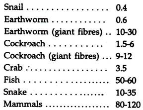
The speed of nerve impulse varies with homeotherms and poikilotherms, slow and first moving animals and also with ordinary and giant fibres. Speed of nerve impulse in the motor nerves of some animals-metre/second.
Type # 6. Reproductive Tissue:
New individuals are formed from the preexisting individuals and the species is perpetuated. No specialised reproductive tissues are involved in asexual reproduction. Sexual reproduction involves gametes. In majority of the metazoans, the gametes are produced in definite structures, the gonads, the sperms in the testis and the ova in the ovary.

The testes of vertebrates are made of thousands of sperm tubules (Fig, 5.15). The walls of the tubules are lined with primordial germ cells, the spermatogonia. The ovaries bear many primordial germ cells, the oogonia, which are enclosed in follicles in mammals (Fig. 5.16). The number of eggs produced by a female is much less than the number of sperms produced by a male.

The spermatogonia and oogonia divide many times and primary spermatocytes and primary oocytes are produced. Each of them undergoes two divisions, one of which is meiotic, and four sperms and one ovum with three nonfunctional polar bodies are formed.
Sperms:
A spermatozoon is an intricate motile cell, which moves to the egg to fertilize it. The sperms are minute in size bearing an ovoid head piece connected to a narrow, long tail piece by a middle piece (Fig. 5.17). Deviation from this is found in some animals.
They vary extremely in shape and size but their essential constituents remain unaltered. Some gastropods produce two types of sperms—pear-shaped, functional eupyrene and worm-like nonfunctional oligopyrene forms. The sperm is spirally twisted in passerine birds.

The head piece is nearly round in shrimp and hook-shaped in bats and mouse. The tail is specialised for movement. The terminal end of the tail piece is termed flagella. The structure of the tail piece vary in different animals.
It is lacking in some parasitic round worms and crustaceans. In sea-urchin tail piece, nine fibres are arranged in a circle with two more in the middle extending further. In some animals, viz. rabbit and others, the fibres with similar arrangement has been termed axial filament.
Ova:
The ova are large, usually round and contain yolk for the development of embryo. The shape and size are variable, the latter depending on the amount of yolk.
In addition to plasma membrane and vitelline membrane, in most cases, the ova are protected with additional coverings, shell membrane and an outer softer leathery and hard or calcareous shell depending on the hazards they are expected to meet after laying. The additional membranes are secreted by accessory glands following fertilization.