
In this article we will discuss about the integument of various chordates:- 1. Fishes 2. Amphibians 3. Reptiles 4. Birds.
1. Fishes:
In bony fishes, dermal scales do not actually pierce the epidermis, but they are so close to the surface they give the impression that the skin is hard (Fig. 2.4). The epidermal covering includes a basal layer of cells. Above this layer are stratified epidermal cells. As they move toward the surface, the epidermal cells undergo cytoplasmic transformations, but they do not become keratinized.

Two types of cells are present in the epidermis of fishes — epidermal cells and specialised unicellular glands. The epidermal cells form the stratified epidermis. Unicellular glands are single, specialised and interspersed among the epidermal cell population (Fig. 2.5). There are several types of unicellular glands, e.g. club cell, granular cell, goblet cell, sacciform cell etc.

These gland cells secrete mucus over the scales as mucous coat. This mucous coat, termed as mucous cuticle, resists penetration of infectious bacteria, contributes laminar flow of water across the surface, makes the fish slippery to the predators and sometimes contains chemicals that are repugnant, alarming, or toxic to enemies.
The dermis gives rise to dermal bones, and dermal bones give rise to dermal scales. Sometimes surface of the fish scales is coated with hard acellular enamel of epidermal origin and a deeper dentin layer of dermal origin.
Scales in fishes:
The skin of vertebrates is rarely naked and is provided with protective structures. In case of fishes the protective structure is scales.
ADVERTISEMENTS:
Divisions of scales:
The scales are classified on the basis of structure and shape. On the basis of shape these are divided into four types:
ADVERTISEMENTS:
(a) Placoid scales:
Plate-like scales with each plate carrying a small cusp (Fig. 2.6). This type of scale is present in elasmobranchs. In these fishes dermal bone is absent, but placoid scales as surface denticles are present.

(b) Rhombic scales:
Diamond shaped scales (Fig. 2.7) present in Acanthodii group of fishes. This type of fishes was found in fresh water during Ordovician to the Permian period.

(c) Cycloid scales:
Disc-like, thin and more or less circular in outline (Fig. 2.8). The centre of the scale is thicker and gradually thins towards the margin. Each scale has concentric lines of growth, called circuli. This scale lacks enamel, dentin and a vascular bone layer. Only lamellar bone remains, which is acellular and mostly non-calcified. The scales are situated in the dermal pockets. This scale is present in teleosts, like rohu, catla etc.

(d) Ctenoid scales:
ADVERTISEMENTS:
It is round-shaped but the free posterior margin possesses horny comb-like projections (Fig. 2.8). Each scale acquires new circuli, like rings of tree-with the growth. The basal end of this type of scale is usually wavy and embedded in small dermal pocket. This scale is present in teleosts, e.g., bhetki, koi, lata etc.
On the basis of structure, the scales are divided into five types:
(a) Placoid scales:
It develops in the dermis but projects through the epidermis to reach the surface. It has an ectodermal cap, which coats the spine (Fig. 2.9). The cap and spine is usually made up of enamel-like substance, called dentine.
The pulp cavity of spine communicates with the pulp cavity of basal plate by a pore situated at the base of the spines. Each scale has a disc-like basal plate made up of calcified tissue and remains embedded in the skin, e.g., shark. The basal plate of placoid scale is attached in the dermis by Sharpey’s fibre and other fibres from stratum compactum.

(b) Cosmoid scales:
This type of scales have three distinct layers:
(i) Innermost layer is called isopedine layer of perforate bony substance with many vascular lacunae,
(ii) The next layer is non-cellular and hard — it is known as cosmine.
(iii) The outermost layer has enamel-like consistency, e.g. primitive sarcopterygians (Fig. 2.10).

(c) Ganoid scales:
In ganoid scale the outermost layer is a thick hard enamel substance called ganoine, without an underlying layer of dentine (Fig. 2.11). Dermal bone forms the foundation of the ganoid scale. It is double layered, vascular and lamellar in palaeoniscoid fishes, and a single layer of lamellar bone in other primitive actinopterygians. These scales are shiny.

(d) Bony ridges:
It is typically thin and translucent, lacking both dense enameloid and dentinal layers. The outer surface of such a scale is marked with bony ridges that alternate with valley-like depressions. In this scale a nuclear or central zone is present, which is called focus of the scale. It is found in most of the cycloid and ctenoid type of scales. The focus is the first part to develop. The grooves or radii radiate from, or near the focus toward one or more of the margin of the scales.
(e) Elasmoid scales:
They are derived from ganoid scales of lepidosteroid type and are restricted to teleosts. The ancestral ganoine is absent and in its place there is a thin surface whose glaze is derived from the enamel organ. These scales are thin and imbricate. Isolated dermal denticles are confined to elasmobranchs.
Modification of scales:
In sharks, teeth are modified placoid scales. In saw fishes ‘saw’ are modified placoid scales. In Terodon and Diodon the scales are transformed into protective spines. In Acanthurus the scales at the base of the tail transformed into sharp cutting blade. In basking shark the gill rackers are modified scales.
2. Amphibians:
Generally, the skin of frogs and salamanders includes two types of dermal glands — mucus and poisonous glands. They open to the surface through connecting ducts piercing the epidermis (Fig. 2.12). Numerous chromatophore cells are also present.
In salamanders, the skin of the aquatic larvae includes a dermis of fibrous connective tissues, consisting of superficial loose tissue over a compact deep layer. Scattered Leydig cells of the epidermis secrete substances, which resist entry of bacteria or viruses. In terrestrial adults the Leydig cells art absent.

During breeding season nuptial pads may develop on the digits or limbs of male salamanders or frogs. Nuptial pads are raised calluses of cornified epidermis that help the male to hold the female during mating.
3. Reptiles:
Maximum keratinization in the epidermis is the characteristic feature for the dry terrestrial atmosphere. Skin glands are very few in reptiles in comparison to amphibians. The epidermis is generally demarcated into three regions — stratum basale, stratum granulosum and stratum corneum (Fig. 2.13). The dermis is composed of fibrous connective tissue.
Integumental glands are restricted to certain areas of the body of some reptiles. Many lizards possess rows of femoral glands along the underside of the hind limb in the thigh region. Scent glands are present in crocodiles and turtles. Most of the glands in reptiles are believed to be associated with reproductive behaviour.

Horny scales and plates in reptiles:
The reptilian scale is a folding in the surface epidermis; thus it is epidermal scale. In the junction between adjacent scales there is a flexible hinge (Fig. 2.14). Large and plate like scales of reptiles are called scute. Sometimes scales are modified into crests, spines or horn like processes. The rattles of the rattle snakes are modified scales which are not shed off.

In many reptiles dermal bone is present, known as gastralia e.g. crocodiles, Sphenodon. These are collection of bones in the abdominal area. Sometimes dermal bones support the epidermis, they are known as osteoderms. Plates of dermal bone located under the epidermal scales, i.e. osteoderms, are found in crocodilians, some lizards, etc. Some bones of the turtle shell are probably modified osteoderms.
Moulting or ecdysis:
It is a process of shedding off epidermal cornified layer of the skin at regular intervals. As moulting begins, the stratum basale, which has given rise to the strata granulosum and corneum, duplicates the deeper layers of granulosum and corneum, pushing up under the older layers (Fig. 2.13).
White blood cells invade the stratum inter-medium, a temporary layer between old and new skin. These WBCs are thought to promote the separation and loss of the old superficial layer of the skin. This process is clearly influenced by the anterior pituitary and thyroid gland activity.
Difference between reptilian and fish scales
Fish Scales
1. Built around bone of dermal origin.
2. It develops from the dermis and subsequently covered by different chemicals and minerals.
3. The scales do not pierce the epidermis; instead, it remains covered under epidermis.
Reptilian Scales
1. Lacks the bony under-support or any significant structural contribution from the dermis.
2. It is an epidermal surface folding.
3. As the scales are epidermal, no question of piercing.
4. Birds:
The epidermis comprises of the stratum basale and the stratum corneum. In between them there is a transitional layer of cells transformed into the keratinized surface of the corneum (Fig. 2.15). The feathers are epidermal in origin. Birds have few glands, like uropygeal gland and salt gland.

The dermis, especially near the feather follicles, is richly supplied with blood vessels. During brooding season, the dermis in the breast of some birds become heavily vascularized, forming a brood patch in which warm blood can come into close association with the incubated eggs.
Exoskeleton of birds:
In most of the members of the phylum Chordata the integument is soft and contains no hard skeletal parts, but many members have bony elements, derived from the dermis, present in the skin. Dermal scales of integumentary origin provide a protective armour, and are present in most fishes, in a few amphibians, in crocodilians, and in turtles.
The term dermal skeleton is used in referring such structures and their derivatives. Sometimes the dermal skeleton is called the exoskeleton, but this term is more properly applied to the skeleton of invertebrates. In birds, what we are referring as exoskeleton, are the derivatives of epidermis of the integument.
Feather of birds:
Structure of a typical feather:
A typical feather consists of several structural peculiarities. The seapus or stem is a stiff axial rod, running the whole length of the feather (Fig. 2.16). The stem has several zones along its length. The calamus is the tubular semi-transparent proximal portion, the base of which is inserted into the skin. At the terminal point of the calamus, there is a small aperture called the inferior umbilicus.
At the opposite end of the inferior umbilicus of calamus, there is another pore, called superior umbilicus. The distal part of the stem is solid rod like structure, called rachis or shaft. It is a tapering, flexible, elastic rod, square in transverse section. It has a ventral longitudinal groove called umbilical groove.

The vexillum or vane is the flattened portion of the feather, attached along the sides of the rachis. It is made up of barbs and barbules. The barbs are series of narrow elastic lamina, attached by their base along the two sides of the rachis.
The barbules are much smaller processes which form fringe along the sides of the barbs, bearing hooklets. These hooklets binds with the hooklets of other barbs and so give the feather a consistent structure.
Varieties of feather:
According to the shape and distribution on the body the feathers are divided into three major types.
Various types of feathers are tabulated in the following chart:
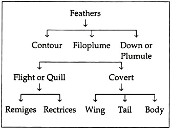
A. Contour feather:
These feathers determine the shape of the body of a bird, as they cover the body in a definite way. The barbules are well-developed and interlocked with others.
These are of two types:
1. Flight or Quill feathers:
Flight feathers constitute the major locomotor surfaces. These are the large feathers of wing and tail.
Accordingly they are defined differently:
(a) Remiges or wing quill, and
(b) Rectrices or tail quill.
(a) Remiges or wing quill:
The feather of the wings is called as remiges. This has the inner half of the vane much broader than the outer half (Fig. 2.16). The narrow half of each feather overlaps the broad half of the one anterior to it. The effect of this is to allow air passage between the feathers in the down-stroke.
The number of remiges is about twenty- three. The remiges are divided into two groups, on the basis of the limb bones on which it is attached. The primary quills are attached on the manus. In pigeon, they are eleven in number, of which six are attached to the metacarpals (meta-carpal quills), one ad- digital is attached to the phalanx of the post axial digit, two mid-digital attached to the middle proximal phalanx of the middle digit, and two pre-digital, one of which is small, attached to the distal phalanx of the middle digit. The secondary quills are attached to the ulna. They are twelve in number.
(b) Rectrices or tail quill:
These are present on tail. These feathers have two halves of the vane about equal in size. These are twelve in wild rock pigeon, but may be more in domestic breed. These feathers are distributed in a half-circle along the tail.
2. Covert feather:
These are medium sized feathers covering the whole body. These are similar in appearance to the flight feathers but smaller in size.
According to their distribution they are divided into three types:
(a) Wing covert:
These are covering the wings, Upper coverers are called upper wing coverts and lower coverers are called lower wing coverts.
(b) Tail covert:
There are upper and lower tail coverts covering the upper and lower part of the tail.
(c) Body covert:
Except wings and tail these feathers cover other parts of the body. They give the body a smooth outline.
B. Filoplumes:
These are minute rudimentary feathers left in the skin after the bird has been plucked. Each consists of a hair like long stem, with a very rudimentary vane at its apex and few barbs devoid of interlocking barbules (Fig. 2.16).
C. Down feather or Plumule:
These are generally short, soft and woolly feathers, covering the body of young newly hatched birds (Fig. 2.16). They have short calamus. Bunch of long, soft barbs are at the tip of the calamus. There are no hooks on the barbules. Usually they are not present in an adult bird.
D. Other feathers:
(a) Powder down feather:
These are down feathers, which do not mature after development. Instead, these are broken down to powdery materials. Their definite function is not known. It is believed that they maintain the texture of the feathers as talcum powder.
(b) Rectal bristle:
These are modified filo plumes. They have small calamus and rachis with rudimentary barbs. Generally found at the base of beak of flycatcher and goatsucker.
(c) Vibrissae:
These are hair-like feathers. Generally present at the base of beaks and around the eyes. These are tactile receptors and well developed in the nocturnal birds.
Arrangement of feathers on the body:
The feathers of a bird are distributed over the body in certain definite areas (Fig. 2.17). The distribution of feather is called pterylosis. The area where feathers are arranged, is called pterylae, the intervening tracts where feathers are not present, is called apteria. Only filo plumes are present in the apteria.

Feather develops during embryonic period from feather follicle. Feather follicles are invaginations of the epidermis that dip into the underlying dermis (Fig. 2.18). The root of the follicle in association with the dermal pulp cavity begins to form the feather. The feather itself grows outward in a sheathed case. Within the sheath, the central axis is divided into a distal rachis and a proximal calamus.

Functions of feather:
Feathers are insulating the body of birds from heat. Flight feathers are highly modified for efficient flying through the air. Sometimes colour of the feather match efficiently with the ambient colours concealing the bird both from predator and prey. In some birds sexual dimorphism is clearly visible from the feathers. Feathers help in sexual behaviour in some birds, e.g. tail feather of peacock.
Beaks of birds:
A hard keratin layer covers the upper and lower jaws of birds. These covered jaws are called beaks. The coverings of the upper and lower beaks are known as maxillary and mandibular rhamphotheca, respectively. These rhamphotheca are actually modified skin.
Beaks act for food collection, nest building, protection and maintenance of feathers by polishing with the secretions of uropygeal gland. There are varieties of modified beaks found in nature; this is supposed to be due to different types of food habits. Nature of beak is an important taxonomic criterion in birds.
Scales of birds:
Epidermal scales are confined to the shank and the feet of birds (Fig. 2.19). In some birds scales are found at the base of the beak. These scales are arranged in an overlapping fashion. In some species males possess horny scaly covering over the bony projections of tarsometatarsus, this is called spur.

Claws of birds:
Claws are situated at the open end of the digits of the foot (Fig. 2.19). These are composed of hard keratin and are modified scales. A claw has a dorsal plate called unguis and a ventral plate called sub-unguis. Claws are used mainly for grasping food and in perching mechanism. In addition it is used for self-defence and aggression.