The fundamental structure of skin in all the vertebrates is the same but there are certain variations in different classes.
1. Protochordata:
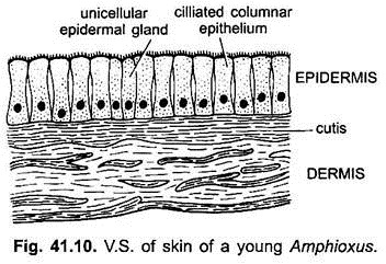
In Branchiostoma the skin is simple without keratin. The epidermis is single layered made of tall or columnar cells. These are ciliated in Balanoglossus. Epidermis has numerous unicellular gland cells which secrete a thin cuticle in Branchiostoma. Dermis (corium) is gelatinous in Amphioxus.
2. Cyclostomata:
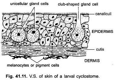
Epidermis is multilayered (stratified) but has no keratin. It has three types of unicellular gland cells- mucous glands secrete mucous, club cells probably scab-forming cells, and granular cells are of unknown function. Below the epidermis is the cutis formed of collagen and elastic fibres. Star-shaped pigment cells are also present in the cutis.
3. Pisces:
ADVERTISEMENTS:
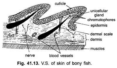
The epidermis has several layers of simple and thin cells, but there is no dead stratum corneum. The outermost cells are nucleated and living. The stratum Malpighii replenishes the outer layers of cells which have some keratin. Unicellular goblet or mucous gland cells are found in the epidermis, as in all aquatic animals.
The mucus makes the skin slimy reducing friction between body surface and water, protects the skin from bacteria and fungi, and assists in the control of osmosis. Multicellular epidermal glands like poison glands and light producing organs (photophores) may also be found. The epidermis rests on a delicate basement membrane.
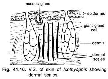
The dermis contains connective tissue, smooth muscles, blood vessels, nerves, lymph vessels, and collagen fibres. The connective tissue fibres are generally not arranged at right angles, but run parallel to the surface. Scales are embedded in the dermis and projected above the epidermal surface.
ADVERTISEMENTS:
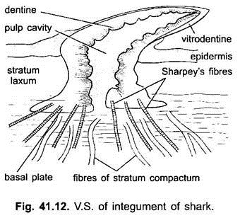
These are of five types. The elasmobranchs have placoid scales, Chondrostei and Holostei have ganoid scales, while most Teleostei have cycloid and ctenoid scales lodged in pouches of the dermis. Extinct Crossopterygii had cosmoid scales.
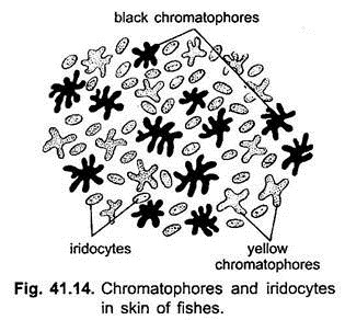
Many bony fishes show more brilliant colours than any other group of animals. The colours of fishes are due to chromatophores and iridocytes.
(a) Chromatophores in the dermis are derived from neural crest cells. They contain pigments which not only produce colours but also cause variations of colours. Chromatophores containing brown or black pigment are known as melanophores and those containing red, yellow, or orange pigment are collectively called lipophores.
(b) Iridocytes or guanophores are reflecting cells. They have no pigment but contain crystals of guanin. They lie in the dermis and cause irides-cence. Iridocytes reflect light from guanin crystals to produce white or silvery colours if the iridocytes are below the scales, if the iridocytes are above the scales they cause iridescent hues. By combinations of chromatophores and iridocytes various colours are produced, e.g., blue is produced by reflection from iridocytes, the blue combines with yellow pigment to produce green.
4. Amphibia:
ADVERTISEMENTS:
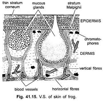
The epidermis is multilayered; the outermost layer is a stratum corneum made of flattened, highly keratinised cells. Such a dead layer appears first in amphibians, and is best formed in those which spend a considerable time on land. The stratum corneum is an adaptation to terrestrial life; it not only protects the body but prevents any excessive loss of moisture.
In ecdysis, the stratum corneum is cast off in fragments or as a whole in some. The dermis is relatively thin, it is made of two layers, an upper loose stratum spongiosum and a lower dense and compact stratum compactum. Connective tissue fibres run both vertically and horizontally.
There are two kinds of glands, they are multicellular mucous glands and poison glands in the dermis, but they are derivatives of the epidermis. The mucous glands produce mucus which not only forms a slimy protective covering but also helps in respiration.
The poison glands found in toads as parotid glands produce a mild but unpleasant poison which is protective, keep the enemies away. In the upper part of the dermis are chromatophores which have black melanophores and yellow lipophores, these produce the colour of the skin.
The ability of the skin for changing colour to blend with the environment is well developed. Skin of labyrinthodontia, the stem Amphibia had a armour of dermal seales. Bony dermal scales are found embedded in the skin of some Gymnophiona (Apoda) and a few tropical toads. These scales are absent in modern Amphibia.
The skin is sensitive to light in amphibians, especially in cave-dwelling forms. It is an important organ of respiration, and also enables the frog to respire under water for long periods, during hibernation or aestivation it is the only organ of respiration.
The skin is loose being attached to muscles only at certain places by connective tissue septa which mark the boundaries of subcutaneous lymph spaces.
5. Reptilia:
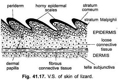
The integument (Fig. 41.17) is thick and dry. It prevents any loss of water. It has almost no glands, this is an adaptation to prevent evaporation of water.
The epidermis has a well-developed stratum corneum which makes the skin dry and prevents any loss of body moisture, thus, well adapted to a terrestrial life. The epidermis produces horny scales. Scales are shed periodically in fragments or cast in a single slough, as in snakes and some lizards.
The scales often form spines or crests. Below the epidermal scales are dermal bony plates or osteoderms in tortoises, crocodiles, and some lizards (Heloderma). These are retained for life and are not shed off. These may form dermal bones in the skull lying superficially or they may be found in the dermis.
The dermis is thick and has an upper loose connective tissue layer and a lower layer tella subjunctiva and separating the two is a horizontal layer of fibrous connective tissue. The upper layer has an abundance of chromatophores in snakes and lizards like fishes and amphibians. Leather of high commercial value is made from the skin of lizards, snakes, and crocodiles.
Many lizards and snakes have elaborate colour patterns, for concealment or as warning colours. There is marked colour change in certain lizards, such as Chameleon, the colour may change with the environment for concealment, or it may change in courtship or threat. In Calotes, the chromatophores have no nerve fibres, they are controlled by hormones of the posterior lobe of the pituitary. In Chameleon, chromatophores are controlled by the autonomic nervous system.
Reptiles essentially lack skin glands. Many lizards have glands near the cloaca. Their openings called femoral or pre-anal pores are generally smaller in female and found only in the male in some species. They are most active in the breeding season. Musk glands in the throat and cloacal opening of crocodilians function during courtship. Generation glands found recently are associated with periodic shedding of the skin.
6. Aves:
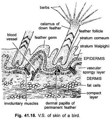
The integument (Fig. 41.18) is thin, loose, dry and devoid of glands except a uropygial gland at the base of the tail whose secreted oil is used for preening the feathers, especially in aquatic birds. The stratified epidermis is delicate, except on shanks and feet where it is thick and forms epidermal scales. The claws, spurs and horny sheaths of beaks are also the modifications of stratum corneum of epidermis.
Claws and beaks are variously modified in birds according to habitat. The rest of the body has a protective covering of epidermal feathers which are evolved from epidermal scales. Feathers protect and insulate the body, i.e, keep the body warm. The dermis is thin and has interlacing connective tissue fibres, abundant muscle fibres for moving feathers, blood vessels and nerves. The dermis forms an upper vascular and spongy layer and a lower compact layer.
The dermis also contains fat cells. The skin has no chromatophores. Pigment found in melanocytes migrates into feathers and scales. Colour patterns of birds are vivid; they are for concealment, recognition, and sexual stimulation. The colours are mainly produced by reflection and refraction of light from surface of feathers.
7. Mammalia:
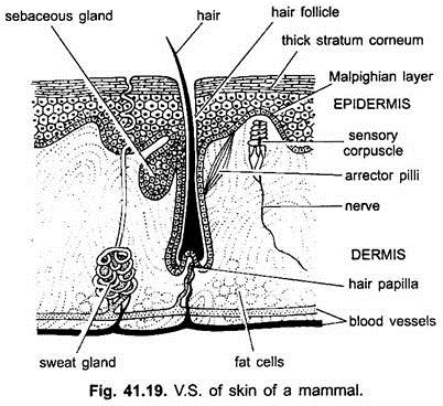
The skin (Fig. 41.19) is elastic and waterproof and is much thicker than in other vertebrates, especially the dermis is very thick and tough and is used for making leather. The epidermis is thickest in mammals and is differentiated into five layers- stratum corneum, stratum lucidum, stratum granulosum, stratum spinosum and stratum germinativum or Malpighian layer.
The outer layer of stratum corneum containing keratin, its cells lose their nuclei, but the cells are not dead as believed before. They secrete several hormones, one of which represses the mitotic activities of the Malpighian layer. In places of friction, such as soles and palms, the stratum corneum is very thick.
Stratum corneum is variously modified in various mammals to form epidermal scales, bristles, hairs, claws, nails, hoofs and horns etc., Below the stratum corneum is a refractive stratum lucidum in certain regions only. The stratum lucidum is now known as a barrier layer because the electron microscope has shown that its cells become compact and closely united to form a region which prevents passage of substances into or out of the body.
Stratum lucidum contains a chemical known as eleidin. Keratohyalin and eleidin are intermediate products in the formation of keratin. Below this is a stratum granulosum which is having darkly-staining granules of keratohyalin. Below the stratum granulosum is a stratum spinosum whose cells are held together by spiny intercellular bridges, each bridge has two arms in close contact, one arm arising from each cell.
Lastly there is a stratum germinativum or Malpighian layer which rests on a thin basement membrane. The Malpighian layer forms new cells continuously which move towards the surface and become flat and keratinised till the stratum corneum has flat, cornified cells made only of keratin.
This layer is sloughed off continuously and replaced by new cells. There are no mucous glands in the epidermis of mammals. The keratin from the epidermis at ends of digits forms claws, nails or hoofs.
The dermis is best developed in mammals. The upper part of the dermis in contact with the epidermis is the papillary layer which is made of elastic and collagen fibres with capillaries in between. It is thrown into folds to form rows of dermal papillae, especially in areas of friction. The greater lower part of the dermis is a reticular layer having elastic and collagen fibres.
In both layers there are blood vessels, nerves, smooth muscles, certain glands, tactile corpuscles, and connective tissue fibres extending in all directions. Below the dermis the subcutaneous tissue has a layer of fat cells forming adipose tissue which helps to maintain body heat. In making leather only the dermis is used. Dermal scales are not found in mammals except armadillos.
In the lowest layer of the epidermis are pigment granules but there are no pigment-bearing chromatophores in mammals. In man some branching dendritic cells or melanoblasts lie between the epidermis and dermis, they contain pigment.
The epidermis forms hairs, sudorific glands, sebaceous glands and mammary glands. Hairs form an epidermal covering. Shafts of hair project above the skin and their roots are embedded in hair follicles, into each of which opens a branching sebaceous gland. Hairs form an insulating layer which prevents a loss of body heat, thus, hairs keep up the body temperature. Sebaceous glands are outpushings of the wall of hair follicle and produce an oily substance which keeps the hair supple and prevents its wetting in water.
It also lubricates the skin. In the dermis are present coiled sudorific or sweat glands, which occur all over except lips and glans penis. Mammary glands are modified sebaceous glands, but in monotremes they are modified sudorific glands. They are functional only in females for producing milk for the young. Mucous glands are not found in mammals.
