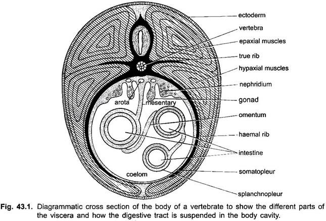
In vertebrates the dorsal part of the mesoderm is epimere which is segmented into somites, the middle part is mesomere which forms the urinogenital organs, and the lower part is the hypomere or lateral plate mesoderm which is never segmented and its cavity is the coelom or splanchnocoel.
The outer wall of the lateral plate is the somatic or parietal layer, which becomes the lining of the body wall and is known as parietal peritoneum. The inner wall of the lateral plate is the splanchnic or visceral layer, which becomes the covering of the alimentary canal and other viscera, and is called the visceral peritoneum.
The kidneys lie outside the coelomic cavity and so only ventrally covered by the peritoneum. The coelom is the space enclosed within the parietal and visceral peritonium.
Mesentery:
The parietal and visceral peritoneum is in contact with each other above and below the intestine forming a double-layered mesentery. The dorsal mesentery which is above the intestine persists, but the ventral mesentery disappears leaving only a few ligaments. In some regions the alimentary canal is joined to an organ by a remnant of the ventral mesentery, which is then known as an omentum or ligament. The coelom is divided longitudinally by the dorsal mesentery.
ADVERTISEMENTS:
a. Pericardial Cavity or Coelom:
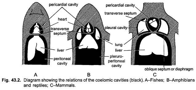
Behind the heart a partition, called transverse septum is formed, which divides the coelom into a small anterior pericardial cavity containing the heart. The peritoneum lining the pericardial cavity is a pericardial membrane inside which is a clear pericardial fluid. The peritoneal cavity is lined with parietal and visceral peritoneum which secretes a coelomic fluid.
b. Abdominal or Peritoneal Cavity:
ADVERTISEMENTS:
The large posterior cavity which contains the rest of the viscera is called the abdominal or peritoneal cavity.
Position of Pericardial Cavity in Various Vertebrates:
In fishes and urodeles the pericardial cavity is anterior to the peritoneal cavity, the transverse septum lying straight transversely. In anurans and amniotes the pericardial cavity comes to lie ventral to the anterior part of the peritoneal cavity, because the transverse septum becomes oblique.
In that part of the peritoneal cavity which lies dorsal to the pericardial cavity lungs are present in tetrapoda, consequently the peritoneal cavity containing both lungs and the viscera is now called a pleuroperitoneal cavity. This condition is found in anurans and most reptiles.
Separation of Thoracic and Abdominal Coelomic Cavities:
In crocodilians, birds and mammals a new fold descends from the dorsal body wall and fuses with the transverse septum to form a new partition. This partition in birds is membranous with some muscles and is called an oblique septum, but in mammals it is a highly muscular dome-shaped, called the diaphragm.
ADVERTISEMENTS:
The oblique septum or diaphragm is formed immediately behind the lungs dividing the pleuroperitoneal cavity into thoracic and abdominal portions.
Pleural Cavities:
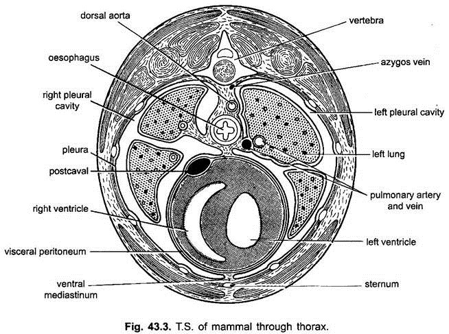
The thoracic part has two pleural cavities, each enclosing a lung. The peritoneum of pleural cavities is called pleura. The two pleural cavities are separated from each other by the pericardial cavity lying in the middle between the ventral portions of the pleural cavities.
The abdominal portion of the cavity lies posterior to the oblique septum or diaphragm and is called a peritoneal or abdominal cavity, which contains the rest of the viscera. Thus, crocodilians, birds, and mammals have four compartments of the coelom, a pericardial cavity, two pleural cavities, and a peritoneal cavity. This arrangement greatly increases the efficiency of lungs for respiration.
Mediastinal Space:
In mammals the two pleural cavities grow ventrally below the pericardium, separating it from the ventral body wall. They grow inwards so that their median walls meet below the pericardial cavity to form a mediastinal septum. The two parts of the mediastinal septum spread apart dorsally to form a mediastinal space which contains the pericardial cavity and the enclosed heart, major blood vessels, oesophagus, and trachea.
Greater Omentum:
In many mammals the mesentery supporting the stomach is drawn out into a double-walled bag, called greater omentum, which protects the abdominal viscera and has fat deposited in it.
ADVERTISEMENTS:
It is convenient to speak of the viscera as lying inside the coelom, but actually the visceral organs and alimentary canal are covered by visceral peritoneum and are outside the coelom (because coelom is the space between parietal and visceral peritoneum). They have pushed into the coelom carrying the peritoneum of the coelomic wall before them, thus, they appear inside the coelom.


