Lymph vessels or lymphatics arise as spaces in the mesenchyme of an embryo. They arise near the large veins but considerably later than and independently of the blood vessels. The lymph spaces unite and then branch to form anastomosing lymph vessels running throughout the body.
The bordering mesenchyme cells form the flat endothelial cells lining the lymph vessels. In certain places in some vertebrates connective tissue covers plexuses of lymph capillaries to lymph nodes. Connections are made between the lymphatics and certain veins.
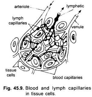
The lymphatic system differs from the blood vascular system in being an open system having lymph spaces between tissue cells, and it does not form a complete circuit. Lymphatic system includes the lymph capillaries, lymph vessels and lymph nodes. Since lymph flows only in one direction that is from the tissues towards the heart it has only capillaries and lymph vessels which are equivalent to veins. It has blind lymph capillaries lying between the tissue cells.
They are interwoven with but independent of blood capillaries. Lymph capillaries form an anastomosing network in all the body organs except in the nervous system. The diameter of lymph capillaries is larger and less uniform than that of blood capillaries.
ADVERTISEMENTS:
At their commencement the closed ends of lymph capillaries have small swollen knobs which help in collecting lymph from tissues. The lymph capillaries unite to form larger lymph vessels or lymphatics which finally discharge their contents into veins. Thus, lymphatics are efferent vessels draining from lymph capillaries into veins.
Structure:
Lymphatics are made of connective tissue fibres with some unstriped muscles and are lined with endothelial cells.
ADVERTISEMENTS:
The largest lymphatics have three-layered walls:
(i) A tunica externa,
(ii) A tunica media, and
(iii) A tunica interna just as in blood vessels.
ADVERTISEMENTS:
The lymphatics have valves arranged in pairs. The valves are more numerous than in veins, and prevent a backflow. Lymphatics in the intestine are called lacteals which absorb emulsified fats.
Lower Vertebrates:
Many lower vertebrates, except cyclostomes and cartilaginous fishes, have large lymph sinuses, the lymphatics communicate not only with lymph sinuses but also with the coelom by minute apertures or stomata. In lower vertebrates there are muscular lymph hearts which pump the lymph into veins. In the path of some lymphatics of higher vertebrates (mammals) are some lymph nodes (wrongly called lymph glands).
They are masses of lymph capillaries enclosed in connective tissue and contain lymphocytes and macrophages. Lymph nodes produce lymphocytes whose function is not clear, probably they transport DNA to those body cells which are unable to form this substance. The macrophages are phagocytic destroying foreign bodies and bacteria. Tonsils, thymus, spleen and Peyer’s patches are also lymph nodes.
Lymph:
Filtration pressure causes the plasma and leucocytes of blood to escape from the blood capillaries, into tissues. Most of this fluid is absorbed by blood capillaries, but some of it passes by diffusion into lymph capillaries and is then known as lymph. Lymph is a colourless fluid; it is a part of the blood which has filtered out from blood capillaries.
Actually it is blood minus its red blood corpuscles and plasma proteins. There is less calcium and phosphorus in lymph than in blood. Lymph has the power to coagulate slowly. Lymph fills body spaces and bathes the tissue cells where it is known as tissue fluid. It picks up colloids, broken tissue cells and bacteria. It is a medium of exchange between the blood and tissue cells.
It carries food and oxygen to the cells and takes away water and waste substances from the cells. The lymph reenters the blood through the lymphatics opening into veins, but some lymph enters the venous capillaries by osmosis.
Pisces:
ADVERTISEMENTS:
Lymph vessels are well developed in fishes. They are below the skin, in the muscles and viscera. They ran along the larger veins and extend into the head, tail and fins. The lymph vessels open into veins in the anterior, middle and posterior parts of the body. Lymph hearts are present only in some fishes but lymph nodes are lacking.
Amphibia (Frog):
In the tissues are lymph spaces from which arise lymph capillaries. The lymph capillaries unite to form lymph vessels, some of which dilate to form large lymph sinuses. Between the skin and muscles are spacious subcutaneous lymph sinuses separated from each other by fibrous membranes. Around the dorsal aorta is a subvertebral sinus which also encloses the kidneys.
From this diffuse lymphatic system, lymph is pumped into veins by two pairs of lymph hearts:
(i) An anterior pair just behind the transverse processes of the third vertebra, opening into the subscapular veins and
(ii) A posterior pair of lymph hearts on either side at the end of the urostyle, which can be seen if the skin is removed. They open into the femoral veins.
Reptilia:
The lymphatic system has lymph spaces in tissues from which lymph capillaries arise and form numerous lymphatics. The main lymphatic, called sub-vertebral trunk divides in front into two branches, each of which enters a precaval vein. There is only one pair of lymph hearts opening into the iliac veins posteriorly.
Aves:
There is an extensive distribution of lymph vessels, which finally form two thoracic ducts (homologous with subvertebral trunk) opening into precaval veins. In the embryo, there is a pair of lymph hearts in the sacral region. They are almost always lost so that the adult has no lymph hearts.
Mammalia:
The lymphatic system is highly developed having an extensive network of lymph capillaries and lymph vessels in the entire body. The larger lymphatics are interrupted at different places by lymph nodes.
Lymph nodes occur in the head, neck, arm-pits, groins, close to large blood vessels, and they also form the tonsils and Peyer’s patches on the intestine. Lymph nodes contain anastomosing lymph capillaries, lymphocytes, reticular cells, and macrophages, which are phagocytic engulfing bacteria and foreign bodies. Thus, they are responsible for bodily protection.
The lymph capillaries coming from intestinal villi are called lacteals, which carry emulsified fats so that their lymph becomes milky-white in appearance and is called chyle.
The lymphatics coming from the left side of the head, neck, arm, and thoracic region open into a thoracic duct lying below the vertebral column. It runs in front and opens into the left external jugular vein near its junction with the subclavian vein. The lower end of the thoracic duct, below the diaphragm, is expanded to form a receptaculum or cisterna chyli.
The lymphatics coming from the legs and lower trunk region, and the lacteals from the intestine pour their lymph into the cisterna chyli. In some mammals, but not in all, there is a right lymphatic duct which receives the lymphatics from the right side of the head and neck, right arm, and right side of the thorax. It opens into the right external jugular vein, where it joins the right subclavian vein.
There are no lymph hearts in mammals but lymph is propelled slowly through the lymph vessels and nodes by the movements of the body muscles and by the osmotic pressure in the lymph capillaries.
Spleen:
A spleen is a dark red organ found in all vertebrates except cyclostomes and Dipnoi. The spleen is haemolymphatic in character and it is interposed in the blood stream and blood, not lymph, filters through it. In an embryo the spleen is formed from mesenchyme cells as a thickening in the dorsal mesentery suspending the stomach. Blood vessels arise in it and bring about a differentiation of mesenchyme cells to form splenic cells and pulp.
Structure:
The spleen is covered by an outer capsule from which trabeculae pass into the substance of the spleen, dividing it into compartments or lobules. The capsule and trabeculae are made of dense connective tissue containing elastic fibres and unstriped muscles, they also contain blood vessels, lymph vessels and nerves.
Only blood vessels pass beyond the trabeculae to enter the parenchyma of the spleen where they form capillary network of arteries and veins, and many sinusoids connecting the arteries and veins directly. The splenic lobules have splenic cells of different sizes which may be pigmented.
They also have red and white pulp. The red pulp is made of diffuse lymphatic tissue of reticular cells and fibres, macrophages, lymphocytes and blood corpuscles. The white pulp consists of compact lymphatic tissue around small arteries. In section, it appears as isolated circular masses, called splenic nodules or corpuscles. The spleen is regarded as a haemolymph organ.
Functions of Spleen:
1. The spleen functions as a haemopoietic organ. In an ordinary embryo it forms both erythrocytes and leucocytes. It continues to do so in the adult, except in mammals where it forms only non-granular leucocytes.
2. The red pulp acts as an efficient blood filter. The old erythrocytes coming into the spleen are destroyed by phagocytic macrophages. Their iron is sent to the bone marrow for being used again, and the hemoglobin goes to the liver to form bile pigment.
3. The spleen acts as a storehouse for blood, one-sixth of the total blood can be stored in the spleen. Large volumes of erythrocytes are stored in the splenic sinusoids. Contractions of the spleen force out the red blood corpuscles into the blood stream when there is a demand for them.
4. The spleen produces antibodies from the cells received from the thymus gland. These cells serve as a defence mechanism against bacteria and some diseases. Though the spleen is extremely important, it is not essential for life. If it is removed then its functions are taken over by other haemopoietic tissues.
