
The nervous system is formed of nerve cells, neurons, which are surrounded by neuroglia, a tender network of connective tissue. Neurons are the distinct units of nervous tissue and are of ectodermal origin.
Structure of Neuron:
A neuron has a cell body or cyton from which arise two types of fibres.
(i) Cyton:
ADVERTISEMENTS:
A cyton has a nucleus and numerous basophilic Nissl’s granules. These granules are made of ribonucleic acid. They produce new cytoplasm in neurons and are also concerned with protein synthesis in nerve cells. The cytoplasm of cyton has fine threads or neurofibrils, forming a network. Mass of nerve cell bodies found in the brain or spinal cord is called nucleus and a similar mass found outside the central nervous system is known as a ganglion.
(ii) Nerve Fibres:
A neuron has a number of processes or fibres projecting from the cell body.
These fibres on the basis of movement of nervous impulse are sense organ of two types:
ADVERTISEMENTS:
(a) Dendrites:
These (receptor) are generally several short branching processes having Nissl’s granule, and close to the cell body. They are afferent because the neuron receives impulses through them.
(b) Axon:
Axon or axis cylinder is a single process which is generally long and without Nissl granules. It has terminal branches, called axon endings, which have small swellings at their ends. Axons are efferent because impulses are sent out through them.

Dendrites lie in contact either with receptors or with the axon endings of another neuron, from either of which they receive stimuli. Neurofibrils found in the cytoplasm of cyton extend both into the dendrites and axons and their branches. They probably transmit impulses.
An axon is covered with one or two sheaths and is then called a nerve fibre. A nerve fibre has a central thin cytoplasmic strand, the axis cylinder. It is continuous with the cell body or cyton. The nerve fibres outside the brain and spinal cord are covered by a thin continuous sheath, known as a neurilemma which is a tough membrane of protein and is made of flat sheath cells or Schwann cells.
Each sheath cell has a thin layer of cytoplasm with a flat nucleus. In nerve fibres there is a second sheath, called a myelin sheath or medullary sheath between the axis cylinder and its neurilemma. Myelin is a shining white substance made of the bodies of sheath cells wrapped around the axon many times. It has alternate layers of proteins and fats.
The myelin sheath, unlike neurilemma, is interrupted at intervals by circular constrictions, called nodes of Ranvier. The part of a nerve fibre between two nodes of Ranvier is called an internode, a single Schwann cell covers an internode.
Nerve fibres having a thick layer of a myelin sheath are called myelinated or medullated fibres and they appear white in colour. While the nerve fibres having a thin layer of a medullary sheath are non-medullated fibres and they appear gray in colour. Medullated fibres conduct impulses much faster than non-medullated ones.
Collateral branches may be given off by the axon at right angles. Nerve fibres lying in bundles are known as fibre tracts when present in the central nervous system, but when the bundles of nerve fibres are found outside the brain or spinal cord, then they are called nerves.
Synapse:
Nerve impulses are relayed from the axon endings of one neuron to the dendrites of another, they are arranged in such a way that the axon endings of a neuron rest on the dendrites or even on the cyton of the next neuron. This linking of neurons is called a synapse or synapsis.
The electron microscope has shown that each axon terminates in a tiny swelling, called a terminal button, which is full of mitochondria. They are very close to each other but have no organic connection with the terminal branches of another neuron.
ADVERTISEMENTS:
This very small gap between these two opposite neurons or terminal branches of axon of one neuron and dendrites of another neuron is called the synapse. Thus, the whole nervous system forms a chain of neurons with a synapse.
Neuroglia:
Besides the neurons in the brain and spinal cord, other cells of ectodermal origin are called neuroglia. They are also present as packing cells between the neurons. They have branching processes and they take the place of connective tissue, but true connective tissue is not present. Lining the cavities of the brain and spinal cord are some ependymal cells, which are like epithelial cells and are often ciliated.
In the brain and spinal cord the nerve cell bodies, their dendrites, neuroglia cells and the proximal unmyelinated portions of axons form a gray matter, which appears gray in colour, while the bundles of myelinated nerve fibres form the white matter.

Divisions of Nervous System:
The vertebrate nervous system has three divisions:
(i) A central nervous system comprising the brain and spinal cord. Its function is to receive the stimulus from the receptors and transmit its response to the effectors. Thus, it coordinates all the functions of the body.
(ii) A peripheral nervous system consisting of cranial and spinal nerves arising from the brain and spinal cord respectively. It forms a connecting link between the receptors, central nervous system (CNS) and effectors.
(iii) An autonomic nervous system made of two ganglionated sympathetic nerves, ganglia in the head and viscera, and their connecting nerves. The autonomic nervous system is often regarded as a part of the peripheral nervous system because the two are connected. But all the three divisions of the nervous system are connected intimately both structurally and functionally.
Nerve:
A nerve is formed of a number of fibres (axons or dendrites) outside the central nervous system. These are bound together in a smaller bundle by white connective sheaths, called perineurium. A number of such smaller bundles of nerve fibres are externally surrounded by another coat of fibrous connective tissue, called epineurium, which has blood vessels.
Functional Divisions of the Nervous System:
The body functions of the nervous system are somatic or visceral. Somatic functions are those which are carried out by the voluntary muscles, skeleton, and the skin and its derivatives. Visceral functions are those which are performed by the digestive, circulatory, excretory, endocrine, and the urinogenital system. Both somatic and visceral functions are divided into two components each- (i) afferent or sensory, and (ii) efferent or motor.
Thus, the nervous system has four functional divisions:
1. Somatic Sensory:
The somatic sensory fibres carry impulses from the skin, sense organs, voluntary muscles, and joints to the central nervous system.
2. Somatic Motor:
The somatic motor dorsal root fibres carry impulses from the central nervous system to the voluntary muscles.
3. Visceral Sensory:
The visceral sensory component carries sensations from the viscera and organs of taste and smell to the central nervous system.
4. Visceral Motor:
The visceral motor component carries impulses from the central nervous system to the involuntary muscles, branchial muscles, and glands.
Association or Adjustor Neurons:
These are located in the brain and spinal cord. They link the afferent and efferent fibres through synapses.
The dorsal half of the central nervous system is sensory and the ventral half is motor, this is especially marked in the medulla oblongata and the spinal cord where the four functional components are arranged from the dorsal to the ventral side in the order, somatic sensory, visceral sensory, visceral motor, and somatic motor.
Typically each peripheral nerve is also made of all four, functional components. This condition is typical of amniotes, but in an amniotes the visceral motor component has its fibres passing through both dorsal and ventral roots (Fig. 46.3).

Kinds of Neurons:
On the basis of number of processes arising from a neuron, these are of 3 types:
(i) Unipolar:
Only single fibre or two fibres arising very closely from the cyton.
(ii) Bipolar:
Two fibres, one axon and one dendrite, arising from the opposite sides of the cyton.
(iii) Multipolar:
A number of processes, i.e., one axon and a number of dendrites arise from the cyton.
Neurons are the longest cells in the body. Axons of the cytons may reach every part of the body, near or distant.
Nerve Impulse:
Impulses do not travel across synapses but a fresh impulse is set up on the other side of a synapse. When an impulse reaches the terminal buttons, they give off a small amount of acetylcholine, while terminal buttons of postganglionic fibres of the autonomic nervous system give off sympathin.
On being released the acetylcholine or sympathin contact the dendrites of the next neuron in which they cause a fresh impulse. After causing an impulse the acetylcholine or sympathin are immediately destroyed by enzymes. The impulses travel in only one direction in neurons, from the dendrites to the cell body and then through the axon.
Development of the Central Nervous System:
In a vertebrate embryo the mid-dorsal strip of ectoderm forms a longitudinal thickening, known as a medullary or neural plate. The neural plate sinks downwards but its edges grow upwards to form neural folds. The neural folds grow towards each other and eventually fuse to form a hollow neural tube of several layers of cells above the notochord.
The lateral ectoderm re-forms above the neural tube. The cells of the neural tube produce two kinds of cells, the neuroblasts and spongioblasts. The neuroblasts develop into neurons, while the spongioblasts form the neuroglia and ependymal cells lining the neural canal.
The neural tube at first is open at both ends, having a neuropore in front communicating with the exterior, posteriorly the neural tube communicates with the archenteron cavity by a very short neurenteric canal, but both openings are soon closed.
The anterior end of the neural tube enlarges forming the brain, while the remaining neural tube will give rise to the spinal cord. The cavity of the neural tube will become the ventricles of the brain and central canal of the spinal cord.
Development of Brain:
The anterior thickened, enlarged end of the neural tube known as encephalon is the embryonic brain. It undergoes differential growth and acquires two constrictions, which divide it into a series of three lobes, the primary cerebral vesicles. The primary cerebral vesicles are a forebrain or prosencephalon, a midbrain or mesencephalon, and a hindbrain or rhombencephalon.
This embryonic brain is bent downwards between the forebrain and midbrain in the region of the mesencephalon and this curvature is known as cranial flexure or cephalic flexure. In later development the cranial flexure is lost in cyclostomes, fishes, amphibians and reptiles, but persists in birds and mammals.

1. Forebrain:
The forebrain or prosencephalon is further sub-divided by a constriction into an anterior telencephalon and a posterior diencephalon. The anterior wall of the telencephalon is known as lamina terminalis which marks the original anterior boundary of the brain. The telencephalon grows beyond the lamina terminalis and forms paired cerebral hemispheres.
The roof of the cerebral hemispheres becomes the pallium and its ventro-lateral thicker walls form the corpora striata. Each cerebral hemisphere has a cavity, the lateral ventricle. From the antero-ventral part of the telencephalon paired olfactory lobes (rhinencephalon) grow out towards the nose. Each olfactory lobe has a cavity, the rhinocoel, which communicates with the lateral ventricle of its side.
The diencephalon or thalamencephalon has an upper epithalamus, a middle thalamus, and a lower hypothalamus. Dorsally the diencephalon gives out a median unpaired paraphysis, which is well developed in fishes but is much reduced in reptiles and birds and is absent in mammals.
Behind the paraphysis the diencephalon gives out two more unpaired evaginations, an anterior parietal body and a posterior pineal body or epiphysis. The parietal body forms an eye in lampreys, some fishes, and many reptiles, in others it is lost in the adult.
The pineal body forms a simplified eye in lampreys but in all other vertebrates it becomes glandular and is believed to be an endocrine gland, but there is no real evidence for this, most probably the pineal body is a vestigial organ of no functional significance. Between the thalami of diencephalon is a third ventricle which communicates with each lateral ventricle by an aperture called a foramen of Monro.
The thalami give rise to optic vesicles which form parts of the eyes. The roof of the diencephalon is a thin ependymal layer which fuses with the vascular piamater to form a tela choroidea. Vascular folds of the tela choroidea project into the third ventricle forming an anterior choroid plexus. The lower part of the diencephalon known as hypothalamus forms a ventral infundibulum.
An ectodermal evagination of the roof of the stomodaeum forms a Rathke’s pouch or hypophysis, which fuses with the infundibulum to form a pituitary body or hypophysis cerebri, which loses its nervous character and becomes an important endocrine gland. Except in a few lower forms the Rathke’s pouch loses its connection with the stomodaeum. The fibres of the optic nerves cross in front of the infundibulum to form an optic chiasma.
2. Midbrain:
The midbrain or mesencephalon has a thick roof. Its dorso-lateral walls form two optic lobes (four in mammals). The optic lobes are hollow, each having an optocoel except in mammals where they have no cavity and are solid. The ventral wall of mesencephalon forms thick crura cerebri, which are tracts of nerve fibres joining the diencephalon with the hindbrain.
Passing through the mesencephalon from the third to the fourth ventricle is a narrow iter or aqueduct of Sylvius, which in non-mammals is also connected to the optocoels.

3. Hindbrain:
The hindbrain or rhombencephalon forms a metencephalon from its anterior dorsal part. The metencephalon enlarges dorsally to give rise to a cerebellum. The surface of the cerebellum has a layer of gray matter, called cerebellar cortex. In birds and mammals there is thick white matter below the cerebellar cortex.
In lower vertebrates the cerebellum has an extension of the fourth ventricle known as cerebellar ventricle or metacoel. The remaining part of the rhombencephalon forms a myelencephalon, which becomes a thick medulla oblongata having a cavity, the fourth ventricle.
The roof of the medulla has a posterior choroid plexus formed in the same way as the anterior choroid plexus. In lower vertebrates the floor of the metencephalon and myelencephalon does not differ, but in higher forms the floor of the metencephalon thickness due to many tracts of nerve fibres, and in mammals it forms a conspicuous pons Varolii.
Commissures:
The two similar halves of the brain are connected by transverse tracts of nerve fibres, called commissures. In most vertebrates there are only anterior and posterior commissures, the anterior commissure originates in the lamina terminalis and lies in front of the third ventricle, it joins the two corpora striata.

The posterior commissure lies in the roof of the diencephalon at its junction with the midbrain. In mammals there are several other commissures. The commissures make bilateral integration possible.
Spinal Cord:
Nerve cord or spinal cord is formed from the neural tube behind the brain. The nerve cord is a cylindrical tube somewhat flattened dorso ventrally. Its anterior end is wide where it is continuous with medulla, the posterior end generally tapers to a fine thread, the filum terminale. In fishes it extends to the posterior end of the tail.
It extends the full length of the vertebral column in amphibians, reptiles and birds, but in mammals it is short and does not extend into the tail. In cyclostomes, fishes and limbless amphibians the nerve cord has a uniform diameter, but in tetrapoda it has two enlargements called cervical (brachial) and lumbar enlargements. They are formed due to a larger number of nerve cell bodies whose fibres form nerves going to limbs.
Histology:
The nerve cord has a thin central canal lined with ciliated ependymal cells. On its mid-ventral side the nerve cord acquires a deep ventral fissure, mid-dorsally is a slight dorsal sulcus from which a dorsal septum extends into the interior. The dorsal septum and ventral fissure divide the nerve cord incompletely into two halves connected across the middle by commissures.
There is an outer white matter with neuroglia and nerve fibres, and an inner gray matter having nerve cell bodies, non-medullated fibres, and neuroglia. In fishes and amphibians the gray matter is rounded or quadrangular in section, but in amniotes it is H-shaped forming dorsal and ventral horns or columns. The gray matter divides the white matter into dorsal, lateral, and ventral funiculi.
Meninges:
The brain and nerve cords are surrounded by protective connective tissue membranes called meninges. Their complexity increases with the advance in the scale of evolution.
In cyclostomes and fishes there is generally a single meninx, the meninx primitiva around the brain and spinal cord, it has a network of blood vessels. Between the skull or vertebral column and the meninx primitiva is a perimeningeal space filled with fat and connective tissue fibres.
In amphibians, reptiles and birds the meninx primitiva splits to form two meninges- an outer tough fibrous duramater and a thin vascular piamater covering the brain and nerve cord, between these two meninges is a subdural space having a small amount of fluid. Between the enclosing skeleton and the duramater is an epidural space having connective tissue, fat, and blood vessels.

In mammals there are three meninges, an outer duramater, a middle non-vascular arachnoid membrane, and an inner highly vascular piamater. Between the enclosing skeleton and the duramater is an epidural space in the region of the vertebral column only, but in the brain region the duramater fuses with the lining of the skull (endorachis) so that there is no epidural space.
Between the duramater and the arachnoid is a subdural space, between the arachnoid and piamater is a subarachnoid space having a network of fine fibres, filled with cerebrospinal fluid. The piamater and the arachnoid being joined by fibres are together known as leptomeninges.
The arachnoid forms arachnoid villi which return the cerebrospinal fluid to the venous blood. The cerebrospinal fluid is formed by the choroid plexuses, which fills the ventricles, central canal, subdural space, and subarachnoid space.
In the roof of the medulla are three apertures, a median formen of Magendie, and two lateral foramina of Luschka, through these foramina the cerebrospinal fluid passes into the spaces between the meninges so that the surface of the central nervous system is bathed by the fluid.
Neural Crest:
After the formation of the neural tube, masses of ectoderm cells appear on each side between the neural tube and ectoderm, these cells are neural crests which extend on each side along the whole length of the neural tube. The neural crests form segmental dorsal root ganglia of spinal nerves and ganglia of the autonomic sympathetic nerves.
They also form parts of the ganglia of sensory cranial nerves. Some neural crest cells form those mesenchyme cells which give rise to visceral arches. While other neural crest cells form the piamater and arachnoid membrane of the central nervous system. Still other neural crest cells form chromatophores in lower vertebrates. Chromaffin cells of the medulla of adrenal glands arise from neural crest cells.
Comparative Account of Brain in Vertebrates:
1. Cyclostomata:
The cerebral hemispheres are barely indicated. Two olfactory lobes are prominent. Cavities of cerebral hemispheres (lateral ventricles) are rudimentary. From the dorsal wall of diencephalon two median eye-like structures may arise.
The anterior one is the parietal body and the posterior one is the pineal body. These two are vestigial in Eptatretus and absent in Myxine. The ventral part of diencephalon gives rise to the infundibulum of the hypophysis as well as the posterior lobe. The optic lobes are not well differentiated.
Cerebellum is poorly developed due to sluggish and slow moving habit. Medulla oblongata is very well developed in all vertebrates since it serves as a centre for many vital body activities (respiration, heart action, metabolism, etc.). It also serves as a relay centre and in fishes is a centre for lateral line system and inner ear.
2. Elasmobranchii:
The brain is large and different from that of cyclostomes and bony fishes. In fishes, telencephalon is primarily olfactory in function and olfactory lobes are large in elasmobranchs. Each olfactory lobe has a rhinocoel cavity.
The cerebral hemispheres (cerebrum) are better developed. Cerebrum in fishes, however, consists of a basal ganglionic mass known as corpus striatum and a thin, dorsal, epithelial layer called the pallium composed essentially of non-nervous tissue and the centre of brain activity is back in the mesencephalon.
The diencephalon is narrow with a thin roof having an anterior choroid plexus below which is the third ventricle. Its lateral walls form thick thalami which serves primarily as a relay centre for olfactory and visual impulses and is associated with several sensory and glandular structures.
Dorsally the diencephalon has a long-tubular pineal body and the floor of the diencephalon give rise to the stalk or infundibulum of the hypophysis as well as the posterior lobe. A thin-walled vascular sensory organ, succus vasculosus is attached to hypophysis. It is connected with the cerebellum by fibre tracts. The midbrain in fishes is the centre of nervous coordination.
Dorsally it develops two prominences known as the optic lobes concerned with visual reception. Cerebellum is a centre of muscular coordination, it is larger than that of cyclostomes and dipnoans. Medulla is well developed and at its antero-lateral margins are present ruffle-like restiform bodies, which assist the cerebellum in the maintenance of equilibrium.


3. Osteichthyes (Bony Fish):
The brain is more specialised than in elasmobranchs. Olfactory lobes are large, having no olfactory peduncles and are closely applied in front of the cerebral hemispheres. Each contains a rhinocoel continuous with the lateral ventricle.
The cerebral hemispheres are better developed. The roof or pallium of the cerebral hemispheres is thin and non-nervous, consequently it is not equivalent to the pallium of other animals. The corpus striatum is very thick. The lateral ventricle is undivided. Diencecephalon is small hidden dorsally by the midbrain. Only the pineal body with stalk is present.
Parapineal body is not found in modern bony fishes. Melatonin has been identified in the pineal gland of certain fishes. It has an inhibitory effect on gonadal activity. In immature Pacific salmon (genus Oncorhynchus) melatonin content is 6 times more than that of adults. On the ventral side the diencephalon has an infundibulum to which the pituitary gland is attached below.
Optic lobes are very large. Cerebellum is larger than that of elasmobranchs. Restiform bodies are also present in some bony fishes. Vagal lobes are the bulgings from the antero-lateral sides of medulla, and are found in bottom feeders which have taste buds on their body surface.
4. Amphibia:
The nervous system of amphibians is still basically like that of fishes. The pallium of cerebral hemispheres is invaded for the first time in vertebrates by nerve cells due to which the cerebral hemispheres become enlarged in accordance with more complex activities of locomotion, hibernation, breeding, etc.
The olfactory lobes are fused together in the median plane. The corpora striata form a thick floor of the cerebrum. The diencephlaon is short but optic fibres end in it. The pineal body is found in all amphibians, but only anurans possess a parietal body or a pineal end organ.
The optic lobes are large and are the centre of brain activity like fishes. The members of this class are notably slow and sluggish in their movement, so the cerebellum is very small, like a simple narrow transverse band. Medulla is also small. Except in caecilians, flexures are lacking in amphibian brain.
5. Reptilia:
The brain shows many features which are characteristic of the mammalian brain. In all anamniotes the midbrain is the centre of brain activity, but in reptiles for the first time there is a shift in the nerve centre to the cerebrum.
This is correlated with a marked increase in the size of the cerebral hemisphere as a result of invasion of the pallium by many nerve cells so as to form the neopallium. Two long olfactory lobes are larger than that of amphibians due to greater thickness and enlargement of corpora striata. Parietal eye is found in Sphenodon and some modern lizards, but absent or vestigial in some other reptiles.
Behind the optic lobes are present a pair of auditory lobes which are not hollow. The third ventricle is reduced and forms a narrow cerebral aqueduct. Cerebellum is relatively larger than that of amphibians. However, it does not attain the size of sharks, and not near the size of birds or mammals. This is correlated with relatively limited locomotive powers of most reptiles.
6. Aves:
The brain of birds shows a considerable advance over that of reptiles. In shape it is short, broad, and rounded. The olfactory lobes are extremely small and degenerate due to poor sense of smell. The cerebral hemispheres are very large and cover the diencephalon and optic lobes.
Its size is due to enlargement of the corpus striatum rather than the cerebral cortex. The cerebral cortex is smooth. The size of corpora striata makes the lateral ventricles small. The diencephalon is covered dorsally by the cerebral hemispheres and cerebellum. It has a small dorsal pineal body, an anterior choroid plexus and a narrow third ventricle. The thalami are highly developed and are connected by fibres to the corpora striata and spinal cord.
The midbrain is highly developed with exceptionally large rounded optic lobes. This seems to be correlated with the remarkable powers of sight that birds possess.

The cerebellum is larger than in reptiles and is deeply fissured, although it is not as large as in mammals. Ventral to cerebellum, the avian brain shows the beginning of the development of a pons. In the cerebellum masses of nerve cell bodies form nuclei so that it has outer layers of gray matter and an inner mass of white matter, forming a branching pattern called arbor vitae.
A small projection of the fourth ventricle extends into the cerebellum. It controls equilibrium and movements in all places during flight. The medulla oblongata has a posterior dorsal choroid plexus and a fourth ventricle, which does not extend into the cerebellum.
7. Mammalia:
The brain reaches its highest development in mammals with better integration and mastery over the environment. The cerebral hemispheres reaching the status of a dominant integrating part of the brain and acting as coordinating centres of the brain.
The cerebral hemispheres in Prototheria are smaller and smooth like that of reptiles. In Metatheria, they are larger and smooth. In most eutherians, the cerebral hemispheres are immense, projecting forwards above the olfactory lobes and backwards above the diencephalon and midbrain and divided into lobes. They have convolutions on the surface so that there are ridges or gyri and depressions or sulci.
The outer layer or cortex of the cerebrum is composed of gray matter. The right and left cerebral hemispheres are connected with one another by a broad white commissure, called the corpus callosum. The olfactory lobes are small compared with those of lower vertebrates.
The diencephalon consists of a dorsal epithalamus, a lateral thalamus, and a ventral hypothalamus. A pineal gland is present on the roof of the diencephalon, but it shows no eye-like structure. Thalamus is an important relay centre.
Hypothalamus is very important in mammals and consists of four parts. These are the infundibulum forming the stalk and posterior lobe of the pituitary, the optic chiasma, where right and left optic nerves cross enroute to the brain; and the mammillary bodies, which integrate olfactory impulses.
The hypothalamus controls a great many mammalian functions including blood pressure, sleep, water content, fat and carbohydrate metabolism, body temperature and possibly rhythmic activities such as moulting, migration and pituitary secretion.
The midbrain in mammals is of less importance than in lower vertebrates, in which it is really the brain centre. It is divided into four prominences, called the corpora quadrigemina. The two superior lobes are concerned with sight, whereas the two inferior lobes are probably concerned with hearing. The III ventricle or iter of midbrain is laterally compressed vertical passage called the cerebral aqueduct.
The cerebellum, which is the centre of control of body movement, is most highly developed in mammals. Its surface is convoluted and it is divided into a number of lobes- central lobe or vermis, two lateral lobes, and two outer floccular lobes. Beneath the cerebellum is a typical mammalian structure the pons. This relay centre is a conspicuous feature of the ventral metencephalon. Pons Varolii have crossing or decussating fibres connecting opposite sides of the cerebellar cortex. These are the transverse nerve fibres.
Besides pons, there is a short tract of transverse fibres, called corpus trapezoideum relaying impulses for sound.
The medulla oblongata lies ventrally and is much thickened. It has a dorsal posterior choroid plexus below which is the fourth ventricle joined in front to the iter and behind to the central canal. The medulla has centres for regulation of digestion, respiration and circulation.
Peripheral Nervous System:
The peripheral nervous system consists of nerves connected to or arising from the central nervous system, it has cranial and spinal nerves. All the nerves arise in pairs. Cranial nerves originate from brain and emerge through foramina of the skull. Except the first four pairs of cranial nerves, the rest arise from the medulla oblongata.
There are ten pairs of cranial nerves in amanita (cyclostomes, fishes and amphibians), and twelve pairs in amniota (reptiles, birds and mammals). There is a paired nervus terminalis or number zero nerve arising from the cerebral hemisphere in all vertebrates except birds, it goes to the organ of Jacobson.
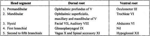
The cranial nerves do not show a clear metameric arrangement, yet they represent a regular series of segmental dorsal and ventral roots of the segments of the head, but the dorsal and ventral roots do not join. Table 46.2 summarises the cranial nerves of vertebrates showing their serial numbers, names, origin from brain, distribution, nature and functions.
The head segments and their dorsal and ventral roots are given below:

Spinal Nerves:
The spinal nerves arise from the spinal cord. They are also paired segmental structures emerging through intervertebral foramina of the vertebral column. Their number corresponds approximately to the number of vertebrae present.
Each spinal nerve is formed by two roots:
(i) A dorsal root (sensory) attached to the dorsal horn of the gray matter. The dorsal root has always a ganglion containing nerve cell bodies of sensory fibres.
(ii) A ventral root (motor) arising from the ventral horn of the gray matter. The nerve cell bodies of motor fibres are always located in the brain or spinal cord.
The roots have different embryonic origin- the dorsal roots arise from neural crests, while the ventral roots arise from the gray matter of the spinal cord. In an amniota, the dorsal roots contain somatic sensory, visceral sensory and visceral motor fibres.
In amniota the dorsal roots have only somatic sensory and visceral sensory fibres. The ventral roots have visceral motor and somatic motor fibres in all. Except in some cyclostomes, a dorsal and a ventral root of each side unite to form a spinal nerve. In cyclostomes, the sensory and motor roots do not join together to form a common trunk. In lampreys, they remain separate and emerge alternately from the spinal cord, whereas in hagfishes there may be an incomplete union.
In all other fishes the dorsal sensory root and ventral motor root unite with each other outside the vertebral column to form a common trunk. In amphibians dorsal and ventral roots of the spinal nerves unite in their passage through the intervertebral foramen rather than outside as in most fishes or within the neural canal as in amniotes (reptiles, birds and mammals).
In mammals, complicated plexuses, resulting from the intermixing of fibres from the ventral branches of spinal nerves are found. These are differentiated into cervical, brachial, lumbar and sacral plexuses.
Each spinal nerve divides into three branches or rami, a dorsal ramus supplying the skin and muscles of the back, a ventral ramus going to body muscles and skin of ventral side, and a communicating ramus communicants or visceral ramus going to the viscera and a ganglion of the autonomic nervous system.
A ramus communicans has two parts, a white ramus and a gray ramus. There are no gray rami in elasmobranchs. The white ramus has medullated visceral sensory and visceral motor fibres. The axons of visceral motor fibres go to autonomic ganglia and form synapses with non- medullated fibres of the gray ramus.
The medullated visceral motor fibres are called preganglionic fibres because they terminate in autonomic ganglia where they form synapses with non-medullated postganglionic fibres of the gray ramus.
These fibres enter the spinal nerves and pass into the dorsal ventral rami to supply structures under involuntary control. In brief the white ramus of ramus communicans carries medullated visceral sensory fibres and medullated preganglionic motor fibres, the gray ramus carries only non-medullated postganglionic autonomic motor fibres (Fig. 46.10).

Autonomic Nervous System:
The autonomic nervous system is so called because it is partly independent and not under voluntary control, though it is involuntarily controlled by the nerve centres located in the central nervous system. It is also connected to spinal nerves and some prevertebral ganglion ramus, cranial nerves.

Cranial and spinal somatic nerves mainly innervate the voluntary (skeletal) muscles of the animal. While the autonomic nerves and ganglia innervate the smooth or involuntary muscles of the viscera (alimentary canal, heart, etc.) and glands, and, thus, control the internal environment of the body. Little is known about the autonomic nervous system in cyclostomes although sympathetic ganglia are present. The digestive system and heart are supplied by the vagus nerve.
In elasmobranchs, an irregular series of sympathetic ganglia lies along the body wall. Fibres from these ganglia connect both to the spinal cord and to the smooth muscles of the digestive and circulatory systems. In bony fishes autonomic nervous system is more advanced i.e., the sympathetic ganglia are arranged in a chain extending forward to the trigeminal nerve.