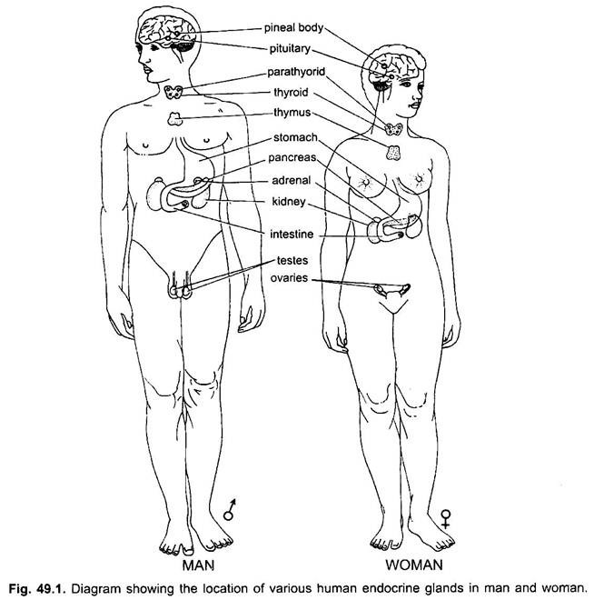
The following diagram (Fig 49.1) shows the approximate position of the important endocrine glands in the body of a man and woman.
The chief endocrine glands of vertebrates are:
1. Thyroid,
2. Parathyroid,
ADVERTISEMENTS:
3. Adrenal,
4. Pituitary,
5. Thymus,
6. Pineal,
ADVERTISEMENTS:
7. Pancreas,
8. Mucous membrane, and
9. Gonads.
1. Thyroid Gland:
Development:
ADVERTISEMENTS:
Thyroid gland arises in an embryo as a diverticulum of the floor of the pharynx which proliferates to form follicles and loses its connection with the pharynx. In lower vertebrates it arises mid-ventrally from between the second and fourth visceral-clefts, but in higher forms it develops between the first and second clefts.
Structure:
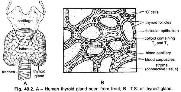
The thyroid has small follicles of cubical cells. The lumen of follicles is filled with an iodine-rich colloid. The follicles are enclosed in a capsule of connective tissue.
Fish-like vertebrates have well developed thyroid glands. It is unpaired in elasmobranchs but paired in adult amphibians, not only regulates metabolic activity in the body but is believed to be important in influencing the periodic moulting of the outer layer of the skin. In amniotes the two lobes of the thyroid are joined by a narrow isthmus of thyroid tissue.
The thyroid is homologous with endostyle of lower chordates. In the ammocoete larva of Petromyzon and in Amphioxus there is an endostyle as an open groove in the floor of the pharynx and its cells contain iodine, but in gnathostomes its major part atrophies and the remaining part becomes the thyroid gland of the adult.
Hormones and their Functions:
i. Thyroxin:
ADVERTISEMENTS:
The hormone of thyroid is a protein, called thyroglobulin which breaks down into a number of molecules of an amino acid known as thyroxin which contains 65% iodine. It increases the rate of metabolism for release of energy, also affects other endocrine glands and is affected by them. In young animals it stimulates growth and in lower vertebrates it is responsible for metamorphosis.
Tadpoles fed on thyroid extract quickly metamorphose into frogs without growing up. Deficiency of thyroxin in a young child causes a condition known as cretinism in which growth is retarded, intelligence is low, sex organs do not develop, legs are bent and the tongue and abdomen protrude out. If the thyroid degenerates in adult life, it results in a disease called myxoedema in which there is sluggishness, swelling of subcutaneous tissue mainly of the face and hands, body temperature is low, and the mind becomes dull.
Hypersecretion:
Excessive production of thyroxin causes exophthalmic goitre in which thyroid is enlarged, eyes protrude, metabolic rate is high, heart beat is rapid and the body is wasted away. Deficiency of iodine in thiyoxin causes simple goitre by a pathological enlargement of thyroid.
ii. Calcitonin:
Secretion of thyroid also contains a hormone, called calcitonin which keeps the bones healthy and prevents an undue loss of calcium from them.
2. Parathyroid Glands:
Development:
Parathyroid glands arise as ventral or dorsal outgrowths of the third and fourth visceral-clefts.
Position:
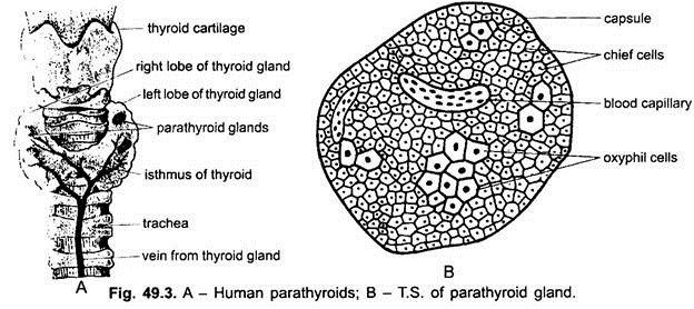
In cyclostomes, cartilaginous and bony fishes parathyroid glands have not been found as compact glands but are probably represented by diffuse cells behind the last pair of visceral clefts. In tetrapoda they form one or two pairs of compact glandular masses lying close to the thyroid glands or partially embedded in the thyroid or thymus glands. In reptiles the parathyroid glands are usually situated posterior to the thyroid gland, which is unpaired, at least in adult reptiles. In man they form four minute yellowish glands attached to the dorsal side of thyroid.
Hormones of Parathormone:
The hormone of parathyroids is a protein, called parathormone. Its chemical nature is not known but it is concerned with maintaining the normal amount of calcium and phosphorus in the blood. It plays an important part in bone formation, a function it shares with vitamin D. In birds it plays an active role in regulating the formation of the calcareous shell of eggs. Hyperactivity- Over-secretion of parathormone causes calcium and prosphorus to go out of bones and teeth, making them soft and weak.
Hypoactivity- Under-secretion lowers blood calcium retarding growth of bones in the young and causes excitability of the nervous system. Extirpation of parathyroids results in a condition, known as muscular tetany in which a spasmodic contraction of limb muscles occurs, calcium content of the blood is reduced and finally results in death.
3. Adrenal Glands:
Adrenal glands occur in fishes as two separate sets of tissues called suprarenal which are homologous with the mammalian adrenal medulla and inter-renal which are homologous with the cortex in mammals. The suprarenal bodies or chromaffin bodies are bright yellow, small, paired masses of cells along the sympathetic ganglia. The inter-renal bodies are pink-coloured paired or unpaired masses of cells between the kidneys. In amphibians, reptiles and birds the two components of the adrenals, the cortex and medulla are joined together rather being separate in fishes.
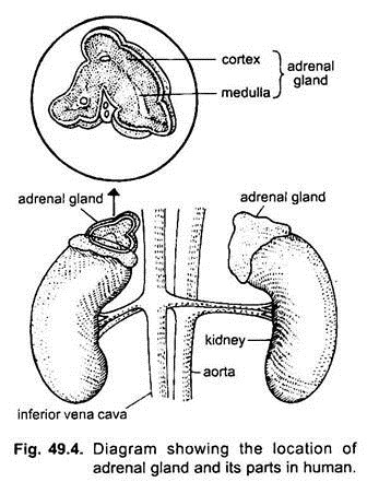
Adrenal glands lie close to the kidneys. In mammals the adrenal glands have two portions, an outer mesodermal adrenal cortex homologous with the inter-renal bodies, and an inner ectodermal adrenal medulla homologous with the suprarenal bodies or chromaffin bodies. The adrenal glands are attached in front of the kidneys.
Hormones of Cortex:
Secretions of the cortex or inter-renal bodies are essential for life. They are concerned with carbohydrate metabolism, balance of salts in the blood, balance of sodium and fluids in the body, maintenance of the volume of circulating blood, control of sexual maturity, and with a resistance to many diseases.
i. Cortin:
Several hormones secreted by the cortex are collectively called cortin which maintains a balance between the salts in the blood and those in the bones.
ii. Cortisone:
A steroid cortisone from the cortex of mammals is concerned with carbohydrate and protein metabolism. It is now used in treating arthritis and certain types of leukemia.
iii. Aldosterone:
The cortex also produces aldosterone, which controls sodium and potassium excretion.
Hyperactivity:
Over-secretion of the cortex causes marked increase of male characters in both sexes. Removal of adrenal cortex results in death.
Hormones of Medulla:
The hormone of the medulla or suprarenal bodies is epinephrine or adrenalin which is regularly present in the blood. It maintains the tone of involuntary muscles keeping the blood vessels contracted and blood pressure normal. It controls the conversion of glycogen from the liver and muscles into blood sugar.
The total effect of epinephrine is to provide the animal with a suitable respiratory rate and heart beat ensuring sufficient oxygen and blood sugar to meet the needs of the body in time of stress. Epinephrine is also secreted in response to emotional factors, such as anger, fear or pain, then it prepares the animal to fight or to retreat.
When extra epinephrine is injected into the blood it increases the rate of heart beat, enhances blood pressure, dilates the air passages to the lungs, increases the amount of blood sugar by breaking down glycogen, and dilates the pupils. These effects of epinephrine are the same as those produced by stimulation of the autonomic nervous system, both affecting the same organs. A substance called sympathin is similar to epinephrine and is produced by the ends of the sympathetic nerve fibres.
4. Pituitary Gland:
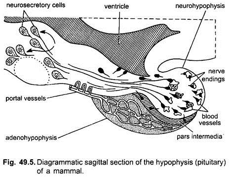
Pituitary gland lies below the diencephalon in a depression of the basisphenoid bone, called sella turcica.
Development:
The pituitary is a compound organ. It is formed in the embryo by an ectodermal evagination of the stomodaeum, called Rathke’s pouch which finally fuses with a ventral down growth of the infundibulum to form a pituitary gland or hypophysis cerebri. Except in a few lower forms the Rathke’s pouch loses its connection with the stomodaeum. Some authorities use the term hypophysis for Rathke’s pouch, while others use it for the entire pituitary gland, here the latter nomenclature is followed.
Structure:
The pituitary gland is well developed in fishes and other vertebrates. The pituitary gland or hypophysis is generally made of four parts- called pars anterior, pars tuberalis, pars intermedia and pars nervosa. The pars anterior and pars tuberalis are together known as the anterior lobe of pituitary, pars intermedia as intermediate lobe, and pars nervosa as the posterior lobe.
The anterior and intermediate lobes are formed from the Rathke’s pouch, while the posterior lobe comes from the infundibulum. The lobes of the pituitary are made of compact masses of cells with a hypophysial portal system, but the different lobes are unrelated functionally. Each lobe producing different hormones.
The hormones of the pituitary gland not only affect other organs of the body but they also influence other endocrine glands. The pituitary is also influenced by other endocrine glands so that the members of the endocrine system cooperate in an adaptive fashion for the general welfare of the animal.
Hormones of Anterior Lobe (Adenohypophysis):
The anterior lobe of the pituitary gland produces about ten hormones which are as follows:
i. Growth Hormone or Somatotropic Hormone:
The growth hormone causes normal growth of the bones and the body of the animal. It probably stimulates the thyroid. Over-secretion of the growth hormone during young stages before bones have stopped growing leads to gigantism in which the bones and body become excessively large. If over-secretion of the growth hormone occurs after the long bones stopped growing then acromegaly occurs in which the bones of the head, jaws, hands and feet become abnormally large. Long bones become thick, and the spine is curved.
ii. Diabetogenic Hormone:
A diabetogenic hormone maintains the balance between the blood sugar and liver glycogen. If this hormone is secreted in excess, then glucose will not be stored in the liver as glycogen but will remain in the blood to raise the sugar level which will cause diabetes. But the diabetogenic hormone is controlled by insulin of the pancreas.
iii. Adrenotropic Hormone:
An adrenotrophic hormone stimulates and exerts a nutritive influence on the adrenal cortex or inter-renal bodies so that the body responds to counteract the harmful effects of stresses, such as starvation, burns and haemorrhage. The hypothalamus secretes a potent substance which acts on the anterior lobe of the pituitary to produce its hormone.
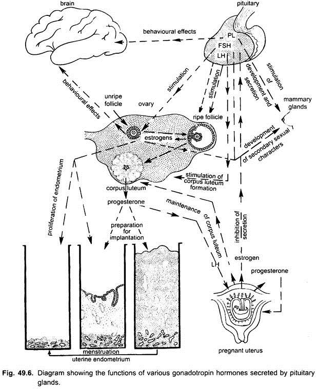
iv. Gonadotropic Hormone:
(i) A follicle-stimulating hormone controls the development of ovarian follicles of the females and maturation of sperms in the males.
(ii) A lactogenic hormone or prolactin initiates and maintains secretion of milk by the mammary glands of female mammals and by the crop in pigeons. It also influences the activity of the corpora lutea in the ovary. In conjunction with hormones of gonads it causes development of mammary glands in female mammals.
(iii) A luteinizing hormone is responsible for ovulation and formation of corpora lutea in female mammals. It stimulates the endocrine cells of the ovaries and testes to produce their secretions.
v. Thyrotropic Hormone:
A thyrotrophic hormone stimulates and exerts a nutritive influence on the thyroid gland for bringing about normal growth.
Hormones of Intermediate Lobe:
The intermediate lobe produces intermedin which controls the expansion of black pigment melanophores in elasmobranchs, amphibians, and many reptiles so that the skin becomes darker due to spreading of pigment granules. In birds and mammals the intermediate lobe is much reduced though intermedin is secreted but it has no effect on their pigments. The secretion of intermedin is due to a response to visual stimuli of the eyes which stimulate the intermediate lobe to secrete.
Hormones of Posterior Lobe (Neurohypophysis):
The posterior lobe of the pituitary produces pituitrin made of two hormones:
(i) Vasopressin or pitressin and
(ii) Oxytocin or pitocin.
But it has been shown recently that the hormones of the posterior lobe are actually produced by the hypothalamus, and the posterior lobe only stores these hormones and is not a true endocrine gland.
(i) Vasopressin regulates blood pressure and acts on urinary tubules to reduce the amount of water production in urine.
(ii) Oxytocin causes contraction of muscles of the uterus, to hasten child birth. Injury to the posterior lobe leads to excessive secretion of dilute urine and a consequent thirst. The posterior lobe increases in size in females during pregnancy.
5. Thymus Gland:
Thymus gland, like the parathyroids and the thyroid, is derived embryologically from the foregut. In mammals this gland migrates posteriorly during development close to the anterior part of the heart. Thymus gland is formed from one to five pairs of visceral-clefts in different vertebrates. In mammals it is formed largely from the third pair.
The second pair of visceral- clefts form tonsils which are found only in mammals as masses of lymphoid tissue. The fifth pair of visceral-clefts form ultimobranchials or postbranchial bodies in tetrapoda. Only the left ultimobranchial is present in lizards, and their occurrence in birds and mammals is doubtful. Ultimobranchials may be lymphoid tissue but their function is unknown.
Structure:
The thymus gland forms a pair of large glandular masses in front of the heart in young mammals. In fishes it forms separate diffuse patches along each gill-cleft. In amphibians it forms one pair of glands found behind the tympanic membrane. In reptiles it forms a single or double mass in the neck. In birds it forms a pair of elongated bodies in the neck. Thymus has many lobules of epithelial cells with a rich blood supply.
Functions:
It is claimed that the thymus hastens sexual maturity. Its removal from a young animal delays the development of the reproductive system. In tadpoles fed on thymus growth is accelerated and metamorphosis is delayed. This effect is the opposite of that obtained by feeding the thyroid extract.
The thymus is comparatively large in young animals but it becomes very small before reaching adult life. It is, thus, inferred that the thymus fulfills some function related to sexual development. In birds it controls the formation of a hard egg shell. The bursa Fabricii of birds has the same histological structure as the thymus, and it is significant that both become atrophied before sexual maturity.
The thymus probably has no endocrine function and it is concerned with conditioning the lymphatic system to form antibodies and reject graft tissues. Mice, rats and hamsters that are thymectomised within a week of birth exhibit a greatly reduced ability to synthesise antibodies and will successfully accept skin grafts from other individuals. In the young it produces the basic lymphocyte cells of lymph nodes and spleen, and once it has done this work it begins to atrophy. Its removal after a certain age causes no ill-effects.
6. Pineal Gland:
In mammals, pineal gland or epiphysis lies on the dorsal surface of diencephalon, but it lacks any photoreceptive elements such as are found in some of the lower vertebrates.
However, it is concerned with circadian rhythms in the synthesis of serotonin, melatonin and certain other chemicals which persist in continuous darkness and disappear with exposure to light. Melatonin is regarded as a hormone and has an inhibitory effect on the gonads. Serotonin is an amine from which melatonin is synthesised.
In cyclostomes, it forms an eye-like structure but in others it loses its nervous character and becomes a pigmented and highly vascular gland. It is larger in the young than in adults. Within the pineal gland of frogs and lizards is a small body whose structure is like rods and cones of the retina.
There appears to be some inter-relationship between the pineal gland and the reproductive organs. Removal of the pineal gland induces enlargement of the gonads, adrenals, and pituitary glands, whereas extracts of the pineal retard the growth of the gonads. Its abnormal development or its removal produces a precocious sexual maturity.
7. Pancreas:
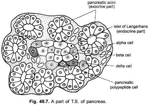
Pancreas is both an exocrine and an endocrine gland with two distinct kinds of tissues. The endocrine part has prism-shaped cells forming several isolated groups, called islets of Langerhans. Their cells develop from the midgut of the embryo which also forms the rest of the pancreas. In cyclostomes pancreas is not definitely represented. Pancreas is present in cartilaginous and bony fishes and possesses well marked islands of Langerhans.
In some bony fishes the islets of Langerhans are grouped together outside the pancreas but in others they lie within the pancreas as anastomosing cords alternating with sinusoids forming rounded masses with no ducts.
Hormones:
The islets of Langerhans have two types of cells known as alpha-cells and beta-cells. The beta-cells produce a hormone called insulin .In a normal animal insulin controls carbohydrate metabolism and stimulates the liver and striated muscles to store glycogen and make the tissues oxidise glucose as a source of energy.
Deficiency of insulin causes a disease called diabetes mellitus in which there is an excess of unused sugar in the blood which is excreted with urine, so that the sugar level becomes low in the blood. The level of glycogen in liver and muscles decreases, and there is also an increased formation of carbohydrates from body proteins and fats so that the body is wasted away. There is muscular weakness and finally coma sets in.
The alpha-cells of the islets of Langerhans produce another hormone, known as glucagon which is controlled by a hormone from the anterior lobe of the pituitary and acts as an antagonistic agent to insulin increasing the amount of sugar in the blood by breaking down glycogen of liver into glucose. A diabetogenic hormone of the pituitary maintains the balance between blood sugar and liver glycogen.
If the diabetogenic hormone is produced in excess, then sugar is not stored in the liver as glycogen but remains in the blood and causes diabetes. Thus, diabetes can be caused in two ways, either by under-production of insulin by the pancreas, or by over-production of diabetogenic hormone by the pituitary. But the diabetogenic hormone is controlled by insulin.
8. Mucous Membrane:
Mucous membrane of the alimentary canal contains digestive glands which produce digestive juices and also certain hormones which are as follows:
i. Gastrin:
The mucous membrane of the stomach is stimulated by gastric juice to produce a hormone gastrin which passes into the blood and then returns to the stomach causing the gastric glands to secrete gastric juice.
ii. Enterogastrone:
The wall of duodenum secretes enterogastrone which inhibits gastric secretion.
iii. Secretin:
The mucous membrane of the duodenum produces a hormone secretin which goes through the blood to the pancreas and causes secretion of pancreatic juice.
iv. Cholecystokinin:
The mucous membrane of the duodenum also produces a hormone cholecystokinin which makes the gall bladder to release its stored bile.
9. Gonads:
Gonads are both cytogenic (producing sex cell) and endocrine in function. The gonads of fishes, like those of other backboned animals are partly endocrine in function and are responsible for various secondary sexual characters that usually manifest themselves during the breeding season.
i. Testes are made of seminiferous tubules between which are found groups of interstitial cells or cells of Leydig which are endocrine in function and produce testosterone, the male hormone. Substances having properties of testosterone are called androgens. Some androgens are found in the male urine.
Testosterone is a liquid and it brings about normal development and functioning of accessory sex organs, such as intromittent organs, reproductive ducts, and associated glands. Androgens prevent the development of Mullerian ducts in the males. Testosterone is responsible for development of secondary sexual characters which distinguish the male from the female such as antlers of stags, brighter plumage of male birds, hair on the face, deeper voices, larger body size and strength, and particularly male behaviour in mating.
If a mammal is castrated before sexual maturity then its masculine characters and structures fail to develop, if it is castrated after attaining sexual maturity then its accessory sex organs are reduced. Testes mature under the influence of a hormone from the anterior lobe of the pituitary.
ii. Ovaries:
The hormones produced by the ovary are as follows:
a. Estradiol or Estrogens:
The follicle cells around developing ova are responsible for secreting a female sex hormone estradiol. Substances having properties of estradiol are called estrogens. Androgens and estrogens are present in both sexes but androgens predominate in the males and estrogen in the females.
Ovarian estrogens are partly responsible for development of female accessory sex organs, such as development of genital organs, i.e., uterus, fallopian tubes, associated glands, and development of mammary glands, fat deposition beneath the skin, changes in body contour, broadening of pelvis and beginning of menstrual cycle. Estrogens differentiate the Mullerian ducts of the embryos to become oviducts and uteri.
b. Progesterone:
The space vacated by an ovum after ovulation is filled with a yellow corpus luteum formed by the proliferation of cells of the membrane granulosa. Its formation is stimulated by a luteinising hormone of the anterior lobe of the pituitary. The corpus luteum is a true but a temporary endocrine gland eventually getting reduced and finally absorbed in the ovary.
Corpus luteum produces a hormone, the progesterone. Progesterone suspends ovulation during pregnancy, prepares the uterus for pregnancy and implantation of the embryo. It maintains pregnancy and completes the formation of milk in the mammary glands. If, however, no pregnancy occurs the corpus luteum degenerates and disappears.
iii. Placenta:
When the placenta is formed in early pregnancy, it also produces two hormones, estrogens and progesterone, which are also produced by the ovary. They maintain the pregnancy, and proper development of the embryo. Placenta and ovary also produce a relaxin hormone in some mammals (e.g. in rabbit). It relaxes the pelvis at the time of young ones birth for its easy passage through vagina.
