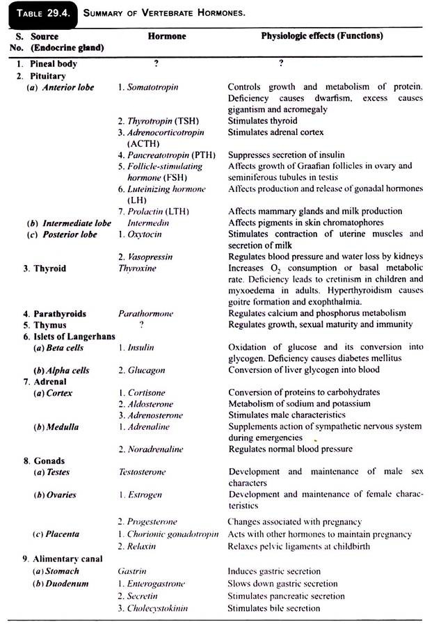
Generally two types of glands are found in the body of all the vertebrates-the exocrine and endocrine.
A. Exocrine Glands:
The exocrine glands are those which liberate their secretions through the ducts in an organ, such as salivary glands, pancreas, liver, etc.
B. Endocrine Glands:
ADVERTISEMENTS:
The endocrine glands are those which liberate their secretions directly into the blood and are without any duct. Therefore, these glands are also known as ductless glands. The secretions of these glands are known as hormones which assist the nervous system in coordinating or integrating various body activities. Their secretions are of vital importance for various metabolic processes and physiological activities to continue in the body.
Hormones:
The term hormone was given by E.H. Starling (1905), a British physiologist. The hormones are the chemical agents or messengers secreted by the ductless glands and are carried by the blood to the different parts of the body called target organ where they perform definite functions. Many endocrine glands cannot secrete their hormones unless they are stimulated to do so by other hormones secreted by other glands.
The amount of the various hormones released in blood is maintained at a proper level. Hormones are chemical messengers and bring about chemical coordination of different parts of the body. It is opposite to nervous coordination which is done by the nervous system. The action of endocrine system is slow and of long duration, whereas the action of nervous system is quite fast.
ADVERTISEMENTS:
The hormones produced by the endocrine glands are classified on the basis of their activities, such as:
(a) Metabolic hormones are those which stimulate the metabolic activities in the body, e.g., insulin stimulate carbohydrate and fat metabolism.
(b) Regulatory hormones are those which control and regulate the rate of secretion of other endocrine glands, e.g., the hormones of pituitary gland.
(c) Morphogenetic hormones are those which affect the rate of development of the parts of the body, e.g., the hormones of the thyroid gland.
ADVERTISEMENTS:
The position of endocrine glands in the body and their secretions are roughly the same in all vertebrates.
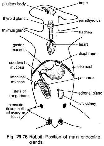
The various endocrine glands in rabbit are as follows:
1. Pituitary or hypophysis
2. Thyroid
3. Parathyroid
4. Adrenal
5. Islets of Langerhans of pancreas
6. Gonads
7. Placenta during pregnancy
ADVERTISEMENTS:
8. Gastro-intestinal tract
9. Thymus
10. Pineal body.
In addition to these, the thymus and pineal body were previously considered as endocrine, but now they are not considered as endocrine glands.
1. Pituitary Gland:
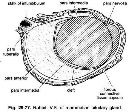
It is a small nut-like gland situated on the ventral side of the diencephalon of the brain, attached with the hyopthalamus by infundibulum. The hormones secreted by this gland regulate the functions of many other endocrine glands and also play a vital role in the chemical coordination of the body. Therefore, this gland is also known as master gland or bandmaster of endocrine orchestra.
Mammalian pituitary consists of three lobes of different embryological origin. The anterior and intermediate lobes arise from the roof of the mouth and together known as adenohypophysis. The posterior lobe develops from hypothlamus of diencephalon and is called neurohypophysis.
The adenohypophysis secretes many chemically distinct hormones which are named by adding the suffix “trophic” or “tropic” or ‘in’ to the name of the part of the body affected by them, such as growth hormone (GH) is called somatotrophic hormone (STH) or somatotropin.
(i) Somatotrophic Hormone (STH) or Growth Hormone (GH):
It controls the protein metabolism and growth of the skeleton. An excess secretion of this hormone results in gigantism (production of giants with abnormal heights). Its deficiency results in dwarfism. Sometimes this gland does not begin over-secretion till maturity, then instead of giants, gorilla-like man with huge head, hands, feet and protruding jaws are the result, which is known as acromegaly.
(ii) Thyroid Stimulating Hormone (TSH):
This hormone stimulates the thyroid gland to secrete thyroxine.
(iii) Adreno-Corticotropic Hormone (ACTH):
It controls and stimulates the adrenal cortex to secrete its hormone.
(iv) Gonadotropic Hormone (GTH):
Three types of gonadotropic hormones are known, e.g.. follicle stimulating hormone (FSH), luteinizing hormone (LH) and luterotropic hormone or prolactin (LTH). FSH hormone affects the development of gonads and formation of gametes. LH stimulates the production of sex hormones from the gonads. LTH stimulates the mammary glands to secrete the milk after the birth of the young ones.
(v) Melanocyte Stimulating Hormone (MSH) or Chromatotropic Hormone (CTH) or Intermedin:
This hormone stimulates the distribution and concentration of the melanin pigment in the chromatophores of the skin.
(vi) Diabetogenic Hormone or Pancreotropin Hormone:
This hormone affects the metabolism of carbohydrates. It converts reserved glycogen into glucose, i.e.. reverse to insulin.
The neurohypophysis or posterior lobe of pituitary secretes only two important hormones:
(a) Oxytocin:
It stimulates the contraction of some smooth muscles of the uterus at the time of childbirth and also stimulates the secretion of milk from the mammary glands.
(b) Vasopressin or Antidiuretic Hormone:
This hormone regulates the reabsorption of water by the renal tubules.
It also causes the construction of small arteries of the body which causes the general increase in the arterial blood pressure. Its deficiency causes a disease, called diabetes insipidus, which is an abnormality characterised by excretion of large amount of urine and, thus, the animal feels great thirst due to which it drinks more and more water.
2. Thyroid Gland:
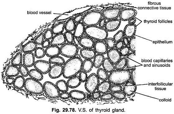
It is the largest of all the endocrine glands and bilobed, situated on the either side of the trachea below the larynx. Both the lobes of thyroid are connected together by a connective tissue, called isthmus. Histologically, it consists of a number of small thyroid vesicles or follicles in the form of closed sacs separated from one another by connective tissue with blood and lymph vessels.
Each follicle is lined by cuboidal epithelial cells. These cells are richly provided with active secretory agents which secrete yellow colloidal secretion into the cavities of follicles. This colloidal secretion is a globular protein, known as thyroglobulin, which is also known as iodothyronines or thyroid hormones. It consists of nearly four most important hormones of which thyroxine is one.
Thyroxine regulates metabolism, i.e., heat production and energy liberation in all the organ systems and also controls the rate of growth. The under-secretion of this hormone during childhood lowers the rate of metabolism and causes the animal to become feeble minded, growth is retarded, heart rate is also lowered and sexual development is retarded.
This condition is known as cretinism. The deficiency of thyroxine in adults stimulates the formation of new gland tissue resulting in an enlargement of the thyroid called goitre. The goitre is actually caused by the deficiency of iodine in the diet. Hypothyroidism causes myxoedema in which person becomes fatty and sluggish due to poor rate of oxidation.
The over-secretion of this hormone increases the rate of oxidation and is responsible for the quick consumption of food, therefore, fat is not stored. Over-secretion also results in the bulging of eyeballs and irregular heart rate, etc. It also causes swelling of the thyroid gland resulting into the goitre.
3. Parathyroid Glands:
These are two small, oval glands in rabbit, situated antero-laterally on the lobes of thyroid. In other mammals these are four in number. These glands are either found embedded in the thyroid or located close to them. Parathyroid secretes two hormones, parathormone and calcitonin.
These are responsible for regulating the amount of calcium and phosphorus in the blood. Parathyroid glands are necessary for the growth of bones, muscle tone and nervous activity. Over-secretion of these hormones results in tumors on the gland and causes calcium to be withdrawn from the bones, thus, producing a high calcium level in the blood.
In such conditions the bones may become soft, porous and weak. Its deficiency lowers calcium level in the blood due to which normal functions of muscles, bone growth and teeth formation are retarded. It has been seen experimentally that the removal of parathyroid results in muscular tremors, cramps and finally convulsious. This is called tetany which results in the death of the individual.
4. Adrenal Glands:
They are a pair of small ovoid white or yellowish glands situated just above each kidney. These are also known as suprarenal glands. This gland consists of outer cortex derived from mesoderm and inner medulla derived from ectoderm.
(i) Cortex:
The cortex secretes a group of hormones collectively known as cortin. These all are steroids and synthesised from cholesterol. Corticosterone and Cortisol constitute the major portion of the total corticosteroid production. These two hormones are mainly active in carbohydrate and protein metabolism and are physiologically classified as glucocorticoids.
The third is aldosterone which is mainly concerned with electrolyte balance, i.e., retention of sodium and release of potassium by the kidney. It is physiologically classified as mineralocorticoid. Their production is under the control of ACTH with the usual negative feedback from adrenal cortex to pituitary via hypothalamus.
Production of aldosterone is not dependent upon ACTH. Deoxycorticosterone and aldosterone are highly potent in correcting the imbalance in the metabolism of sodium and potassium Cortisone and Cortisol (hydrocortisone) have profound influence on protein and carbohydrate metabolism, and suppression of inflammatory reaction.
Over-production of aldosterone causes hypertension with sodium retention, low serum potassium, and excess aldosterone in urine. The deficiency of cortin results in muscular weakness, nervous depression, low blood pressure, appetite is lost, the metabolic rate falls below normal etc. This condition is known as Addison’s disease. Over-production of androgens leads to virilism, hirsutism and precocious masculinisation with enhanced protein synthesis.
(ii) Medulla:
The medulla produces a very important hormone known as adrenaline which is responsible for controlling the involuntary muscles and blood pressure. It increases the heart beat increases the blood sugar level, thus, increasing the metabolic rate, dilates the bronchi and pupil. The adrenaline is, however, used in the treatment of asthma and similar respiratory troubles. Noradrenaline regulates the blood pressure under normal conditions and constricts the blood vessels.
5. Islets of Langerhans:
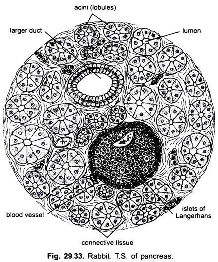
Clusters of specialised cells, (Fig. 29.33) described by Paul Langerhans (1869). These are found distributed in the pancreas, which are endocrine in nature.
These clusters are formed of two types of cells:
(i) Beta-cells, which constitute the maximum part of the cluster and secrete the hormone insulin, and
(ii) The alpha-cells constituting nearly 20% of the cluster and believed to secrete a second hormone called glucagon.
C-cells and D-cells are in very low percentage and their function is not known. Insulin hormone is necessary for the conversion of excess sugars into glycogen in liver and muscles and glucagon function is conversion of glycogen back into glucose, when the sugar contents of the blood fall below the normal level. Deficiency of insulin results in diabetes mellitus in human beings.
6. Gonads:
The gonads also perform endocrine function by producing sex-hormones in addition to their normal function of producing gametes.
(i) Testis:
Testis secretes testosterone or male sex-hormone. The hormone testosterone is actually secreted by the interstitial cells or cells of Leydig situated in the connective tissue of the testes between seminiferous tubules. This hormone is responsible for the development of secondary sexual characters in male, e.g., deep voice, beard on the face and the distribution of hairs on the body in man, etc.
(ii) Ovary:
Ovary secretes estrogen (oestradiol), a female sex-hormone produced by the cells surrounding the Graafian follicles. It is responsible for the mature growth, development and maintenance of the female reproductive system. It stimulates the secondary sexual characters and also contributes to the sexual desire of the female.
Progesterone:
It is produced by the corpus luteum of the ovary formed after ovulation. It suspends further ovulation during pregnancy. It maintains pregnancy and prepares the uterine wall for the attachment of embryo with the uterine wall.
7. Placenta:
The placenta also produces estrogen and progestin (chorionic gonadotropin). These hormones augment the action of pituitary luteinising hormone in maintaining the corpus luteum during pregnancy. These hormones work along with the hormones of ovary. In rabbit, etc., the placenta as well as the ovary secrete relaxin hormone. This relaxes pelvic ligaments for the easy birth of the child.
8. Gastro-Intestinal Tract:
Some hormones like gastrin, secretin, enterogastrone and cholecystokinin are also produced in the different parts of gastro-intestinal tract which stimulate gastric glands to produce gastric juice, secretin stimulates pancreas to produce pancreatic juice into duodenum, enterogastrone inhibits the gastric secretion and cholecystokinin causes contraction of gall bladder to expel bile into duodenum.
9. Thymus:
It is a large bilobed gland in the upper chest region in front of heart and below the base of trachea. In many mammals it remains as such throughout the life, while in some it disappears altogether at the time of puberty. Its secretion stimulates metabolism, growth and helps in attaining sexual maturity. Its removal from the embryo hampers the development of reproductive system. It is regarded as lymphatic gland and, hence, its endocrine nature is not definitely known.
10. Pineal Body:
It is situated on the roof of the diencephalon of the brain on a small pineal stalk. The nature and physiological role of the secretion produced by pineal body is not definitely known. However, its removal retards growth, and quickens the maturity or has no effect.
