In a vertebrate embryo the endoskeleton is at first, cartilaginous, which is replaced by bone in most adult vertebrates. In cyclostomes and elasmobranchs, the skeleton does not pass beyond the cartilage stage. In higher vertebrates the endoskeleton is largely bony.
Types of Bones:
Bone exists in cancellous and compact form.
(i) Spongy or cancellous or endochondral bone consists of small pieces joined together irregularly, the small spaces between the pieces contain bone marrow.
ADVERTISEMENTS:
(ii) Compact or periosteal bone is a hard, solid mass of bone without any apparent spaces.
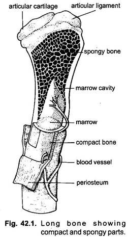
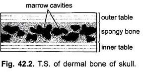
The broad ends of long bones are made of spongy bone underlying a layer of compact bone. The shafts of long bones are made of compact bone enclosing a marrow cavity. The skull bones have a layer of spongy bone between two layers of compact bone called inner and outer tables.
Bone Formation:
ADVERTISEMENTS:
Bone is formed in two ways, by a replacement of pre-existing cartilage with bone, called cartilage bone, and secondly by direct ossification of mesenchyme or connective tissue without an intervention of cartilage, in which case the bone is known as membrane or dermal bone. In both types of bones the method of formation is similar.
1. In formation of cartilage bones known as endochondral bone formation, the original cartilage cells become arranged in rows and then they die. The matrix becomes calcified and invaded by blood vessels and connective tissue eroding the cartilage to make intercommunicating channels between bars of calcified cartilage. The connective tissue fibroblasts become differentiated into osteoblasts.
The osteoblasts secrete ossein of bones in concentric layers around bars of cartilage. The original perichondrium becomes the periosteum. Several osteoblasts fuse to form new cells called osteoclasts, which reconstruct the forming bone and dissolve away unwanted portions.
ADVERTISEMENTS:
2. In formation of membrane bones mesenchyme cells collect and form an interlacing network and in between the cells of the network are present collagen fibres. An amorphous matrix is formed between the cells and fibres. Bone is secreted by cells which are now called osteoblasts, and the matrix gets impregnated with calcium. The osteoblasts now known as osteocytes lie in spaces in the matrix. Later the bony tissue matures.
The cartilage bones and membrane bones have the same structure. They differ only in embryonic origin, and many bones contain tissues of both kinds. Dermal bones are laid down layer by layer forming compact bone, while cartilage bones generally form cancellous bone, though an admixture of the two is found in many bones.
Heterotopic Endoskeleton:
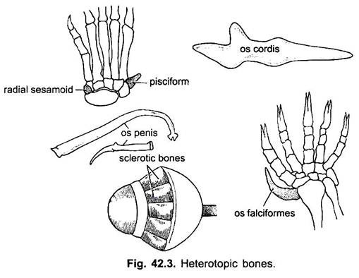
In some animals certain bones may be present which do not belong either to the axial skeleton or to the appendicular skeleton, they are spoken of as heterotopic bones.
These are:
1. Sesamoid bones are formed by ossification of a tendon where the tendon moves over a bony surface, such as patella or knee cap which appears first in some lizards, and two sesamoid bones are commonly present in the wrist (pisciform bones) of tetrapoda. They are a radial sesamoid anterior to the radiale, and a pisciform posterior to the unlnare.
2. Os cordis is a bone in the interventricular septum of deer and bovines.
3. Os penis or os priapi is a bone giving rigidity to the penis in marsupials, insectivores, rodents, Carnivora, Chiroptera, Cetacea, walrus and lower primates.
4. Os rostralis is a bone in the snout of some ungulates, such as pigs.
ADVERTISEMENTS:
5. Os falciforme is an additional bone in the palm of moles which helps in digging.
6. Sclerotic bones form a ring in the eyeball of lizards and birds.
7. Os clitoridis, present in the clitoris of otters, rabbits and several rodents.
8. Pessulus is found in syrinx of birds.
9. Epipubic is found in ventral abdominal wall of monotremes and murspials.
Structure and Formation of Long Bones:
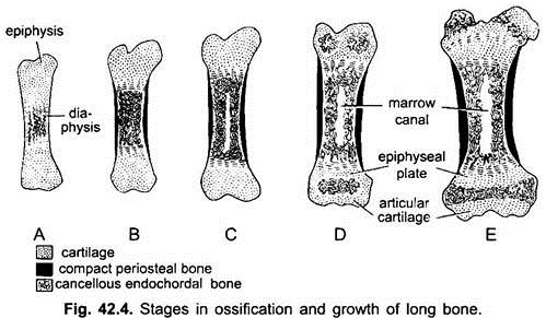
In long bones, such as the femur, the main shaft is diaphysis made of cartilage. A ring of bony tissue around the diaphysis form the compact bone which surrounds spongy bone. Ossification begins in the middle of the diaphysis and extends towards the ends. The ends remain cartilaginous for some time and continue to grow, increasing the length of the bone.
In mammals, and to some extent in reptiles, the long bones and vertebrae are formed from three centres of ossification: a central diaphysis and two terminal epiphyses. Between the diaphysis and each epiphysis arises a new cartilaginous pad, the epiphyseal plate where increase in length takes place. Eventually epiphyseal plates are replaced by cancellous bone and epiphyses are fused to the diaphysis, after which no further growth in length can take place. Ends of long bones are covered with articular cartilage.


