In this article we will discuss about the receptor organs in vertebrates with the help of suitable diagrams.
Sensory systems consist of peripheral receptor cells and integrating neurons in the brain that provide a biologically relevant image of the environment to an organism. The receptor elements range from single cell, such as tactile corpuscles, to complicated structures, such as the eye.
A vertebrate has receptors or sense organs for touch, smell, taste, sight, and hearing, which are stimulated by the environment. These sense organs are termed external receptors or exteroceptors. There are other sense organs found in the body, which detect temperature, pain, hunger, thirst, fatigue, and muscle position. They are spoken of as internal receptors or interoceptors.
Besides these two, third is proprioceptors, which are stretch receptors found in the muscles, joints, tendons, connective tissue and skeletons. All receptors are closely associated with the nervous system and respond to external or internal stimuli. Impulses are transmitted from receptors by sensory fibres to the central nervous system where they are interpreted as sensations or messages, which are sent to effector organs through efferent or motor nerve fibres, for responding in an appropriate manner.
ADVERTISEMENTS:
List of Common Senses:
1. Touch. It includes contact, pressure, heat and cold, etc.
2. Taste. Receive stimulus by chemicals in solution.
3. Smell. Receive volatile chemicals and gases in air.
ADVERTISEMENTS:
4. Hearing. Receive sound vibrations.
5. Sight. Receive light waves.
Cutaneous (General) Receptors:
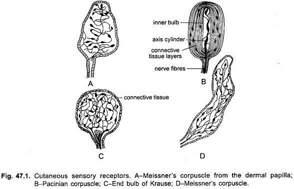
The entire body surface of aquatic vertebrates and the moist parts of the skin of terrestrial vertebrates are sensitive to chemicals. Certain parts of the skin are sensitive to contact and temperature changes. The cutaneous receptors are free nerve endings of peripheral nerves, and some encapsulated nerve endings or tactile corpuscles.
1. Free Nerve Endings:
ADVERTISEMENTS:
Branches of sensory nerves terminate in the epidermis and form patches, cornea of the eye, hair follicles, and mucous membrane. These parts are sensory to pain (algesireceptors), or to touch (tangoreceptors). The nerve endings do not reach the outermost layer of the epidermis so that the stimulus first travels through the outer cells before it reaches the nerve endings.
2. Tactile corpuscles of various kinds are located in the dermis or in the sub-epidermal connective tissue. They have a basic structure of nerve endings or tactile cells enclosed in layers of connective tissue. Tactile corpuscles are found all over the skin, in birds they are found also on the beak, in mammals they occur also on palms, soles, and external genital organs.
The encapsulated corpuscles may be tactile (tangoreceptors), sensitive to pressure, to heat (caloreceptors), or to cold (frigidoreceptors). But even free nerve endings are capable of discerning sensation of touch, heat, cold, and, pain, e.g., the cornea has only free nerve endings yet it can appreciate all these sensations. The parts played by cutaneous receptors are not clear, and it is doubtful if any one of them is sensory to a particular sensation. The cutaneous receptors form a general sensory system.
Chemoreceptors:
Chemoreceptors or organs of chemical sense consist of olfactory organs and organs of taste. Both these organs are stimulated only by chemical substances or odours in air (nostrils) and in solution (tongue). The medium for dissolving substances for taste is water for aquatic animals and mucus for land animals. The olfactory organs can respond to a low concentration of the dissolved substance, whereas organs of taste need a higher concentration of the dissolved substance for a response.
Olfactory Organs in Vertebrates:
Olfactory organs (olfacto-receptors) are a pair of invaginations of the ectodermal cells of the skin forming olfactory sacs on the anterior end of head. Their external openings are called nostrils or nares. In most fishes the olfactory organs consist of a pair of pits lined with folds or ridges of sensory epithelium.
The cyclostomes have a single median olfactory organ. This is a blind pit in the lampreys, but in hagfishes it opens into the pharynx. Dipnoans resemble higher vertebrates in possessing paired nasal passages that open by means of choanae into pharynx. The nasal passages, therefore, have both internal and external openings. The olfactory epithelium within canals appears in the form of folds.
The olfactory epithelium in amphibians is generally smooth and is restricted to the upper part of the nasal passages Within each of the nasal passages, which are elongate in higher reptiles because of the development of a secondary palate, there is a shelf or concha which serves to increase the surface for the olfactory epithelium. In birds, the nasal passages have three shelves or conchae.
ADVERTISEMENTS:
In most birds there are external nostrils leading into the nasal passages, but in certain members of Pelecaniformes these are closed. Generally the nostrils are separated internally from one another by a septum, but in New World vultures as well as some of the Gruiformes, a partition is lacking. Olfactory epithelium in most birds is restricted to the surface of the upper most or superior concha.
This is correlated with the small size of olfactory lobes and poor sense of smell. But kiwi has relatively more developed sense organs. In mammals, the sense of smell is highly developed in many mammals. It is due to the development of the nasal conchae into elaborate scroll-like structures, which greatly increase the surface available for olfactory epithelium. In whales, the olfactory organs are essentially non-functional.
Organs of Jacobson or Vomeronasal Organ:
In many tetrapoda there is a pair of vomeronasal organs which are sac-like chambers lying below the nasal cavities but above the buccal cavity. They have a pigmented epithelial lining like that of the olfactory organs. Each opens by a short duct into the olfactory organ in amphibians, but in others the duct opens into the buccal cavity.
The organ of Jacobson receives nerves from the nervus terminalis, a branch from the olfactory nerve, and a branch from the trigeminal nerve. The organ is believed to aid by smelling the recognition of food held in the mouth, and in lizards and snakes it appreciates the scent introduced into it by the tip of the tongue.
The organ of Jacobson first appears in amphibians as evaginations from the nasal passage and is lined with olfactory epithelium. It is believed to be an aid in tasting food. They may also be important in reproductive behaviour in case of Ensatina (lungless plethodontid salamander) since the first act of male in courtship to nose the females’ head and neck.
It is best developed in Sphenodon, lizards and snakes and is connected with the roof of mouth rather the nasal canal. It is also well formed in monotremes, marsupials, insectivores, and rodents. But in turtles, crocodiles, birds, and many mammals such as Primates and Cetacea, it is found only in the embryo and is absent in the adult.
Gustatory or Organs of Taste in Vertebrates:
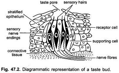
Organs of taste (gustatoreceptors) consist of taste buds which perceive the sense of taste from the dissolved substances. Their structure is fairly uniform in vertebrates. Each taste bud consists of cluster of neurosensory cells and supporting cells arranged to form a barrel-like structure embedded in stratified epithelium.
Each neurosensory cell is long and narrow with a thin taste hair at its free tip and a sensory nerve fibre at its base. Taste hairs project into a depression or taste pore in mammals, in others they project above the surface, there being no taste pore. In man, there are four fundamental sensations of taste- sweet, salty, bitter, and sour.
Taste buds are widely distributed in fishes since they live in a liquid medium, being found in the mouth, pharynx and outer surface of the head. In some fishes they occur on the entire body surface including even the fins.
The taste buds are innervated by the V, VII, IX and X cranial nerves. In amphibians, the taste buds are restricted to the roof of the mouth, the tongue and mucosa that lines the jaws. In most reptiles, the taste buds are restricted largely to the pharyngeal region. Taste buds are lacking on the tongue of most birds, although they are found on the lining of the mouth and pharynx.
In mammals, there are various kinds of papillae on the tongue which possess taste buds except filiform papillae. Some taste buds of vertebrates are not for tasting but for testing substances in the pharynx to cause reflexes which prevent solid particles from entering air passages.
Eyes or Photoreceptors:
Eyes are of two types:
(1) Median and
(2) Lateral.
1. Median Eye:
In amphibians, both the parietal and pineal bodies are known to function as photoreceptors, being sensitive to wavelength and intensity of light. It has been suggested that they may play a role in thermoregulation, compass orientation and circadian rhythm. Pineal eye is also present in tuatara and a few lizards. It aids in regulating the amount of exposure to sunlight.
2. Lateral Eyes:
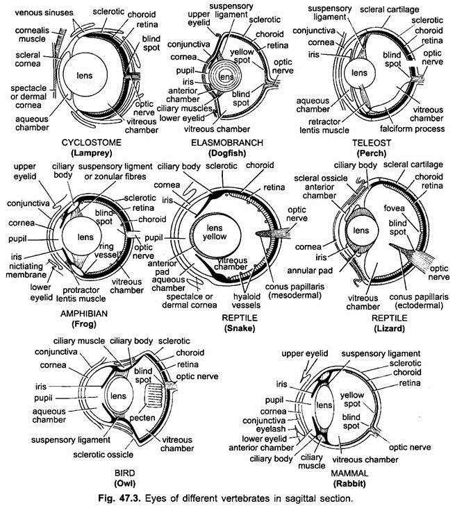
The paired eyes of vertebrates, except for small differences, are remarkably constant in structure, innervation, and extrinsic muscles. The differences occur in eyelids, glands of the eyes, and in the mechanism of accommodation. Fishes have no movable eyelids and tear glands. Cornea remains flat in lower vertebrates and buldging in higher vertebrates.
There is no basic difference between the eyes of fishes and those of other vertebrates. Differences are in the methods of accommodation or special adaptations to a particular mode of life or are the result of degeneration.
Accommodation or the ability of the eye to adjust itself to near and far vision, is accomplished in fishes by moving the lens forward or backward so as to change its distance from the sensitive retina. Sharks are fast moving predatory fishes, usually have distant vision, they move the lens forward to see near objects. In most teleosts, the eyes are normally adjusted to near vision so the lens is moved back toward the retina when distant vision is required.
The eyes of some fishes are highly specialised, as an adaptation to a particular mode of life. South American “four-eyed fish” (Anableps) inhabits quiet water where it floats with the upper half of the eyes above the surface. So that it may see both in the air and in water, presumably at the same time. Its lens is divided into two parts, each part being a different distance from the retina.
Another four-eyed fish Dialommus fuscus (Galapagos blenny) spends considerable time out of water. Its lens is not divided but it has a divided cornea-anterior section for air vision and posterior section for vision under water.
Many deep fishes have very large eyes for vision in regions of low-light intensity. Other deep sea fishes and some cave inhabiting species have degenerated eyes, have lost the power of sight.
In various lower vertebrate groups accommodation to near and distant vision is accomplished by moving the lens forward or backward so as to change the distance between the lens and the sensitive retina. Accommodation in reptiles and most other amniotes is accomplished by changing the shape of lens.
It may be flattened for distant vision or rounded for near vision through the action of the muscles of the ciliary body. Eyelids are present in many reptiles and generally are more movable than those of amphibians. In snakes and some of the burrowing lizards, movable lids are absent, and the eye, which itself is capable of moving, is covered with immovable transparent skin.
These so called “eye plates” are believed to have been derived from the lids and are like scales which shed periodically. Many reptiles possess the nictitating membrane, situated beneath the upper and lower lids and closes the eye by moving backward or outward from the anterior or median margin of the eye aperture. The blood that a horned toad squirts from the eye comes from blood vessels in the nictitating membrane which rupture easily as a result of muscular contraction.
The amphibian eye is basically like that of other vertebrates. The lens is fixed for relatively distant vision but may be moved forward toward the cornea for seeing close objects, by means of small muscles of accommodation.
The pupillary aperture may be vertical, horizontal, three-cornered or four-cornered. Eyelids are poorly represented in aquatic forms but are well developed in many terrestrial species. The lower lid is usually more movable than the upper lid. Lacrimal or tear glands are present in many amphibians, although they are poorly developed. The eye, however, is kept moist by the oily secretion from the Harderian gland.
In birds, eye are highly developed and quite large in proportion to body size. Accommodation is accomplished by action of the ciliary muscles which change the shape of the lens. An ossified sclerotic ring is present and surrounds the ciliary region.
A fan-shaped pecten is present in the eye, which extends into the posterior chamber from the point at which the optic nerve emerges from the retina. Pecten provides nourishment to avascular parts of the eye. As in other groups of vertebrates, in the retina of diurnal birds, cones predominate, whereas rods are much more numerous in nocturnal species.
The mammalian eye is basically like that of most other vertebrates, with certain modifications correlated with habits. As in birds, nocturnal mammals have a preponderance of rods in the retina, while in diurnal species cone cells predominate. Land mammals have an emmetropic condition in air but become hypermetropic in water.
Aquatic or marine mammals have developed emmetropic vision under the water. In pinnipeds this is accomplished by a permanent change in the curvature of the lens. They also have special dioptric mechanisms for air vision. In terrestrial mammals, the strong sphincter muscles in eye are present in the iris that changes the shape of the lens to preserve visual acuity when under water.
In fossorial species such as moles (Talpidae), the eye may be of little importance. This is also true of the Gangetic dolphin (Platanista gangetica) living in very turbid water. Retina is more sensitive to light, lens is devoid of a crystalline body and probably serves no function.
Lateral Line System:
Lateral line system is found in cyclostomes, fishes, aquatic amphibians and aquatic larval stages of terrestrial amphibians. It is not found in terrestrial vertebrates. It is related to an aquatic mode of life. The receptor organs are sensory papillae or neuromasts which are arranged in rows on the body.
In most fishes there is a row on each side of the body which branches into three parts on each side of the head. The neuromasts may be on the surface of the skin (cyclostomes and a few fishes) or they may be in covered canals which open to the outside through small pores (cartilaginous and bony fishes).
A lateral line system, similar to that of fishes, is present in larval amphibians and even in the adult stage of certain aquatic species. Lateral line organs are lacking in reptiles and other amniotes. Lateral line receptors detect pressure changes and currents in the water.
Ears or Statoacoustic Organs:
The vertebrate ear is commonly regarded only as an organ of hearing (phonoreceptor), but at least in higher vertebrates, it serves the dual function of equilibrium and hearing.
The ear of a fish, is very different from the ear of a mammal. In mammals ear consists of an external pinna which funnels sound waves into the external auditory canal. At the inner end of this canal these waves strike the ear drum or tympanic membrane and are then transmitted across the middle ear or tympanic cavity by means of a chain of small bones to the cochlea or ventral part of the inner ear.
From this complicated structure sensory impulses pass to the brain by means of the auditory nerve. The dorsal part of the inner ear consists primarily of three semicircular canals which are essentially organs of balance or orientation. The ear of fish, however, is not suited for the reception of sound. There is no external or middle ear, nor is a cochlea present.
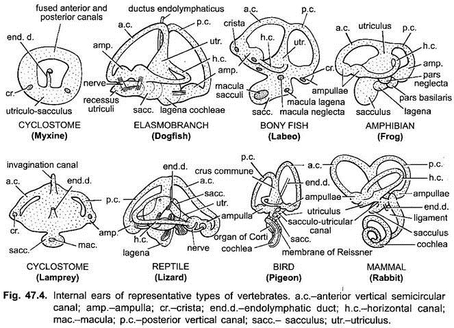
The inner ear of a fish consists of a dorsal utriculus, which is connected with the semicircular canals, and a ventral enlargement known as the sacculus. It is from the sacculus that the lagena of amphibians, reptiles and birds as well as the cochlea of mammals arises to permit hearing in true sense. While certain fishes are definitely able to detect vibrations in the water. The ear of a fish is primarily an organ of balance, enabling it to maintain its equilibrium.
In cyclostomes, the membranous labyrinth is degenerate with only one (Myxine) or two (Petromyzon) semicircular canals. In fishes the membranous labyrinth is complete with three semicircular canals, and in many teleosts it is connected to the air bladder by a duct or a chain of bony Weberian ossicles.
Amphibia:
There is considerable variation in the auditory apparatus of amphibians. Salamanders and their relatives lack any middle ear, although it is believed that they can detect vibration. Most frogs and toads, however, have a middle ear and an external ear drum. Sounds are transmitted from the ear drum across the tympanic cavity to the inner ear by means of a bone called the columella which is homologous with the hyomandibular element of the gill-arches of cartilaginous fishes.
The tympanic cavity, since it is derived eubryologically from the first pharyngeal or gill-pouch, is concerned with the pharynx by means of the eustachian tube. This permits equalisation of pressure on both sides of the tympanic membrane. From the ventral part of the sacculus of the inner ear there is a ventral out-pocketing called the lagena which is the forerunner of cochlea of mammals. This is believed to be concerned with the reception of sound vibrations.
Reptilia:
There is considerable variation in the structure of the ear in reptiles. Lizards have a well-developed middle ear and in some forms the eardrum has sunk into a depression. Sound waves, thus, pass through a short canal known as the external auditory meatus in order to impinge upon the tympanic membrane. This is the first indication of an external ear in vertebrates.
In snakes the tympanic membrane, middle ear and eustachian tube are lacking. Vibrations are received by snakes are transmitted by way of the quadrate to the columella and then to the inner ear. The lagena is more elongate than in amphibians and in the crocodilians actually forms a cochlear duct rather similar to birds.
Aves:
The ear in most birds lacks an external pinna and has only an internal ear and a middle ear. In the barn owl (Tyto alba), the external pinna is well developed. As in reptiles there is but a single bone, the columella in the middle ear. A cochlea is present although it does not assume the complicated spiral form that is found in the higher mammals.
Barn owls are able to locate small mammals in total darkness by means of extremely acute hearing. Sounds are heard loudest in different sites in each ear, but the sensitivity is greatest along the line of vision. When the bird, by moving its head, hears the sound of moving prey the loudest, it is facing directly toward its potential food.
Mammalia:
The ear in mammals shows several advances over the ears of other vertebrates. The middle ear contains three bony ossicles which transmit the vibrations from the tympanic membrane to the internal ear. An external auditory canal is present, and in the great majority of mammals, a well-developed pinna is present which aids in funneling sounds into the auditory canal. The cochlea is generally coiled to accommodate its increase in length.
The auditory apparatus of some mammals shows remarkable specialisation. Many bats, cetaceans and pinnipeds depend largely upon the echoes of sounds that they themselves produce to detect the presence of objects in their environment as they move about. Bats navigate by means of echo-location and they also come to know the size and shape of the object by echo.
Many kinds of cetaceans also produce underwater sounds with wider range of frequency than bats. In water, because of the similarity in density of the mammalian body and water, sound waves may be conducted through the body. They, therefore, could pass from one ear to another rather than be received independently. Cetaceans have tympanic bone, in which cochlea is located, suspended from the skull by ligaments and surrounded by a cavity filled either with air or foam.
Sound waves, therefore, can only be transmitted in the cochlea by specially modified auditory ossicles. The sound itself is produced in nasal sacs just inside the blowhole, rather than in a larynx as in terrestrial mammals. Recent experiments also indicate that sound reception from the water to the middle and inner ear may be by way of lower jaw.
The mandibles of porpoises, dolphins and other toothed cetaceans are hollow and each is composed of an outer covering of very thin bone around an oil-filled cavity, which would favour sound transmission. The back of the mandible is very close to the middle ear.
Internal Receptors or Interoceptors:
Interoceptors are directly affected by stimuli arising within the body itself. Those found in the walls of the alimentary canal and viscera are called interoceptors, and those located in the tendons, joints, and voluntary muscles are spoken of as proprioceptors.
1. Interoceptors are connected to the visceral sensory nerves but they do not form definite sense organs. They are stimulated by conditions in the digestive system and supply information to the central nervous system. Interoceptors detect appetite, nausea, hunger, and thirst. Hunger is not to be confused with appetite.
Hunger is a nutritional deficiency in the blood, while appetite is a memory of food causing internal changes in conjunction with sight, colour, or taste. Thirst is a condition of the mucous membrane of the throat due to decrease of water and an increase of salt content in the blood. Some genital corpuscles are present in the dermis of nipples and external genital organs, they are associated with sexual sensations.
2. Proprioceptors are connected to the somatic sensory nerves. They generally form spindle-shaped bodies consisting of free nerve endings enclosed in layers of connective tissue or encapsulated corpuscles enclosing tactile cells. They are found in tendons, joints and voluntary muscles. Proprioceptors are stimulated only indirectly by the environment.
They supply information to the central nervous system regarding visceral pains, position of the body, and coordinated movements of parts of the body. They give an idea of weight of objects, and provide a “muscle sense” making one aware of the degree of tension in a muscle.


