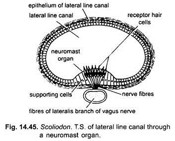
The receptor organs or sense organs of Dogfish (Scoliodon) include the paired olfactory, optic or photoreceptor, statoacoustic organs, the lateral line organs or neuromasts and the ampullae of Lorenzini.
Olfactory Organs (Olfactoreceptors):
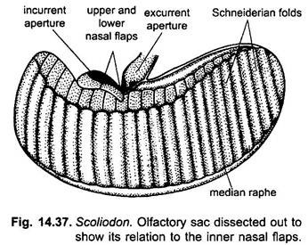
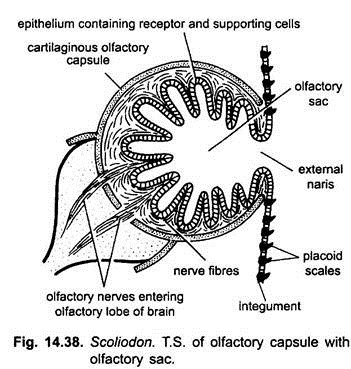
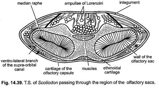
The olfactory organs of Dogfish (Scoliodon) are a pair of blind olfactory sacs lying on the ventral side of the head in front of the mouth. They are invaginations of the ectoderm and do not communicate with the buccal cavity by an internal opening. Each olfactory sac is ellipsoid in shape and proportionately very large. It is covered externally by a thin membrane and is lodged on each side in the cartilaginous olfactory capsule of the skull.
The mucous membrane lining the inner wall of the olfactory sac is thrown into a double series of folds, the Schneiderian folds. The Schneiderian folds are held in place by a median band of connective tissue, the median raphe. Internally olfactory epithelium consists of two types of cells, the olfactory receptor cells and supporting cells.
The olfactory receptor cells bear internally sensory hairs and their basal ends are connected with the fibres of the olfactory nerve which passes from each olfactory sac with olfactory lobe of its side of the brain. The olfactory sacs open outside ventro- laterally by external nares.
Each external nostril is more or less completely divided into two, a lateral incurrent siphon and a median ex-current siphon by three nasal flaps. Of these flaps two project inwards fitting closely, one above the other, while the third hangs down from the dorsal side slightly overlapping the other two.
The median raphe of the Schneiderian folds lies parallel to the long axis of the nasal aperture, and, therefore, the current of water that enters through the incurrent siphon is at first directed against the oval wall of the olfactory sac, then turns internally to the nasal valve in the direction of the median raphe and finally passes out through the median ex-current siphon. The current of water enters at the lateral end of the nasal aperture and, thus, takes a zig-zag course through the olfactory sac finally leaving by the mesial end of the nasal aperture.
The great size of the olfactory sacs is characteristic of elasmobranchs and is correlated with a highly developed sense of smell. The highly developed sense of smell enables them to detect food. If the external narial openings are plugged the dogfish will swim over food material without detecting it, on the other hand with one nostril open it will detect even concealed food.
Eyes (Photoreceptors):
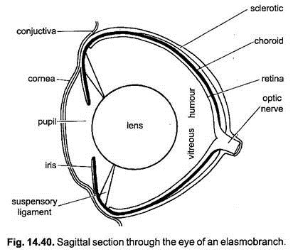
In Dogfish (Scoliodon), the eyes are very large and housed in orbits on the lateral sides of cranium. Each eye is elliptical and supported not only by the recti and oblique muscles but also by a cartilaginous optic pedicel which connects the eye-ball to the orbit. The eye-ball consists of three concentric layers, viz., sclerotic, choroid and retina. The sclerotic is cartilaginous and opaque and forms the outer protective layer of the eye.
ADVERTISEMENTS:
The sclerotic of the exposed part of the eye is transparent and is known as cornea through which the light enters. Cornea is flat and fused with an outer conjunctiva that is continuous with the living of immovable eyelids. The sclerotic is lined internally by a connective tissue coat, the choroid. The choroid is richly supplied with blood vessels and is pigmented. It is continued in front as a strongly circular curtain, the iris, with a median vertical slit, the pupil. Pupil cannot be dilated or contracted.
The retina is the innermost layer of the posterior region of the eye-ball and forms its sensitive portion. It is made up of special elongated nerve cells called rods. Rod cells are connected with the fibres of optic nerve. Closely attached to the posterior surface of the iris behind the pupil lies the spherical crystalline lens which divides the eye into two unequal chambers, each filled with a semifluid substance.
The chamber in front of the lens is filled with a saline liquid, the aqueous humour, while the chamber behind the lens contains a denser fluid, the vitreous humour. The lens is kept in place by a suspensory ligament. In elasmobranchs, choroid possess guanine crystals bearing cells which form a silvery light-reflecting surface called tapetum lucidum beneath the rods of retina. This is an adaptation to life in dim light, reflects additional light on the retinal cells.
ADVERTISEMENTS:
Muscles of the Eye:
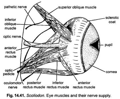
The eye-ball is lodged in the orbit and is held in position by six eye-muscles and a cartilaginous stalk called the optic pedicel arising from the posterior angle of the orbit. The eye-muscles are inserted on the eye-ball in two groups. The first group comprises four recti muscles which arise from the posterior median angle of the orbit and are called the superior rectus, inferior rectus, anterior rectus and posterior rectus muscles.
The superior rectus is inserted on the postero-dorsal surface of the eye-ball and the inferior rectus is inserted on the postero- ventral surface of the eye-ball. These rotate the eye-ball in vertical plane. The anterior rectus is inserted on the anterior surface and the posterior rectus is inserted on the posterior surface of the eye-ball and rotates it in horizontal plane.
The second group comprises two oblique muscles which arise close together from the anterior angle of the orbit and run backward and outward to the eye-ball. The superior oblique is inserted on the antero-dorsal surface of the eye-ball and the inferior oblique is inserted on the antero-ventral surface of the eye-ball in front of the inferior rectus. These muscles twist the eye-ball.
Working:
The eyes of Dogfish (Scoliodon) though large yet are placed apart as to render it impossible for both of them to focus together on a given point. There is, therefore, no binocular vision and each eye has its own range of vision. Further, since little adjustment of the shape of the lens is possible and the pupil is non-contractile, the power of estimation is also poor.
ADVERTISEMENTS:
But the lens can project through the pupil against the rounded cornea, so that even rays of light entering at a very wide angle can be caught and focused on the retina. Since the retina is without cones, the fish is colour blind and the eye cannot discriminate minute details, but the eye is adapted for near vision in dim light and fish can see its prey.
Stato-acoustic Organs (Internal Ears):
Structure:
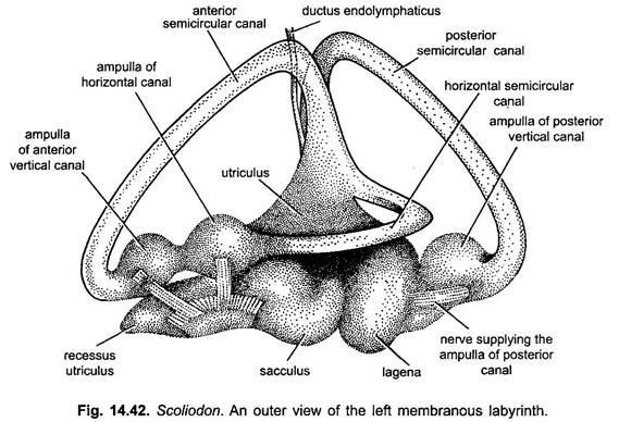
The stato-acoustic organs of Dogfish (Scoliodon) are the internal ears or membranous labyrinths. The internal ear of each side is a closed ectodermal sac, the membranous labyrinth which is enclosed within the cartilaginous auditory capsule one on either postero-lateral side of cranium. It consists of mainly two parts-the central otosaccus and three peripheral semicircular canals. The otosaccus is laterally compressed and is differentiated into a dorsal and anterior part, the utriculus, and a ventral and posterior part, the sacculus.
The posterior outgrowth of sacculus is called lagena cochleae. From the utriculus arises an anterior outgrowth called recessus utriculi. The three slender semicircular canals arise from the otosaccus and also open into it. From the top of the utriculus arise the anterior vertical and horizontal semicircular canals which form their independent ampullae before opening into the middle of the utriculus above the recessus utriculus. The posterior vertical canal arises from the dorsal surface of the sacculus, forms the complete circle, and opens by its ampulla into the lagena cochleae.
The three semicircular canals are arranged at right angles to one another in the three planes of the space. The cavity of the sacculus communicates with the exterior through a long slender tube, the ductus endolymphaticus, which runs upward and pierces the roof of the cranium within the parietal fossa to open to the exterior by a minute pore in front of the commissural occipital canal.
The interior of membranous labyrinth, including the semicircular canals, is filled with a fluid the endolymph which is sea water. The endolymph contains the calcareous particles the otoliths. The space between the membranous labyrinth and the wall of the auditory capsule is the perilymphatic space, filled with a perilymph which is actually the cerebro-spinal fluid. The perilymphatic space opens to the exterior by a large aperture, the fenestra lying immediately behind the opening of the ductus endolymphaticus.
The membranous labyrinth is innervated by the auditory nerve. In utriculus, sacculus, recessus utriculi, lagena cochleae as well as in the ampullae of the semicircular canals, there are groups of delicate receptor cells bearing stiff hairs. Groups of receptor cells located in the utriculus and sacculus are called maculae, and those found in the ampullae are called cristae.
Functions:
In fishes the internal ear is not concerned with hearing but it performs the following functions:
(i) It maintains muscle-tone,
(ii) It detects acceleration of speed, change of direction and orientation,
(iii) It is concerned with balance or equilibrium with regard to gravity. Movements of endolymph and otoliths stimulate sensory nerve endings in vestibule and ampullae, thus, informing the fish position in water and
(iv) It may detect low frequency vibrations of water but there is no proof that fishes can hear. Sacculus and lagena probably receive auditory stimuli.
Neuromast Organs (Rheoreceptors):
The neuromast organs consist of:
(i) Lateral line receptors and
(ii) Pit-organs.
(i) Lateral Line Receptors:
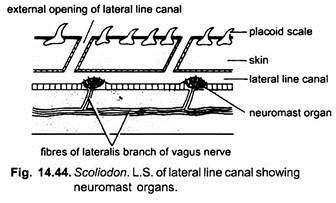
The lateral line receptors lie in the lateral line canal which runs on each side of the trunk and tail. The lateral line canal is a slender mucus filled canal embedded in the dermis. It opens on the surface by minute pores found at intervals through a series of vertical tubes. Each lateral line canal is continued into the cephalic canal on the head. These canals have groups of epidermal receptors or neuromasts embedded in their walls. The neuromasts are innervated by the branches of seventh nerve and the lateralis branch of the vagus.
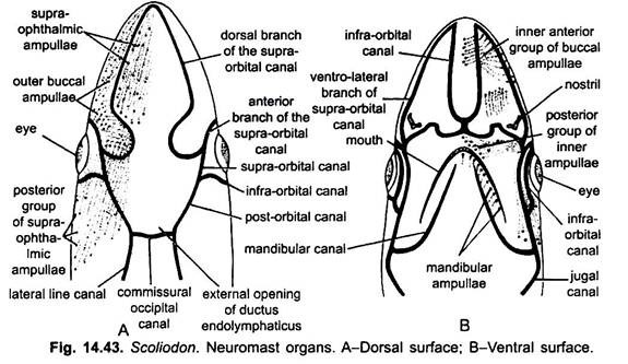
In the head region two lateral canals are joined by a transverse commissural occipital canal above the head, then each lateral line canal runs forward as a post-orbital canal which divides into two branches, a supra-orbital canal above the orbit and an infra-orbital canal below the orbit, both run up to the snout. The supra-orbital canal further divides into anterior and dorsal branches.
The dorsal branch continues as ventro-lateral branch and joins with the infra-orbital canal. Arising from the infra-orbital canal is a jugal canal below the eye which runs back up to the first gill-cleft. The jugal canal gives off a mandibular canal to the lower jaw. The canals are lined with epithelium having many mucous gland cells which secrete mucus. The canals open at intervals on the surface by vertical tubes. The canals are filled with fluid and mucus.
Neuromast Organs:
In the canals are found scattered sensory receptor organs, the neuromasts. Each made of a group of sensory receptor cell and supporting ectodermal cells. Each receptor cell has a stiff sensory hair projecting into the canal and a nerve fibre at the other end. The hairs of receptor cells are tipped with a heavy gelatinous substance. The lateral line neuromasts are current receptors (rheoreceptors) detecting any type of vibrations of water.
(ii) Pit Organs:
Pit organs are found scattered on the dorsal and lateral surfaces of the head. Pit organs consist of sensory hair cells and supporting ectodermal cells innervated by nerve-fibres of seventh cranial nerve. Pit organs are scattered individual neuromasts found in all fishes. They are rheoreceptors.
Ampullae of Lorenzini:
The ampullae of Lorenzini are found in clusters on the dorsal and ventral surfaces of the head embedded below the skin but opening externally on the surface of the skin. Each ampulla has a pore opening on the surface, the pore leads into an elongated mucous-filled tubule which ends in a radially septate ampullary sac lying deeply beneath the skin.
Each ampullary sac consists of eight to nine radially dilated chambers arranged around a central core, the centrum. Two kinds of cells are found in the ampullae- pear-shaped gland cells which secrete the jelly filling the ampullary tubules and the pyramidal cells or sensory hair cells forming the receptor cells. The ampullae of Lorenzini are innervated by branches of the facial nerve.
The ampullae of Lorenzini are arranged in groups and are named according to their position.
The groups are:
(i) Supra-ophthalmic group lies around the supra-orbital canal,
(ii) Outer buccal lies between the supra-orbital and infra-orbital canals,
(iii) Inner buccal lies beneath the infra-orbital canal.
The ampullae of Lorenzini were formerly regarded as neuromast organs but Sand (1938) has proved that these are thermo-receptor organs. The change in the temperature of water is carried to the brain from the receptor cells of ampullary sac.
