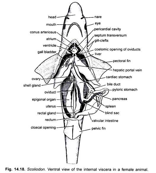
In Dogfish (Scoliodon), the coelom is specious and is divided into two unequal cavities, the small anterior pericardial and the large posterior abdominal, separated from each other by a membranous partition, the septum transversum.
Pericardial cavity is comparatively smaller in size and triangular in shape and lies beneath the pharynx enclosing the heart. It is enclosed between a tightly fitting smooth layer of peritoneum lining the outer wall of the cavity and the inner pericardial layer which adheres closely to the heart itself.
The pericardial cavity contains a clear colourless fluid, the pericardial fluid. It communicates with the abdominal cavity through aperture in the septum transversum called the pericardio-peritoneal canal.
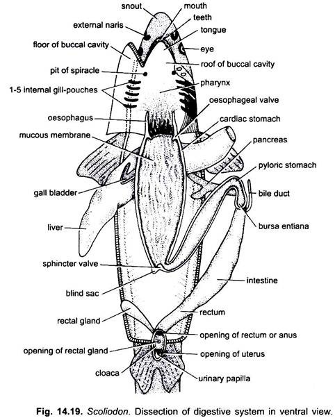
Abdominal cavity is very large surrounding the viscera (alimentary canal, liver, pancreas, spleen and gonads) and communicating with the exterior through a pair of abdominal pores situated on papillae, one on either side of the cloacal aperture. The abdominal cavity is lined with the peritoneum which also covers the surface of viscera. It is also filled with a colourless coelomic fluid.
The alimentary canal is suspended in the body cavity by a double fold of peritoneum with thin connective tissue in between, called mesentery. The mesentery is incomplete, only the anterior and posterior portions of the mesentery are well developed.
The anterior portion of the mesentery forms a large flap, the mesogaster which suspends the stomach, while the posterior portion, the mesorectum suspends the hinder part of the alimentary canal.
There are other membranes which attach different organs to one another and are called omenta (singular omentum). The membrane which connects the stomach with the liver is knows as gastro-hepatic omentum, while the membrane connecting the spleen with the stomach is the gastro-splenic omentum.

