The appendicular skeleton consists of skeletal elements of pectoral and pelvic girdles, and of the appendages. The appendages are median and paired. Median appendages are found in fishes and aquatic tetrapoda, and paired appendages are found in all vertebrates except cyclostomes. In fishes the paired appendages are pectoral and pelvic fins, in tetrapoda they are fore and hindlimbs.
Origin of Appendages:
1. Fin-Fold Theory:
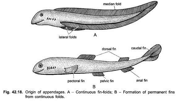
According to the fin-fold theory of Balfour, the ancestral vertebrate (fish) had a pair of lateral fin-folds, one on each side of the trunk, they fused behind the anus with a median ventral fin which extended along the tail to the mid-dorsal side of the body. This hypothetical condition resembles the median fins and metapleural folds of Amphioxus. Persistence of certain parts and disappearance of others of the fin-folds and median fin resulted in the formation of paired and median fins respectively in fishes.
Later on, the limbs of tetrapoda evolved from the paired fins of fishes. Thus, there are two transitions in the evolution of appendages, first from fin-folds to fins, and second from fins to limbs.
ADVERTISEMENTS:
This theory is supported by certain evidences:
(i) Basic skeletal structures of paired and median fins are similar. This shows a common mode of origin.
(ii) In embryos of certain elasmobranchs, a continuous series of muscle buds appear, certain of which later disappear leaving only paired and median fins.
(iii) In some extinct Devonian acanthodian sharks were present a row of numerous small accessory spiny fins on either side between pectoral and pelvic fins. These are supposed to be the remnants of fin folds.
ADVERTISEMENTS:
(iv) In Cladoselache (extinct shark) pectoral and pelvic fins are broad, having no basal notches and are supported by parallel cartilaginous rods, the pterygiophores. Their origin may be like primitive fin-fold. But no primitive fossil fish possessing continuous fin folds is known. Thus, this theory is not widely accepted now.
2. Fin-Spines Theory:
According to fin-spines theory, probably the first fins are formed of metamerically arranged spines with fleshy webs, like extinct acanthodian sharks. Later on, all spines with webs may have been disappeared leaving only an anterior and a posterior pair, that later gave rise to pectoral and pelvic fins.
ADVERTISEMENTS:
3. Ostracoderm Theory:
Certain ostracoderms had lateral fleshy lobes on either lateral side. They may be supposed to be the precursors of pectoral fins. Certain other ostracoderms possessed ventro-lateral rows of dermal spines like acanthodians. Leaving certain spines in the pectoral and pelvic regions, all were lost. This may be supposed to be the origin of paired fins and later limbs.
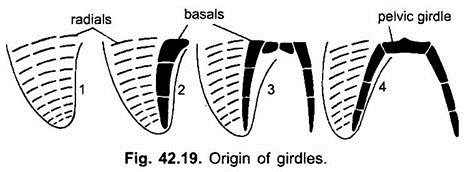
The paired and median fins are supported by a series of endoskeletal pterygiophores; each divided into pieces: those in the basal part are basals, while the distal pieces are radials. Muscles are attached to basals and radials for moving the fins.
The distal part of a fin is vane-like and is supported by integumentary fin rays. The horny unjointed fin rays are ceratotrichia found in elasmobranchs, bony jointed fin rays are lepidotrichia found in most bony fishes, and fibrous and jointed fin rays are campotrichia found in Dipnoi. In elasmobranchs and Dipnoi the paired fins have basal lobes and a vane-like part, these are called lobed fins. In most bony fishes the basal lobe is missing and paired fins have only a vane-like part projecting from the body, they are called rayed fins.
Origin of Girdles:
The basal pterygiophores fuse into larger pieces. The large anterior-most basal of each side extends towards the mid-ventral line, and fuse together to form a girdle made of a bar of cartilage.
Girdles and Limbs in Tetrapoda:
Girdles:
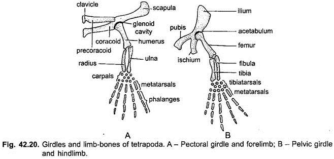
The girdles and paired limbs of tetrapoda are remarkably similar and have homologous parts.
ADVERTISEMENTS:
Pectoral girdle has several cartilage bones. They are a scapula, a suprascaphla, precoracoid, and a coracoid in each half. Between the scapula and coracoid a glenoid cavity appears for the first time. Two dermal bones, the clavicle and interclavicle are added in each half. But there is a gradual reduction or loss of some bones.
Pelvic girdle has three cartilage bones in each half. They are ilium, ischium and pubis. There are no dermal bones like pectoral girdle. All three bones participate in the formation of an acetabulum cavity. A cotyloid or acetabular bone often forms a part of the acetabulum in mammals. Generally a large obturator foramen lies between the ischium and pubis. The two halves of the pelvic girdle often meet midventrally at the pubic or ischiac symphysis. In most tetrapoda the pelvic girdle is attached to the axial skeleton through the ilia articulating with the sacral vertebrae.
Homology:
The bones of the pectoral and pelvic girdles are homologous, the scapula with the ilium, the precoracoid with the pubis and cotyloid, and the coracoid with the ischium. The clavicle is a dermal bone and is not homologous with the pubis which is a cartilage bone.
Limbs:
The limbs of tetrapoda are pentadactyl. They are used not only for locomotion but also to support the weight of the body above the ground. Thus, they are provided with joints. Each has three segments, the stylopodium, zeugopodium and autopodium with joints between the segments. The stylopodium is the upper arm (or thigh), having a single humerus (or femur).
The humerus joins the pectoral girdle at the glenoid cavity, while the femur joins the pelvic girdle at the acetabulum cavity. The zeugopodium is the forearm (or shank). It contains two parallel bones, the radius and ulna (or tibia and fibula). The radius and tibia are lateral, while ulna and fibula are medial. Between the stylopodium and zeugopodium is an elbow joint (or knee joint).
The autopodium has three divisions, a carpus or wrist (tarsus or ankle), metacarpus or palm (metatarsus or sole), and digits forming fingers or toes. The carpus or tarsus in the primitive condition has 10 bones in 3 rows, the first row has a radiale (or tibiale), an intermedium, and an ulnare (or fibulare), in the second row are generally two centralia, while the third row has five distal carpals (or distal tarsals).
The metacarpus (or metatarsus) has five long metacarpals (or metatarsals). The digits are generally five in number or pentadactyl, with the first digit on preaxial side and the fifth on the postaxial side. Each digit has a linear row of phalanges, they are 2, 3, 4, 5, 4 beginning from the first to the last.
There is some evidence that early tetrapoda had seven digits, one on the preaxial side was prehallex (or prepollex), and the postaxial digit was a postminimus. But these were probably additional bones of the wrist or ankle.
Origin of Pentadactyl Limbs:
I. Gegenbaur’s Theory:
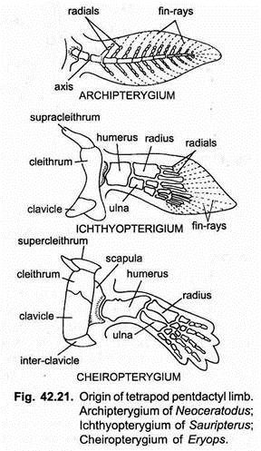
It was erroneously believed by Gegenbaur that Dipnoi were the ancestors of tetrapoda. The skeleton of their lobed fins, as seen in Neoceratodus, consists of a jointed axis of numerous bones from which biserial segmented radials and fin rays arose on both sides for supporting the fin. This type of fin is called an archipterygium. Gegenbaur’s theory deriving the tetrapod limb from the archipterygium is now discarded.
II. From Crossoptergians Fins:
The link between the fins of fishes and limbs of tetrapoda is found in fossil crossopterygians. The lobed fins of extinct Sauripterus and Eusthenopteron were like paddles. They are called ichthyopterygium and suggest the beginnings of tetrapod limbs.
Their fin had a well-developed fleshy lobe covered with scales and a paddle-like fin blade. In the lobe the skeleton had in the first row a single basal comparable to the humerus (femur), in the second row were two radials comparable of the radius (tibia) and ulna (fibula). The rest of the radials are comparable to carpals (tarsals).
Terrestrial animals have acquired jointed limbs for locomotion on land. The limb of an ancient extinct amphibian Eryops is known as a cheiropterygium in which there is a humerus in the first row, radius and ulna in the second row, followed by several carpals in diagonal rows, after which there is a prepollex and five digits which are not present in the fin blade and have been formed as new distal outgrowths.
If the skeleton of an ichthyopterygium is compared to that of a cheiropterygium, it is easy to see how the transformation of a fish fin into an amphibian limb took place. The former shows that even before locomotion on land was adopted the arrangement of its skeleton had already come to resemble that of land animals.
Thus, the crossopterygian fin or ichthyopterygium gave rise to a pentadactyl limb by an elongation of the basal lobe and its skeleton, by a loss of the fin blade and supporting fin rays and by formation and separation of terminal radials to form digits.
Girdles and Limbs in Tetrapoda:
1. Amphibia (Frog):
Pectoral girdle is an arch enclosing the chest. The two halves are united midventrally and closely related to the sternum. (In toads the two halves overlap). Each half has a coracoid, precoracoid, clavicle, paraglenoid cartilage, scapula and suprascapula.
The two precoracoid catilages fuse ventrally. The clavicle is the only dermal bone. It is fused to and reinforces a cartilaginous precoracoid. Glenoid cavity is bordered by coracoid, paraglenoid, and scapula. A partly bony suprascapula reinforces the scapula. On the inner margin of the coracoid is an epicoracoid cartilage. Anura have two kinds of pectoral girdles-(a) firmisternal in which the two epicoracoid cartilages are fused mesially along their entire lengths, (b) arciferal in which the two epicoracoids are fused only along their anterior edges.
A sternum having two separate parts is fused to the middle of the pectoral girdle. The pectoral girdle is reinforced anteriorly by an omosternum and posteriorly by a mesosternum and their cartilaginous expansions, the episternum and xiphisternum respectively.
Forelimb:
Humerus has a prominent deltoid ridge. Radius and ulna fuse into a radioulna. There are five carpals in two rows, in the first row the intermedium and ulnare are fused together, and there is a radiale. There is a single centrale. The three carpals of the second row are fused together. Thus, the number of carpals is reduced. Only four digits are present besides a rudimentary pollex enclosed in the skin.
Pelvic Girdle:
It is V-shaped and has a long ilium on each side. It articulates with the transverse processes of the sacral (9th) vertebra. The ilia act as shock absorbers when the animal lands after a jump. Pubis and ischium are small. The pubis is only partly ossified. The two pubes and two ischia are fused together to form a solid fulcrum.
All three bones join at the acetabulum cavity. Pelvic girdle is different from that of any other vertebrate; with very long ilia it forms a long lever for transferring the force from the hindlimbs to the vertebral column in jumping.
Hindlimb:
The tibia and fibula fuse into a tibiofibula or crural bone. There are five tarsals in two rows, the promixal row has two long bones, an inner astragalus (ulnare) and outer calcaneum (fibulare) which make the ankle very long for jumping.
There are five digits of which the first is called hallux. There is an additional digit called calcar or prehallux enclosed in the skin. It probably represents an additional tarsal and may be regarded as a remainder of a fin ray of the ancestral fin. It helps in stretching the web of the foot.
2. Reptilia (Varanus):
In reptiles the girdles and limbs are well-developed, except in snakes and limbless lizards, but vestigial pelvis and hindlimbs are found in some snakes.
Pectoral Girdle:
It is well formed and joined to the sternum. Each half has a bony coracoid and scapula, between which is a glenoid cavity. There is an expanded suprascapula of calcified cartilage. Between the coracoids of the two sides is an expanded epicoracoid cartilage formed by the fusion of two. In each half there are three fenestrae between the coracoid and epicoracoid.
A T-shaped unpaired dermal bone called interclavicle (episternum) lies midventrally. On the arms of the interclavicle are two clavicles bracing the girdle, but they do not meet ventrally. Clavicles and interclavicle are dermal bones. Attached to the coracoids is a sternum made of cartilage.
In turtles the coracoids and scapulae are slender rods, while the clavicle and interclavicle form dermal bony plates of the plastron.
Forelimb:
It is typical with no unusual feature. Humerus is broad, radius and ulna are separate, carpals are in two rows: four in the first row including a centrale, and five in the second row. There are five digits ending in claws.
Pelvic Girdle:
All three bones are fused inseparably to form a single innominate bone in each half. Ilium points backwards and is firmly united with two sacral ribs of the sacrum so that the articulation is post-acetabular. Both pubis and ischium meet their fellows to form pubic and ischiac symphyses. At the pubic symphysis is a epipubis cartilage and at the ischiac symphysis is a hypoischium cartilage.
The pubis has a small foramen for the obturator nerve. Between two innominate bones is a large cordiform (or ischiopubic) foramen divided by a ligament into two obturator foramina. Acetabulum is bordered by all three bones.
Hindlimb:
It is typical, but the tibia begins to become larger than the fibula. The tarsals are reduced by fusion, the proximal row has two bones and distal row has three, between the two rows of tarsals is an intratarsal ankle joint where bending takes place. There are five digits ending in claws.
In some reptiles, but not in Varanus, two structures appear for the first time in the hindlimbs, first the calcaneum bears a posterior calcaneal process or heel, secondly a sesamoid bone forms a patella or knee cap in front of the tibia.
3. Aves (Gallus):
The appendicular skeleton of birds is remarkably uniform. The pectoral girdles and forelimbs are modified for flight. The pelvic girdles and hindlimbs are modified to support the weight of the body in standing and walking. Bones are tubular, spongy, and pneumatic so that they are light, but lightness is achieved without any sacrifice of strength. The marrow cavity has no marrow but there are supporting bony struts which make the bones strong to bear stresses.
Pectoral Girdle:
It lies far backwards towards the centre of the body. It has a large, stout coracoid attached to the sternum at the coracoid groove by means of a synarthrosis or immovable articulation. Coracoid is joined at its other end at right angle with a sword-like scapula lying over and bracing the ribs. A glenoid cavity is formed by an imperfect union of scapula and coracoid.
It is greatly displaced above the centre of gravity, consequently the coracoids are elongated. In front of and attached to each coracoid is a thin clavicle. The two clavicles are joined ventrally to a small, flat interclavicle forming a furcula or “wishbone”. In flying birds the furcula is joined to coracoids and is well developed. In non-flying birds it is reduced or absent.
The arched clavicles and strong coracoids brace the sternum and resist the inward pressure of the powerful down-stroke of the wings. At the junction of coracoid, scapula, and clavicle is a foramen triosseum through which the tendon of a pectoralis minor muscle passes and is inserted on the humerus.
Forelimb:
Forelimb and its skeleton are modified for flight. The wings are attached high up on the thorax towards the centre of the body for balancing the weight in flying. The forelimb has a stout humerus with a deltoid ridge. Humerus has a pneumatic foramen through which an air sac enters making the bone spongy and light.
Air in pneumatic bones oxygenates blood and adjusts air pressure when a bird descends from a great height. Such bones have no marrow. Radius and ulna are well formed. The ulna is more strongly developed than the radius; the ulna is large for attachment of flight feathers. Carpus has four carpals in two rows-in the first row are radiale and ulnare, but the carpals of the second row fuse to three fused metacarpals to form a carpometacarpus.
Of the three metacarpals the first is much reduced, all three are fused proximally, but distally the second and third are free forming two rods. There are three digits- the first has one phalanx, second has two phalanges, and third has one phalanx. The digits are reduced to almost a one-fingered condition. Muscles of the upper arm are large and strong, those of the forearm are reduced, and those of the hand have atrophied.
Pelvic Girdle:
It has a longitudinal curvature which distributes the body weight evenly in bipedal locomotion. Ilia and ischia have become long and wide and lie longitudinally. Large expanded ilium extends in front and behind the acetabulum, and is fused to the synsacrum along its entire length. Ischium is broad and extends posteriorly. It lies parallel to and is fused with the ilium, an ilioischiatic foramen lies between the two bones.
A slender pubis extends backwards and ends freely. Between the ischium and pubis is an obturator foramen. All three bones are fused together to form an innominate bone which has an incompletely ossified acetabulum with a foramen.
Pubic and ischiac symphyses are absent so that large-shelled eggs can be laid. But in ostrich (Struthio) there is a pubic symphysis, while in Rhea, the South American ostrich, there is an ischiac symphysis.
Hindlimb:
These are modified for bipedal locomotion. A strong femur articulates at the acetabulum. Fibula is much reduced and is short. The tibia has a chemical crest, in front of the junction of femur and tibia. A sesamoid bone forms a patella. The tibia is fused with the proximal tarsals (astragalus and calcaneum) to form a tibiotarsus.
The distal tarsals fuse with the second, third, and fourth fused metatarsals to form a compound bone, the tarsometatarsus. The ankle joint is between the two rows of tarsals is intratarsal in position. The first metatarsal is free and projects near the lower end of the tarsometatarsus.
In the fowl and some other birds a bony spur projects backwards from the tarsometatarsus, it has a horny epidermal covering. The spur is more developed in male birds for fighting. The digits are never more than four, having 2, 3, 4, 5 phalanges. The last phalanx of each digit bears a claw. The first digit (hallux) usually points backward, while the other three digits are directed forward.
4. Mammalia:
The appendicular skeleton of mammals shows much variation from a reptile like condition to a high degree of specialisation.
Pectoral girdle of Ornithorhynchus, (a monotreme), has a scapula, coracoid, and a cartilaginous precoracoid (epicoracoid) on each side and there is an expanded suprascapula. Between coracoid and scapula is a glenoid cavity on each side.
Ventrally the two coracoids are connected with the manubrium of the sternum. Between the precoracoids is a median T-shaped episternum (interclavicle) forming a connection between the girdle and sternum. Two clavicles rest on the episternum and each is attached to a simple acromion of the scapula. Clavicles and episternum are dermal bones. Such a pectoral girdle is like that of a reptile.
In most mammals the precoracoids and interclavicle are lost, and the coracoids are much reduced, the clavicle may be a strong bony arch from the scapula to the sternum or it may be reduced or even lost. When the clavicles are lost all connection between the pectoral girdle and axial skeleton (sternum) disappears.
Pectoral Girdle (Rabbit):
Each half of the girdle has a triangular scapula or shoulder blade, at the end of which is a glenoid cavity. Near the glenoid cavity there is a coracoid process formed by the reduced coracoid. On the outer surface of the scapula is a spine which has two projections, an acromion process and a metacromion process for attachment of muscles.
There is a clavicle, a dermal bone forming a slender rod attached by ligaments to the acromion process and manubrium of the sternum. Clavicles are retained only in those mammals which have an extensive movement of forelimbs, in others they are reduced or lost.
Forelimb:
The limbs of most mammals are capable of extensive rotation, extension and bending. Humerus is stout with a deltoid ridge; the distal end has two expanded condyles between which are a pulleys or trochlea for articulation with ulna. Above the trochlea is an olecranon fossa perforated by a supratrochlear (supracondyle) foramen for the passage of a nerve and brachial artery.
Radius and ulna are separate but tightly bound together. In primates, with prehensile forelimbs, the radius and ulna are not fixed but the radius can rotate over the ulna to turn the hand to either a prone or a supine position.
The ulna is produced into an elbow or olecranon process, in front of which is a sigmoid notch for articulation with the humerus. Carpus has eight bones, three in the first row, then a small centrale and four distal carpals. There are five metacarpals having five digits with 2, 3, 3, 3, 3 phalanges ending in claws.
In primates the first digit or pollex is independent of the others. It can be brought opposite the other digits and palm. This opposable thumb can handle minute objects. This factor has led to a superior position of primates.
Pelvic Girdle:
In each half the ilium, ischium, and pubis fuse to form an innominate bone. The two halves meet by a midventral pubic symphysis. In rabbit and some others there is a small acetabular (cotyloid) bone at the lower margin of the acetabulum. The acetabulum lies where all three bones meet (except the pubis). There is a large obturator foramen between the pubis and ischium on each side. The two ilia articulate with the transverse processes of the first vertebra of the sacrum.
Hindlimb:
Femur is strong with a head for articulation with the acetabulum. There are three trochanters for attachment of muscles, distal end of femur has two smooth condyles. Tibia is strong and has a cnemial crest, above is a sesamoid bone or patella formed in the tendon of triceps femoralis muscle. It forms a knee cap which reinforces the ligaments of the knee joint.
Fibula is reduced and fused distally to the tibia but separate proximally. Tarsus has six bones, proximal row has astragalus and calcaneum, its postior projection, the calcaneal process forms a heel. Centrale lies in front of the astragalus, distal row has three tarsals.
There are four metatarsals bearing digits. Each digit has three phalanges ending in claws. First digit or hallux is absent. In some primates the hallux is opposable in the same way as the pollex of the hand.
Postures of Limbs:
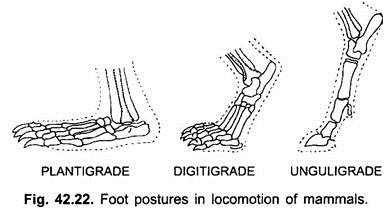
In terrestrial mammals there are three kinds of postures of hands and feet in locomotion.
a. Plantigrade:
Plantigrade locomotion is the most primitive, in which the entire hand and foot are in contact with the ground, as in bears, rabbit, some insectivores, and man.
b. Digitigrade:
Digitigrade locomotion is used for increased speed. In it only the digits are placed on the ground. The wrist and ankle are lifted above, as in dogs, cats and wolves.
c. Unguligrade:
Unguligrade locomotion is an extreme condition in which only the tips of the digits are placed on the ground and the tips are covered with hoofs. This is the most specialised condition, as in deer, horse, and cattle.
