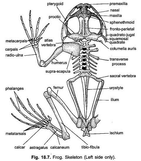
The skeleton that supports the soft parts lies internally and is designated as the endoskeleton. It is chiefly made of bones and cartilages. These two structures are joined with one another to form the internal framework.
The endoskeleton is described under two broad heads:
(i) The axial skeleton and
(ii) The appendicular skeleton.
ADVERTISEMENTS:
The axial skeleton comprises the skull, vertebral column and sternum and the appendicular skeleton consists of pectoral and pelvic girdles, and the skeleton of paired limbs.
(i) Axial Skeleton:
1. Skull:
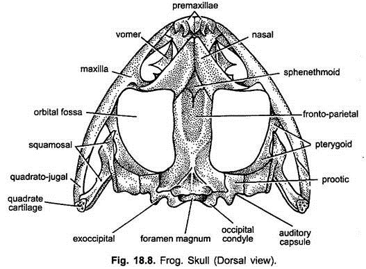
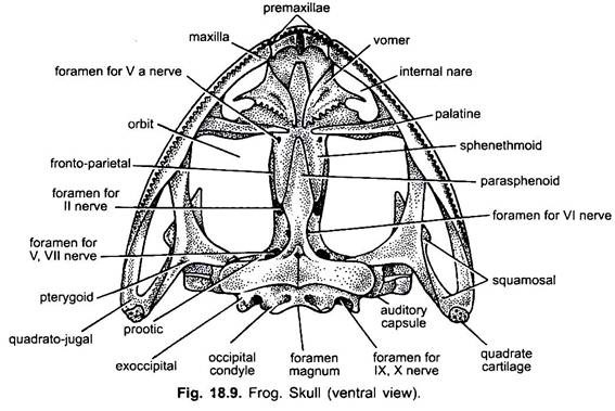
The skull of frog is broad and flat and consists of a narrow cranium or brain box, paired sense capsules, large orbits, the jaws, hyoid and cartilages of larynx.
2. Cranium:
ADVERTISEMENTS:
The cranium is the narrow cavity lodging the brain, hence, it is also called brain box. In the front part of the cranium is a sphenethmoid enclosing the forebrain and olfactory sacs.
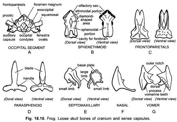
Skull is divided by a transverse partition into an anterior ethmoidal region and a posterior sphenoidal region which encloses the forebrain. The ethmoidal region encloses the olfactory sacs. Sphenethmoid is only exposed laterally. It is dorsally covered by a pair of nasals and a fronto-parietal except a small diamond-shaped area. It is ventrally covered by a dagger-shaped parasphenoid.
Occipital Segment:
ADVERTISEMENTS:
At the posterior end of the cranium is a foramen magnum surrounded by two exoccipitals. Each exoccipital bears at its posterior end a convexity, the occipital condyle which articulates with the concavity of the atlas vertebra.
The auditory capsule is fused on the outer side of each exoccipital. Each auditory capsule has a prootic in front, the capsule has an aperture, the fenestra ovalis into which a cartilaginous stapedial plate fits. The stapedial plate is a part of the columella derived from the hyomandibular.
Olfactory Capsules:
The olfactory capsules have two nasals dorsally and two vomers ventrally, the vomers bear vomerine teeth. A pair of special bones called septomaxillary (ethmoids) form the boundary of nostrils. They are associated with and surround the Jacobson’s organ.
Optic Capsules:
They enclose the eyes and are not fused with the skull.
Visceral Skeleton:
Visceral skeleton includes the upper and lower jaws, the hyoid apparatus, the columella auris and cartilages of larynx.
Upper Jaw:
ADVERTISEMENTS:
The upper jaw has two halves, each half has an anterior premaxilla followed by a long maxilla, both bear teeth. The posterior part of the upper jaw has a small quadratojugal. Its broad posterior end unites with quadrate cartilage, which is a small thin rod forming the suspensorium.
The mandible articulates with the quadrate cartilage. Ventral anterior to the orbit is a slender, rod-like palatine. At the posterior lateral end of cranium is present a large 3-rayed or Y-shaped pterygoid. It articulates anteriorly with the maxilla and palatine and on the inner side with the parasphenoid and auditory capsule, and posteriorly with the quadratojugal and quadrate cartilage.
At the posterior dorsolateral end of cranium is the hammer-shaped bone, the squamosal. It lies above the pteryoid. Its anterior limb or head is free and the short posterior limb articulates with the auditory capsule and prootic. Its handle joins with the quadrate cartilage.
Lower Jaw or Mandible:
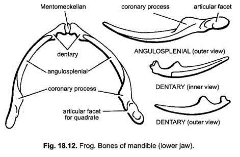
The lower jaw has two rami joined in front by elastic ligament. Each half has a core of Meckel’s cartilage covered over by an angulosplenial forming the inner and posterior portion of each ramus. Its anterior end is tapering and postenor end possesses dorsally a condyle for the articulation with the quadrate cartilage.
Just anterior to the condyle is present a small dorsal projection, the coronary process. Anterior outer surface of Meckel’s cartilage is covered by a small, flat, dogger-like dentary. It extends anteriorly up to Mentomeckelian bone and its posterior part is attached with the outer side of angulosplenial. At the extreme anterior end of Meckel’s cartilage is a small cartilage bone, the Mentomeckelian.
Hyoid:
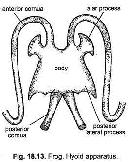
Hyoid has a thin body made of cartilage. It is produced in front into two alar processes and behind into two posterior lateral processes. In front are also two long slender anterior cornua of cartilage, they curve backwards and join the auditory capsules. Posteriorly is a pair of strong bony posterior cornua or thyrohyals between which lies the glottis. The hyoid lies in the floor of the pharynx, beneath the tongue.
Vertebral Column:
The vertebral column or backbone of frog encloses and protects the spinal cord. It is very much short due to the absence of tail. It is composed of nine vertebrae and a terminal rod-like structure called the urostyle. The first, eighth and ninth vertebrae are peculiar, while vertebrae from second to seventh are almost similar in structure.
Typical Vertebra:
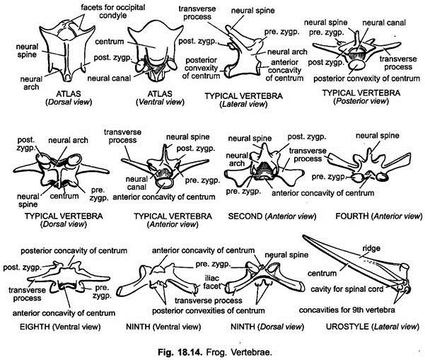
A typical vertebra has a solid cylindrical part known as the centrum. The centrum is procoelous, i.e., it is concave in front and convex behind. On the dorsal side, the centrum bears a ring like neural arch which encloses the neural canal.
The neural arch possesses a backwardly directed spinous process or neural spine. The lateral sides of the neural arch carry transverse processes. The neural arch possesses two articulating processes. The anterior processes having upwardly and inwardly directed articular surfaces are known as the prezygapophyses and the posterior processes having downwardly and outwardly directed articular surfaces are called the postzygapophyses.
The posterior convexity of a vertebra fits into the anterior concavity of the centrum of next vertebra. This mode of articulation of the centra gives flexibility. Between the neural arches of the consecutive vertebrae are spaces called inter-vertebral notches through which the spinal nerves pass.
First Vertebra:
The first vertebra is called the atlas vertebra. It is ring-like in form. Centrum and neural spine are reduced. Transverse processes and prezygapophysis are absent. The neural arch is large. The anterior face of centrum possesses a pair of concave facets for the articulation with the occipital condyles of the skull. The posterior margin of the neural arch bears a pair of postzygapophyses.
Eighth Vertebra:
The centrum of eighth vertebra is amphicoelous, i.e., concave on both the sides. The anterior concavity receives the posterior convexity of the Vllth vertebra. Transverse processes are long, slender and outwardly directed. Prezygapophyses and postzygapophyses are present on the anterior and posterior margins of the neural arch respectively.
Ninth Vertebra:
The ninth vertebra is also known as sacral vertebra. The centrum of ninth vertebra is biconvex, i.e., convex on both the sides (bearing one convexity anteriorly and two convexities posteriorly). The anterior convexity fits into the posterior concavity of eighth vertebra. The posterior convexities fit into the anterior concavities of urostyle.
Transverse processes are cylindrical, stout and backwardly directed. Iliac facet is present at the tip of each transverse process for the articulation of ilium bone of pelvic girdle. Neural spine is inconspicuous, i.e., greatly reduced. Prezygapophyses are well developed along the anterior end of neural arch, while the postzygapophyses are entirely absent.
Functions of Vertebral Column:
The vertebral column serves the following functions:
1. It supports the trunk region.
2. It encloses and protects the spinal cord.
3. In front it supports the head which is held slightly above the ground.
4. It acts as a body-axis from which viscera are suspended in the body cavity by mesenteries.
5. It helps in locomotion by providing attachment in the pelvic girdle and hindlimbs.
Sternum:
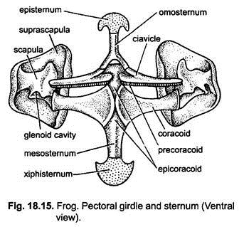
It lies midventrally connected between the two halves of pectoral girdle. It is composed of four parts. Anterior to the clavicle lies an inverted Y-shaped bony omosternum which is anteriorly attached with the rounded, flat cartilaginous episternum. Posterior to the epicoracoid and coracoids is a bony rod-like sternum proper or mesosternum to which is attached a broad cartilaginous xiphisternum posteriorly. Ribs are absent.
(ii) Appendicular Skeleton:
Pectoral Girdle:
The pectoral girdle or shoulder girdle (Fig. 18.15) is present in the thoracic region and provides attachment to the forelimbs and their muscles. It also protects the inner softer parts of the thorax. It consists of two similar halves united mid-ventrally and separated dorsally. Each half is divided into a dorsal scapular portion and a ventral coracoid portion.
Scapular Region:
The scapular portion comprises the suprascapula and scapula. Suprascapula is a thin, flat, somewhat rectangular cartilaginous plate on the dorsal side. It covers dorsally the first four vertebrae. Its free margin is calcified and the lower part articulating with the scapula is bony. At its posterior end is present a glenoid cavity into which articulates the head broad towards the end and narrower in the middle.
Coracoid Portion:
The coracoid portion comprises the clavicle, coracoid, precoracoid and epicoracoid. Clavicle (a slender rod) and coracoid (dumb-bell-shaped) meet mid-ventrally with the sternum and their counterparts of other side by a strip of cartilage, the epicoracoid.
Pelvic Girdle:
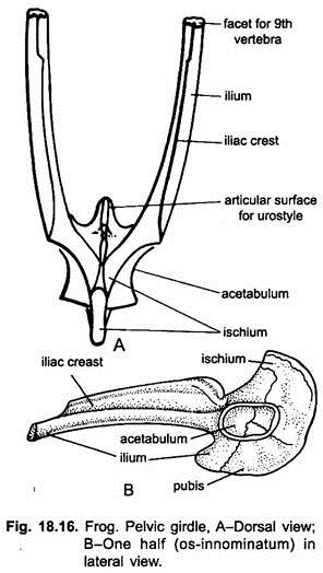
In frog, the pelvic girdle (Fig. 18.16) lies in the posterior region of the trunk. It gives support to the hindlimbs. It is V-shaped and composed of two similar halves, each of which is known as os-innominatum. Each os-innominatum is composed of three bones, ilium, pubis and ischium, which form the disc and the acetabulum.
Ilium is greatly elongated and forms the major part of each os-innominatum. It runs forwards to meet the transverse process of the ninth vertebra. It bears a prominent dorsal vertical ridge, the iliac crest. Both the ilia fuse posteriorly and form the anterior and upper half of the disc and acetabulum.
Publis is much reduced. It is a triangular piece of calcified cartilage, forming the central part of the disc and a small part of the acetabulum. Both the pubes are also fused. Ischium is larger and slightly oval bone and both the ischia fused in the middle and form one- third part of the disc and acetabulum. Thus, the disc is formed by the union of three bones containing a cup-shaped cavity, the acetabulum. In acetabulum the head of femur articulates.
Forelimbs:
The bones of the forelimbs include humerus, radio-ulna and the bones of hand.
a. Humerus:
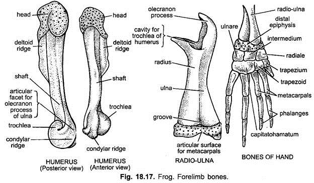
Humerus (Fig. 18.17) is the bone of the upper arm of forelimb. It is a short, stout and cylindrical bone with a slightly curved shaft. Its proximal rounded end is known as the head which fits into the glenoid cavity of pectoral girdle. The head is covered with calcified cartilage. The ridge below the head is known as deltoid ridge to which muscles are attached. The distal end forms a rounded trochlea with a condylar ridge on either side. The trochlea articulates with the groove of radio-ulna.
b. Radio-Ulna:
Radio-ulna (Fig. 18.17) is a compound bone of forearm of forelimb. It is formed by the fusion of radius and ulna bones. Its proximal end has a concavity to receive the trochlea of humerus. The proximal portion of ulna projects into an olecranon process forming the elbow joint.
The distal portion of radio-ulna is somewhat flat having a median groove dividing it into an anterior (radial) part and a posterior (ulnar) part terminating into a facet providing articular surface for the proximal row of carpals.
c. Bones of Hand:
The bones of wrist are called carpals. The carpal bones are six in number and are arranged in two rows of three each. The bones of the proximal row are called ulnare, intermedium (centrale) and radiale. These bones articulate with the radio-ulna. The bones of the distal row are called capitohamatum, trapezoid and trapezium. These bones articulate with the metacarpals.
The hand or manus is provided with five slender metacarpals. The first metacarpal is rudimentary. The digit corresponding to thumb is absent; the remaining four metacarpals are supported by phalanges. The second digit bears two phalanges. The third and fourth digits bear three phalanges each.
Hindlimbs:
The bones of handlimbs include femur, tibio-libula, astragalus-calcaneum and bones of the foot.
a. Femur:
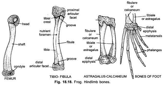
Femur (Fig. 18.18) is the bone of thigh region of hindlimb. It is long and slender having a slightly curved shaft. The proximal swollen end is called the head. Head fits into the acetabulum of pelvic girdle forming a ball and socket joint. The distal end forms a condyle which articulates with the tibio-fibula. The head and condyle are covered by calcified cartilage.
b. Tibio-Fibula:
Tibio-fibula (Fig. 18.18) is a compound bone of shank region of hindlimb. It is formed by the fusion of inner tibia and outer fibula bones forming a single bone called the tibio- fibula. In between the two is a median longitudinal groove. The proximal and distal ends are covered by calcified cartilage. Near the proximal end tibia bears an cnemial or tibial crest. The proximal end articulates with the femur, while the distal end articulates with the astragalus-calcaneum.
c. Astragalus-Calcaneum:
Astragalus-calcaneum (Fig. 18.18) is a compound bone of ankle of hindlimb. The ankle consists of two rows of four tarsal bones. The proximal row consists of two long bones fused together at their proximal and distal ends with a wide gap in the middle.
The inner bone is thinner and slightly curved, called the astragalus or tibiale, while the outer bone is thicker and straight, called the calcaneum or flbulare. The proximal and distal ends are covered by epiphyses of calcified cartilage. Distal row of tarsals has two very small bones.
d. Bones of Foot:
The foot or pes (Fig. 18.18) is supported by five long and slender metatarsals bearing five true toes, having 2, 2, 3, 4 and 3 phalanges respectively. A small preaxial sixth toe composed of 2 or 3 bones is present on the inner side of the first toe or hallux. Preaxial sixth toe is called the prehallux or calcar and does not project from the toe.
