The nervous system includes:
(i) A central nervous system comprising the brain and spinal cord,
(ii) A peripheral nervous system consisting of cranial and spinal nerves arising from the brain and spinal cord respectively and
(iii) An autonomic nervous system made of two ganglionated sympathetic nerves.
ADVERTISEMENTS:
The autonomic nervous system is often regarded as a part of the peripheral nervous system because the two are connected.
Central Nervous System:
A. Brain:
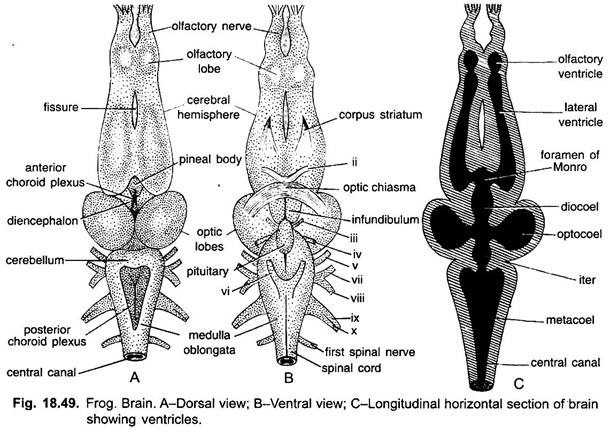
The brain of frog is elongated, bilaterally symmetrical, white coloured structure safely situated in the cranial cavity of the skull. It is surrounded by a thin, pigmented and vascular connective tissue membrane, the piamater, which is closely applied with the brain. Outside this membrane is a tough, fibrous membrane lining the interior of the cranial cavity called duramater. These two membranes are called meninges (singular, menix).
The space between the piameter and duramater is known as subdural which is filled with a kind of shock absorbing watery, clear lymphatic cerebro-spinal fluid. It is also found in the cavities of brain and central canal of spinal cord.
The brain of frog is divisible into three main parts:
ADVERTISEMENTS:
(i) Forebrain or Prosencephalon;
(ii) Midbrain or Mesencephalon;
(iii) Hindbrain or Rhombencephalon.
(i) Forebrain:
ADVERTISEMENTS:
It is the largest part of the brain consisting of a pair of anteriorly directed olfactory lobes, a pair of cerebral hemispheres, and a diencephalon.
(a) Olfactory Lobes:
The olfactory lobes are anterior small, spherical structures which are fused together in the median plane. Each lobe gives off an olfactory nerve and possesses a small cavity rhinocoel or olfactory autricle.
(b) Cerebral Hemispheres:
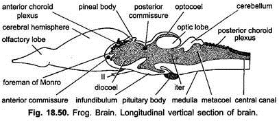
The two cerebral hemispheres are long, oval structures separated from olfactory lobes by a slight constriction. They are wider behind and narrower in front. They are separated from each other by a deep median longitudinal fissure. Each cerebral hemisphere has a large lateral ventricle or paracoel which is continuous anteriorly with the rhinocoel. Posteriorly the lateral ventricles communicate with each other and with the ventricle of diencephalon called diocoel by an opening, the interventricular foramen or foramen of Monro.
The nerve cell bodies form masses around the lateral ventricles and lie in layers. Fibres of the olfactory, tactile and optic impulse reach the cerebral hemisphere which may act as correlating centres but the hemispheres are largely olfactory in function.
On ventro-lateral side of each of the cerebral hemispheres there is a thick fibrous tract called the corpus striatum containing a network of white medullated nerve fibres and nerve cells. The corpora striata of two hemispheres are joined by a transversely running tract of fibres called anterior commissure and above which is another commissure (upper line) partly representing the hippocampal commissure of the brain of reptiles and mammals. They have a thick roof called the pallium in which more nerve cells have moved to the periphery.
(c) Diencephalon or thalamencephalon is a short unpaired structure of the forebrain situated behind the cerebral hemispheres. Its lateral walls are thick called optic thalami (singular thalamus) and its thick floor is called the hypothalamus. Its roof is thin and lined with a vascular membrane, the anterior choroid plexus.
ADVERTISEMENTS:
Behind it arises a hollow, thin-walled stalk, called the pineal stalk which terminates dorsally at the brow spot. The pineal stalk or epiphysis, which originally was continuous with the brow spot, becomes constricted off from it in early larval life. On the ventral side of diencephalon is the optic chiasma or crossing of the optic nerves which go the eyes. Just behind the optic chiasma is a flattened bilobed infundibular lobe or infundibulum extending posteriorly and divided by a median longitudinal groove.
It is formed of nervous tissue and contains a cavity which is continuous with the III ventricle or diocoel of diencephalon. The hypothalamus cerebri or pituitary body lies behind and partly covered by infundibulum. The hypothalamus is an important centre regulating the whole endocrine system as well as other parts of the brain. It is composed of anterior and posterior parts. The anterior part of the hypophysis has no connection with the brain.
(ii) Midbrain:
It is well developed consisting of two dorso-lateral large rounded optic lobes. The optic lobes are centres for impulses coming from the eyes. Their cavities are called optocoel or optic ventricles communicating with each other and the fourth ventricle behind through a narrow cavity, the iter or aqueduct of Sylvius.
Below the optic lobes are present two thick longitudinal bands of nerve fibres, called the crura cerebri. These connect diencephalon and medulla. These form the floor of midbrain. Lying transversely between the diencephalon and optic lobes is a band of nerve fibres called posterior commissure.
(iii) Hindbrain:
It consists of the cerebellum and the medulla oblongata:
(a) The cerebellum is a rudimentary narrow transverse solid band lying dorsally immediately behind the optic lobes. Its function is probably to regulate the vestibulo-oculomotor system controlling movements of the eyes,
(b) Medulla oblongata is short and somewhat triangular structure which is simply a widening of the spinal cord.
Its cavity is also triangular called fourth ventricle or metacoel which is joined in front to the iter but posteriorly it is continuous with the central cavity of the spinal cord. Its roof is thin, vascular and thrown into folds called the posterior choroid plexus.
From the sides of medulla arises several pairs of cranial nerves. Its lower surface is divided by a median fissure which is continuous with the ventral fissure of the spinal cord. On the dorsal surface of the medulla oblongata there is a triangular area of reddish brown colour which is called posterior choroid plexus.
B. Spinal Cord:
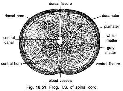
The medulla oblongata continues behind as spinal cord, lying in the neural canal of the vertebral column. It is short, somewhat flattened dorsoventrally and terminates behind the lumbar swelling in a tapering narrow thread called filum terminale lying in the urostyle.
The filum terminale with the nerve roots on either side is sometimes called cauda equina as it looks like a horse tail. It presents two enlargements during its course, one at the level of the forelimbs where nerves for arms arise and one far behind at the level of hindlimbs where nerves for hindlimbs arise.
The anterior enlargement of the spinal cord is known as brachial or cervical swelling, while the posterior as lumbar or sciatic swelling. It is traversed throughout its length by two longitudinal grooves, one on the dorsal side and the other on the ventral side, known as dorsal and ventral fissure respectively. These two fissures almost completely divide the spinal cord into two symmetrical halves.
Like the brain the spinal cord is also formed of gray matter with ganglion cells and non-medullated nerve cells surrounding the central canal, and white matter with ganglion cells and medullated nerve fibres surrounding the grey matter.
Grey matter is produced at the comers into dorsal and ventral columns or horns, while the white matter is divided by grey matter into four zones or funicules, the dorsal, the ventral and two lateral funicules. It is covered by two protective membranes; the duramater lines the neural canal and the vascular, thin and pigmented piamater closely covers the cord.
Functions of Different Parts of the Brain:
Brain is the only centre for the immediate control of all vital activities as it receives impulses from different parts of the body through sensory nerves and issues orders through motor fibres to different parts of the body for appropriate action.
i. Cerebrum:
The pallium of cerebral hemispheres controls the activities of the olfactory, tactile and optic organs, whereas the cerebral hemispheres coordinate the activities of the neuromuscular mechanism of the body, but these are supposed to be the seat of intelligence and voluntary control in higher animals.
ii. Diencephalon region of the brain controls the metabolism of the fats and carbohydrates and also regulates the genital functions.
iii. Optic lobes and optic thalami are supposed to be concerned with the sensation of sight and the control of the movement of the eye muscles.
iv. Cerebellum controls the mechanism of the automatic movements and also brings about coordination in movements of locomotion. It is in correlation with the medulla oblongata and regulates complex muscular movements of the body.
v. Medulla oblongata is an important nerve centre. It has nerve centres of all reflex functions and, thus, regulates particularly those functions of the body which are not directly under the control of the will like heart beating, respiration, swallowing, taste, hearing, sound production and secretions of various digestive juices.
Peripheral Nervous System:
The peripheral nervous system includes the nerves arising from the brain and spinal cord. Those nerves which arise directly from the brain are called cranial nerves, while those arising from the spinal cord are called spinal nerves.
Structure of Nerve:
The nerves are solid structures looking like white threads. Each nerve is composed of several bundles of nerve fibres called the funiculi (singular funiculus) and is covered externally by a sheath of loose connective tissue called the epineurium.
Each funiculus is also being enclosed by a thick covering of dense tissue called the perineurium. In each bundle or funiculus the individual nerve fibre is also covered by a connective tissue sheath called endoneurium which is continuous with the neurilemma of nerve fibres.
Types of Nerves:
A nerve may be afferent or sensory with sensory nerve fibres or efferent or motor with motor nerve fibres or mixed with both sensory as well as motor nerve fibres. Sensory nerves or nerve fibres are those which carry the impulses from the receptors to the central nervous system, whereas the motor nerves or fibres carry the impulses of appropriate order from the central nervous system to the effector organs.
In the body the following four types of fibres are recognised:
(a) Somatic sensory;
(b) Somatic motor;
(c) Visceral sensory; and
(d) Visceral motor.
(a) Somatic Sensory:
These are the fibres or nerves which carry the impulses from receptors such as skin, eyes, nose, body wall, muscles and joints to the central nervous system. The dendrons of these nerve fibres are very long, starting from the receptors pass to dorsal root ganglion in which their cell body lies.
(b) Somatic Motor:
Such fibres or nerves carry impulses from the central nervous system to the effector organs which include mainly the muscles. Their dendrites and cell bodies are always found in grey matter but their long axons pass through the ventral roots to the muscles (effector organ).
(c) Visceral Sensory:
Such fibres or nerves carry sensations from the receptors situated in the wall of alimentary canal to the central nervous system. Their dendrites start from the receptors located in the wall of alimentary canal and pass to the cell bodies situated in the dorsal root ganglia from where the axons pass into the grey matter.
(d) Visceral Motor:
These fibres are able to convey impulses from the central nervous system to the involuntary muscles of the alimentary canal, glands and other visceral organs. Their dendrites and cell bodies are found in the grey matter, while the axons pass out through the ventral root and end in the autonomic ganglia (preganglionic) from where the second neuron starts whose axon extends to the involuntary muscles or glands (post-ganglionic).
Cranial Nerves:
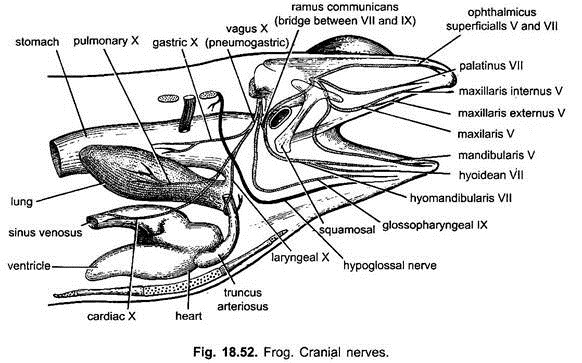
In an adult frog ten brain pairs of cranial nerves are found which emerge from the brain through various foramina of the cranium to supply the different organs of the body. Each cranial nerve originates from the brain by two roots, a dorsal and a ventral root, but these two roots do not unite with each other and, thus, look like separate nerves. These are numbered I to X, some of which are purely motor, some are sensory, while others are mixed.
The cranial nerves with their names are as follows:
I. Olfactory Nerve:
It arises from the anterior end of olfactory lobe and innervates the cells of olfactory sac. It is sensory in nature.
II. Optic Nerve:
Nerve fibres arise from the retina of the eye. The fibres of the two sides generally cross or decussate out the optic chiasma and then enter the optic thalamus of the opposite side, finally terminating in the thalamencephalon. It is also purely sensory.
III. Oculomotor Nerve:
It is small nerve arising from the ventral side of the midbrain (crura-cerebri). It divides into branches which supply the anterior, superior and inferior recti muscles and the inferior oblique muscle of the eye ball. It is exclusively motor.
IV. Trochlear or Pathetic Nerve:
It is also a small nerve arising from the dorsal side of the brain between the optic lobes and cerebellum and going to the superior oblique muscle of the eye ball, it is exclusively motor.
V. Trigeminal Nerve:
It is the largest of the cranial nerves arising from the sides of the anterior end of the medulla oblongata. Before it emerges from the skull it bears Gasserian ganglion.
It divides into three branches:
(a) Ophthalmic superficialis passes along the dorsal border of the orbit and goes to the skin of the snout. It is somatic sensory,
(b) Mandibular and
(c) Maxillary arise from a common stem and then separate.
The mandibular goes to the muscles of lower jaw and the maxillary forms the two branches going to the skin of the upper jaw and upper lip. Maxillary is a somatic sensory, whereas the mandibular is visceral motor nerve. Thus, trigeminal is a mixed nerve.
VI. Abducens Nerve:
It arises from the ventral side of the medulla oblongata and enters the orbit and goes to the posterior rectus muscle of the eye ball. It is a motor nerve.
VII. Facial Nerve:
It arises from the antero-lateral side of medulla oblongata close behind the fifth. It is mixed nerve as having both visceral sensory and visceral motor fibres.
It is divided into two branches:
(a) Palatine going to the roof of the buccal cavity,
(b) Hyomandibular going to the tongue and muscles of the lower jaw.
VIII. Auditory Nerve:
It is somatic sensory arising from the medulla oblongata behind the seventh and goes to internal ear.
IX. Glossopharyngeal:
It is mixed nerve arising from the lateral side of medulla and goes to the tongue, hyoid and pharynx. It does not bear pre-frematic and post-trematic branches to the first gill.
X. Vagus or Pneumogastric:
It is mixed nerve arising from the lateral side of medulla and goes as visceral branch to the larynx (laryngeal), oesophagus and stomach as gastric, heart (cardiac) and lungs (pulmonary).
Spinal Nerves:
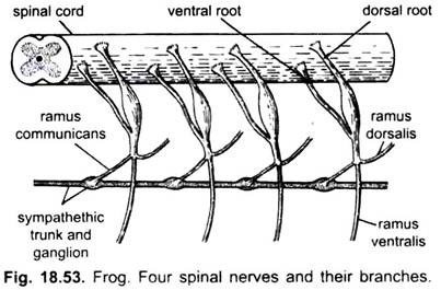
In Indian frog, Rana tigrina usually 9 pairs of spinal nerves are found which arise from the spinal cord by two roots, a dorsal or sensory root and a ventral or motor root. Both the dorsal and ventral roots unite immediately after coming out of the neural canal through intervertebral foramen. Dorsal root has a ganglion of nerve cells.
The dorsal root entirely of afferent fibres which may be somatic sensory or visceral sensory which carry impulses from different body parts towards the spinal cord. The ventral root consists of somatic motor or visceral motor fibres of which the cell bodies together with dendrons are situated in the ventral part of the grey matter of spinal cord.
These carry responses from the spinal cord to the body tissues. Thus, in the spinal nerves these different kinds of fibres are mixed but as the roots are approached, they are segregated.
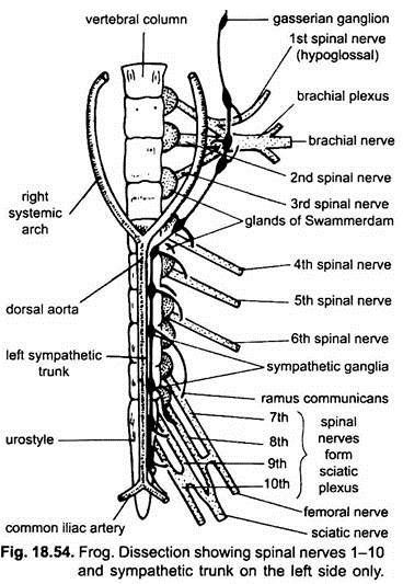
The dorsal root ganglia are covered by white soft chalky masses, calcareous bodies or glands of Swammerdam or periganglionic glands. It is probably the reserve supply of calcium to the body. Each nerve immediately after its origin gives off a short dorsal branch, ramus dorsalis to the dorsal skin and muscles of the back, a large ventral branch, ramus ventralis to the ventral skin and muscles of the body and a very short ramus communicans which unite to the nearest sympathetic ganglion.
Ramus dorsalis contains only somatic sensory fibres, whereas the ramus ventralis contains somatic motor fibres, while ramus communicans contains both visceral sensory and visceral motor fibres.
The first spinal nerve (hypoglossal) comes out between the first and second vertebrae and innervates the tongue and several muscles attached to the hyoid. The second spinal nerve is quite large and emerges between the second and third vertebrae. It receives the branches of the first and third spinal nerves to form a brachial plexus, then it proceeds as the brachial nerve to the skin and muscles of the forelimbs. The fourth, fifth and sixth nerves are small going obliquely to the skin and muscles of the abdomen.
The seventh, eighth and ninth spinal nerves pass almost directly backward and anastomose to form the sciatic or lumbo-sacral plexus from which a large sciatic nerve and several small nerves enter the hindlimb. The most important one is sciatic nerve which divides into tibialis and peroneus.
The tibialis gives branches to the gastrocnemius, tibialis posticus and numerous muscles of the plantar surface of the foot. The peroneus supplies the peroneus muscle tibialis. The tenth spinal nerve is not found in Rana tigrina but in other frogs it is present. When it is present it emerges from a foramen in the anterior part of the urostyle and goes to the cloaca and urinary bladder.
The roots of the last four pairs of spinal nerves are elongated forming a bundle of nerves called Cauda equina which lies inside the vertebral column along the filum terminale.
Autonomic Nervous System:
The autonomic nervous system is partly independent and not under voluntary control. Though it is involuntarily controlled by the nerve centres located in the central nervous system, it is also connected to spinal nerves and some cranial nerves. It is simply concerned with the intestinal regulation of the body with the central nervous system together with its spinal and cranial nerves and is concerned with the external regulations.
The autonomic nervous system consists of two delicate longitudinal chains of ganglia, lying one on either side of the backbone and the dorsal aorta from the brain to the end of the urostyle. Each chain at regular intervals has ten small metamerically arranged sympathetic ganglia. Each ganglion is connected with the corresponding spinal nerve by a small branch called ramus communicans.
Corresponding ganglia of both the cords are also connected together by small transverse commissure. Between the first and second ganglia the sympathetic chain of each side divides to form a loop around the subclavian artery which is known as annulus of Vieussens. Each sympathetic chain enters the skull along with the Xth cranial nerve through the vagus foramen and joins the vagus ganglion and then proceeds forward to join the Gasserian ganglion of the fifth cranial nerve.
Posteriorly each sympathetic chain ends by joining with one or more branches of the 9th spinal nerve. Sympathetic ganglia give nerves to the viscera, where they form plexuses, such as a solar plexus near the coeliaco-mesenteric artery and a cardiac plexus on the heart.
Kinds of Automatic Nervous System:
The autonomic nervous system is simply formed of visceral motor and visceral sensory fibres, which can be classified as follows:
1. The sympathetic;
2. The parasympathetic.
The visceral sensory fibres have their cell bodies in the dorsal root ganglia and their dendrites lie in the organs which are not under voluntary control like the heart, blood vessels, different parts of the alimentary canal. Their axons are extended from the dorsal root ganglia to the grey matter of the central nervous system.
The visceral motor fibres have their cell bodies in the spinal cord forming synapses with neurons whose cell bodies are situated in the sympathetic ganglia.
Because of these synapses the visceral motor fibres are classified under two heads:
1. Preganglionic fibres which arise from the grey matter of the spinal cord and pass through spinal nerves and rami communicants and go to the corresponding sympathetic ganglia are medullated.
2. Postganglionic fibres are those whose cell bodies are in sympathetic ganglia and going in organs they supply are non-medullated.
Thus, sympathetic nerves are made of both visceral sensory and visceral motor fibres which on stimulation secrete a chemical substance called sympathy which generally stimulate the organs.
Parasympathetic system includes parasympathetic nerves and ganglia. The fibres of parasympathetic nerves come from cranial and spinal nerves, while parasympathetic ganglia are situated in the organs innervated by parasympathetic nerves. Their preganglionic fibres are very long and extend from the central nervous system to a ganglion in or near the organ which they innerverate. Here they are connected to the short postganglionic fibres.
These fibres on stimulation secrete a chemical substance called acetylcholine which has an inhibitory effect on the organs. The parasympathetic nerve fibres are included in oculomotor, trigeminal, facial and vagus cranial nerves.
The visceral and some other organs receive fibres both from sympathetic and parasympathetic nerves. These are antagonistic in effect as sympathin of sympathetic fibres brings an action, whereas acetylcholine of parasympathetic fibres retards it.
The autonomic nervous system simply regulates the functions of certain organs which are involuntary. It is not independent because it is intimately bound structurally and functionally with central and peripheral nervous system. Its nerve centres are located in the central nervous system, while its most fibres are parts of the peripheral nervous system.
