In frog various types of receptors or organs of special sense are found which are supplied with nerve fibre and, thus, convey the stimulus to the central nervous system.
These receptors can be grouped under two heads:
I. External or Exteroceptors:
External receptors are those which receive impulses from the external environment.
External receptors can be grouped under following heads:
ADVERTISEMENTS:
(i) Tangoreceptors or organs of touch;
(ii) Olfactoreceptors or organs of smell;
(iii) Gustatoreceptors or organs of taste;
(iv) Photoreceptors or organs of sight;
ADVERTISEMENTS:
(v) Statoacoustic receptors or organs of hearing and balance.
(i) Tangoreceptors or Organs of Touch:
The entire skin serves as organs of touch as it is abundantly supplied with sensory nerve endings situated in the spaces between the cells. At places compact groups of cells form corpuscles which project into papillae of epidermis. These are supplied with sensory nerve endings. Such groups of cells are called tactile organs or patches.
These are very much sensitive to touch and also to temperature. The nerve endings never reach the cells of the outermost layer of epidermis. Consequently the stimuli which produce the sensation of touch are not directly received by the tactile organs. The tactile organs make the skin of the frog sensitive to touch, heat, cold and the effects of the chemicals. In tadpole larva a lateral line system is found which is absent in adult frog.
(ii) Olfactoreceptors or Organs of Smell:
These are the organs which are simply concerned with distinguishing the various kinds of smell given off from different substances or things. They include a pair of olfactory or nasal sacs located in the olfactory capsules of the skull. Each nasal sac communicates with the outside by the external nares and with the buccal cavity through the internal nares.
ADVERTISEMENTS:
These are internally lined with columnar epithelial cells out of which certain are special modified cells called neurosensory or olfactory cells which are bipolar in shape. Their deeper ends are connected to the fibres of the olfactory nerves which are directly connected with the brain (olfactory lobes), while their other ends are produced into sensory hairs projecting into the lumen of the nasal sac.
The mucous lining of the sac also has supporting and mucus secreting cells whose secretion keeps the nasal epithelium moist in order to make it more effective. The odours of a substance are brought to these organs either through the medium of air or water. From these organs the stimuli travel along the olfactory nerves to the brain.
(iii) Gustatoreceptors or Organs of Taste:
Gustatoreceptors or organs of taste are found in the form of taste buds which are confined chiefly over the tongue and the floor and roof of the buccal cavity. Each taste bud is more or less spherical body consisting of a group of barrel-shaped columnar cells, some of which are neurosensory and others are supporting cells.
The neurosecretory cells are slender and elongated and found in the middle and are covered all around by supporting cells. The free ends of neurosensory cells are produced into delicate fine taste hairs projecting above the surface, while other ends are innervated with sensory nerve fibres. The supporting cells are larger in size but lack sensory hairs. Taste buds of frog are supplied by the branches of the VII and IX cranial nerves.
The mucous membrane of the tongue is produced into two kinds of papillae- the conical filiform papillae and rounded knob-like fungiform papillae. The taste buds are confined only in fungiform papillae and stimulates whenever their taste hairs come in contact with a substance in solution.
(iv) Photoreceptors or Organs of Sight:
The organs of sight are two spherical eyes which are situated in the orbital fossae on either side of the head. Their structure and function is like that of other vertebrates. Their one-third part is visible externally but remaining part lies hidden in the orbit. The orbit has no bone at its bottom, therefore, the eyeballs can be seen projecting on the roof of the buccal cavity as spherical prominences. The eyes are protected with two eyelids-the upper is immovable and lower one is movable.
There is a transparent nictitating membrane which covers the eyes when the frog goes inside the water or underground. The nictitating membrane is simply a transparent part of the upper border of the lower eyelid which lies folded beneath the opaque part of the lower eyelid. Its movement is controlled by means of two types of muscles, the retrator bulbi and the levator bulbi.
On the contraction of the retrator bulbi muscles the eyeball is drawn deep into the orbit due to which the eyelids come nearer and the nictitating membrane is unfolded over the eye. On the contraction of the levator bulbi muscles the eyelids are separated that results in the folding back of the nictitating membrane to its original position.
Movement of Eyeball:
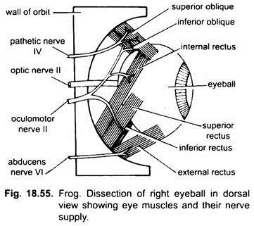
The eyeball is moved by a set of six extrinsic muscles:
(i) Four rectus mucles (anterior rectus, posterior rectus, superior rectus and inferior rectus). These muscles rotate the eyeball forwards, backwards, upwards and downwards respectively,
(ii) Two muscles are superior and inferior oblique muscles.
The superior oblique muscles bring about the rotation of the eyeball along the axis between the optic nerve and cornea, while inferior oblique muscles bring just the opposite movement. The muscles arise from the front part of the orbit.
Harderian Glands:
The surface of the eyes remains moist due to the presence of Harderian glands situated at the lower inner angle of the eye. These glands secrete a liquid which lubricates the eyeball. Excess secretion is drained into the nasal sac through a fine naso-lachrymal duct.
Structure of Eyeball:
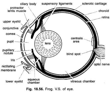
The eyeball is made of three concentric layers or coats, an outermost sclerotic, a middle choroid and an inner retina.
a. Sclerotic:
It is the outermost think protective layer which is made of fibrous connective tissue and cartilage. It is actually the optic capsule which has not fused with the skull but fits closely to the eye. It occupies about two-thirds of the entire circumference of the eyeball and is mostly out of sight being within the orbits. The remaining one-third of it is continued in front as an arched transparent cornea.
It simply maintains the shape of the eyeball and also protects the eyeball and also provides surface for attachment of extrinsic eye muscles. Cornea is covered externally by a thin transparent membrane called conjunctiva which is the continuation of skin. It is also continued with the inner lining of the eyelids. Nictitating membrane which is the continuation of the inner membrane of lower eyelid protects the outer exposed surface of the eye. It has thin blood capillaries.
b. Choroid:
It is the middle vascular and pigmented layer made of loose connective tissue fibres. Towards the anterior side it thickens as a ring-like ciliary body having ciliary muscles and then separates from the sclerotic forming a circular pigmented iris with a central aperture, the pupil, which looks like a black spot. Within the iris are two sets of involuntary muscles, circular and radials, which regulate the opening of pupil.
Immediately behind the pupile iris is a large, circular and transparent crystalline lens which is enclosed in a delicate lens capsule. It is suspended behind the pupil by a membranous suspensory ligament from ciliary body. The iris divides the hollow of the eyeball into a small anterior aqueous chamber and a large posterior vitreous chamber. The aqueous chamber is filled with a watery aqueous humour, whereas the vitreous chamber is filled with gelatinous vitreous humour.
c. Retina:
It is the innermost sensitive layer present only in the posterior part of eyeball behind ciliary body. It is made of several layers. The outermost layer is formed of pigment cells lining the choroid and beneath it is the sensory layer of rods and cones, which is followed by several layers of sensory cells.
The innermost layer is of nerve fibres which are connected to the optic nerve. Towards the posterior side the optic nerve leaves the eyeball through the three coats. This site is called the blind spot which has no rods and cones. Close to it is the yellow spot where a distinct image is formed but at blind spot no image is formed.
Microscopic Structure of Retina:
Microscopically the retina is a complicated coat consisting of several layers of different kinds of sensory cells. Next to the outer pigmented layer of the retina, there is a layer of visual cells consisting of rods and cones followed by a layer of bipolar nerve cells and an outer layer of ganglionic cells. The rods and cones are modified sensory cells which have been so named due to their shape.
These cells are placed at right angle to the surface of the retina and their thin ends are embedded in a layer of pigment cells. The rods are cylindrical cells containing a purple pigment and the cones are conical tapering cells. The rods are meant simply to perceive the amount of light, while the cones for distinguishing colours.
The bipolar nerve cells on one side form synapses with the rods and cones, while on the other side with the ganglionic cells. The axons of ganglionic cells spread over the inner surface of the retina, and converge at the back of the eyeball where they pierce the retina and come together to form the optic nerve going to the brain.
The posterior part of the retina just opposite to the lens is called area centralis or yellow spot which contains only cones and has yellow pigment, the images are normally focused on this area.
Working of the Eye:
Frog has a monocular vision as the two eyes are situated far away from each other over the head and their images also do not coincide. The eyes function like photographic camera. The eyelids function like the shutter of camera, the iris like diaphragm which regulates the amount of light entering the eye through the pupil, the lens like camera’s lens and the sensitive retina like the film or the plates of the camera.
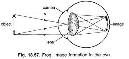
When light from any object in front of the eye falls upon it, the rays pass through the cornea and aqueous humour and reach the spherical lens through the pupil. The cornea and lens both in combination focus the rays on the retina to stimulate the rods and cones. The impulses then pass along the bipolar and ganglionic cells to reach the brain through the optic nerve.
Light rays falling on the retina produce an inverted and reduced image of the object which is rightly corrected by the brain but never reinverted in the brain as it is often supposed. The rays passing through the pupil converge due to their passage through the cornea and aqueous humour which together act as a convex lens but the rays as pass through the crystalline lens are further refracted in order to form an inverted and diminished image of the object on the retina which is rightly corrected by the brain.
During the image formation the iris acts as a diaphragm. If the object is well illuminated the pupil closes down to a pin point in order to check the excess of light intensity inside the eyeball and also to prevent the disintegration of the retinal cells, but when the object is poorly illuminated the pupil is widely open in order to allow as much light as possible to enter the eyeball to form the clear image of the object. In this way the iris helps in the formation of a well-defined image after regulating the amount of light entering the eye.
(v) Statoacoustic Receptors or Organs of Hearing (Ear):
The statoacoustic organ is for hearing and equilibrium. It includes a pair of ears present on the postero-lateral sides of skull enclosed in auditory capsules. The ear of frog includes a middle ear and an internal ear. The external ears are absent.
1. Middle Ear:
The middle ear consists of all those structures which are related simply in transmitting the sound waves to the internal ear which acts as suspensory apparatus. The cavity of middle ear is called the tympanic cavity lined by a membrane. It is an air-filled chamber communicating with the pharynx by a slender eustachian tube; the opening is normally kept closed by a valve.
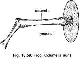
Externally the cavity of middle ear is bounded by a circular patch of dark skin, the tympanic membrane or eardrum which is tightly stretched over a ring of cartilage, the annulus tympanicus, it is simply a modified skin. It is vibratile in nature and from the centre of which a club-shaped rod, the columella auris, is extended across the tympanic cavity and attached internally to a membrane and a cartilaginous small nodule, the stapedial plate fused over a small window-like oval aperture, fenestra ovalis (a hole in the auditory capsule). The columella auris is partly made of bone and partly of cartilage. The columella auris is equivalent to hymandibula of dogfish.
2. Internal Ear:
The internal ear consists of a bony auditory capsule which is cartilaginous in the beginning but later in the adult is bony. It is formed by the pro-otic and exoccipital bones. The auditory capsule is filled with a watery fluid called perilymph in which the membranous labyrinth floats which is partially supported by connective tissue. It is comparable with inner ear of dogfish in structure and function.
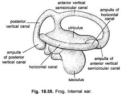
The membranous labyrinth is a sac-like complicated structure consisting of a larger oblong utriculus on the dorsal side and the smaller oval sacculus on the ventral side.
From the posterior side of the sacculus arise two small dilations, the lagena or cochlea and the pars basilaris which is now regarded as a part of lagena. Lagena is the forerunner of cochlea of higher vertebrates.
Dorsal to these is the protuberance of utriculus, called the pars neglecta. From the inner dorsal side of the sacculus arises a narrow tube, the ductus endolymphaticus and terminates in a dilated thin-walled endolymphatic sac present over the hindbrain within skull.
The three semicircular canals arise from the utriculus, are placed at right angles to one another, these are anterior and posterior vertical semicircular canals, while the third is a horizontal semicircular canal. The anterior and posterior canals have their adjacent limbs dorsally united and open into the utriculus through a common opening, whereas the horizontal semicircular canal opens into the utriculus at either end.
Each semicircular canal at its distal end is dilated to form a small round ampulla. The ampullae of the anterior and horizontal semicircular canals are found at their anterior ends and that of the posterior canal is at its posterior end.
The entire membranous labyrinth is hollow and is filled with a fluid called endolymph which contains pieces of calcium carbonate forming otoliths or ear stones. The various parts of the membranous labyrinth are innervated by the fibres of the auditory nerve.
Histology of Membranous Labyrinth:
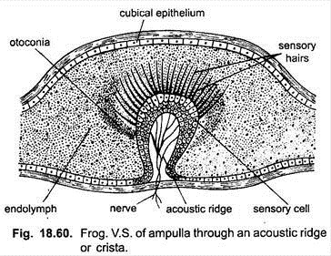
The wall of entire membranous labyrinth is made of dense fibrous connective tissue and is lined with cubical epithelial cells. The epithelial lining is modified at certain places to form receptors of the labyrinth which are called sensory patches or acoustic spots.
There is one acoustic spot in each ampulla, one spot in the utriculus, one in the sacculus and one in the lagena. The acoustic spots of the ampullae are called cristae, while those of the utriculus, lagena and sacculus, maculae.
The macula of utriculus is a large pars neglecta and that of the lagena is called basilar papilla. The cristae and maculae have the same structure as they are made of sensory hair cells and the supporting cells. The sensory cells have stiff and tapering hair-like processes at their inner free ends, while their lower ends are connected with nerve fibres of the auditory nerve.
Working of the Ear:
Ears have two functions, hearing and equilibrium. The membranous labyrinth of the frog functions primarily as the organ of balancing and secondarily as the organ of hearing.
(a) Equilibrium:
The semicircular canals along with their ampullae and the utriculus are concerned with the balancing of the body. On any change in the position of the animal the endolymph and the otoliths start movement and also exert pressure due to which the three cristae and one macula are stimulated. From which the impulses of change of position are transmitted by nerve fibres of the auditory nerve to the brain which sends the impulses to muscles concerned to bring the correct position.
(b) Hearing:
The sacculus and the lagena are the main structures of the membranous labyrinth which are concerned with the hearing. In hearing the sound waves strike the surface of the tympanic membrane and set it to vibrate. The columella which is extended from the tympanic membrane directly to the stapedial plate transmits these vibrations to the perilymph and then to the endolymph of membranous labyrinth.
These vibrations stimulate the sensory hair cells of sacculus and lagena. These sensory cells transmit the impulses to the brain by the auditory nerve, produce the sensation of sound. Thus, columella is meant only for the transmission of sound waves across the tympanic cavity. It also concentrates the sound waves to a point.
II. Interoceptors:
These are certain muscles and tendons of the body and also the alimentary canal which respond to stimuli as they are very much supplied with nerve fibres called interoceptors. The interoceptors found in the alimentary canal and viscera are called interoceptors, whereas those found in the muscles and joints are called proprioceptors.
Interoceptors provide information about the hunger, thirst, pain or comfort in the alimentary canal. Proprioceptors provide information about pains in viscera and tension in the muscles of the body.
