The pigeon, like other vertebrates, has receptors or sense organs for touch, smell, taste, sight and hearing which are stimulated by the environment. These sense organs are termed external receptors or exteroceptors.
The pigeon possesses following exteroceptors:
1. Tactile Organs:
These are poorly developed in birds due to feathery covering of the body. Tactile organs of pigeon remain confined to the bill and tongue of pigeon. The cere is a sensitive soft fold of skin at the base of the upper beak in pigeons, is said to have a stimulating effect during love making. The corpuscles of Grandry in the bill of ducks and other birds are probably tactile receptors.
They are composed of cells with a flattened nerve ending between cells. These are comparable to Meissner’s corpuscles in mammals. Merkel’s corpuscles are also found in many birds. The corpuscles of Herbst, resembling Pacinian corpuscles of mammals, are found in the dermis, are vibration receptors, sensitive to mechanical deformation by rapid pressure changes.
ADVERTISEMENTS:
They are found in great numbers in the beaks of all birds and also in the feather follicles, between tibia and fibula and in the tip of the tongue of woodpecker. Tactile nerves are also present at the base of the feathers, especially those of the wings and tail.
2. Gustatory Organs (Chemoreceptors):
Sense of taste and smell are little developed. The sense organs of taste, the taste buds, occur in limited number on the dorsal surface of tongue. The sense of taste is poorly developed in pigeons.
3. Olfactory Organs:
Birds are usually unable to distinguish delicate odours, and on the whole their sense of smell is very poor, as flying animals cannot depend on smell. The nasal cavity is large but the olfactory epithelium is restricted. Birds use the nose to test air coming from the internal nostrils. In kiwis, olfactory sense is well developed. These birds are nocturnal and terrestrial.
Structure of Olfactory Organs:
ADVERTISEMENTS:
The nostrils, overhung by cere, lead into the small, paired olfactory sacs or nasal chambers in the base of upper beak. The two chambers are separated medially by mesethmoid and bounded externally by ectoethmoid. The ectoethmoid produces inwards three scroll-like turbinal processes to increase the olfactory surface.
The nasal chamber has an anterior non-sensory respiratory part and a posterior sensory part. The non-sensory part or vestibulae contains the anterior turbinal covered by laminated epithelium.
The sensory or olfactory part contains the middle and posterior turbinals invested by the one-layered olfactory mucous epithelium or Schneiderian membrane, which is made of basal cells, supporting cells and elongated neurosensory cells. The neurosensory cells remain connected with olfactory nerve. Both the olfactory chambers remain communicated to the pharynx by the internal nares.
4. Auditory Organs or Ears:
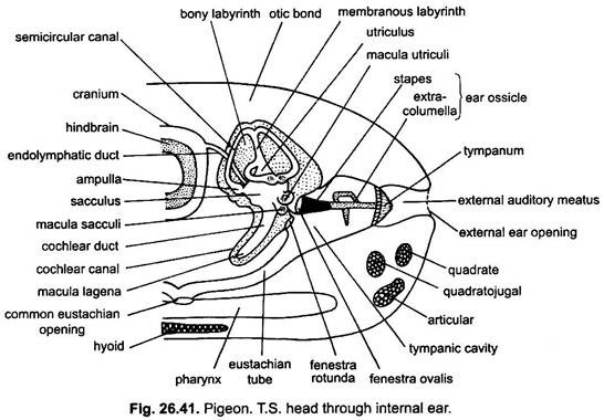
The sense of hearing is acute in most birds. Its auditory sense organs, the ears, serve their dual function of equilibrium and hearing. Auditory organs consist of a fundamental ear, the internal ear or membranous labyrinth and middle ear or tympanic cavity, like mammals. But, unlike mammals they lack external ear.
Internal Ear:
The internal ear or membranous labyrinth lies embedded in a dense ivory like bone in the side wall of the skull and is surrounded with a perilymph fluid. Membranous labyrinth is filled with a dense fluid, called endolymph. It contains a high concentration of potassium as in mammals, maintained by the activity of a tegmentum vasculosum, which also absorbs sodium.
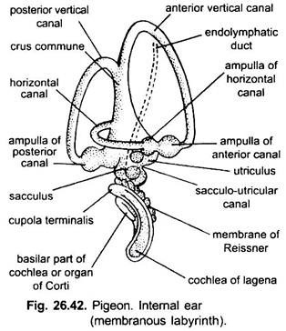
It consists of three semicircular canals (one horizontal, one anterio-vertical and one posterio-vertical), relatively small sacculus and utriculus and a short blind tube, the cochlear duct or lagena. Lagena is larger than in reptiles and less developed than in mammals.
The proximal limbs of anterior and posterior semicircular canals unite to form a crus commune. The sacculo-utricular connection is narrow. From the sacculus arises an endolymphatic duct which ends in the duramater.
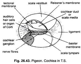
The cochlear duct, with its surrounding bony labyrinth or cochlear canal, is called cochlea. The cochlear duct is a slightly curved tube, surrounded by a perilymphatic space within a bony tube. In a transverse section, the cochlea shows three chambers: an upper scala vestibuli, a middle scala media and a lower scala tympani. The scala media is the actual cochlear duct and contains endolymph, while other two scalae are filled with perilymph.
At the apex of the cochlea (scala media) the scala vestibuli and scala tympani are continuous, this junction is known as the helicotrema, near it the scala media ends blindly. At the inner end of cochlea, scala vestibuli is continuous with fenestra ovalis, while the scala tympani with the fenestra rotunda. The floor of scala media is formed by the basilar membrane and the roof by the Reissner’s membrane. The basilar membrane consists of tall auditory hair cells (40), together constituting an organ of Corti.
The organ of Corti is sensitive to sounds of higher frequencies. The free ends of the auditory cells bear hairs, which are embedded in a tectorial membrane, lying above the organ of Corti. From the basal ends of the cells arise nerve fibres which unite to form the cochlear branch of the auditory nerve. The cochlear duct at its apical end has another set of auditory hair cells, their hairs embedded in a gelatinous cupola terminalis having minute calcareous otoliths.
At the tip of the cochlear duct is the lagena. Groups of auditory cells are called cristae and maculae. Those present in the ampullae of three semicircular canals are called cristae ampullares which possess the sense of direction and equilibrium. One macula is present in utriculus and one in sacculus. One macula is present in the lagena (tip of cochlear duct). Macula lagena is separated from the other maculae by a long cochlear duct. It perceives low frequency sounds.
Middle Ear:
A circular external ear opening lies on each side of the head, behind the eyes concealed beneath auricular feathers. It leads into a short canal, the external auditory meatus, at the base of which lies a thin, transparent, vibrating septum, called tympanum or ear-drum.
The ear-drum is followed by the cavity of middle ear or tympanic cavity. From the tympanic cavity arises a eustachian tube which unites with its fellow to open by a common aperture in the pharynx. The eustachian tube serves to equalise the air pressure on both sides of the tympanum.
Across the tympanic cavity lies a single rod-like bone, the columella auris, which transmits sound waves from tympanum across the tympanic cavity to the fenestra ovalis of the inner ear. The outer end of columella auris has three rayed cartilaginous processes called extra columella connected to the inner surface of tympanic membrane, and its inner disc-like bony part, the stapes, is wholly occluded the fenestra ovalis.
Columella auris (stapes) is derived from the hyoid arch. Sound is transmitted from the tympanum by the columella auris (stapes). Extra columella reduces the amplitude and increase the force of vibrations. There is single middle ear muscle attached to columella and tympanum and innervated by facial nerve. Acoustic vibrations are transmitted to an oval window and so around cochlea to a round window, as in mammals.
The equilibratory and auditory parts of ear of pigeon are well developed. Ability to localise sound in birds is high. Owls and other birds probably find their prey largely by ears. For the purpose of direction-finding they have developed very long cochleas and an asymmetrical arrangement of ear cavities (Strix) or asymmetrical external ears (Asio). Cave birds have the power of avoiding obstacles by echolocation, Steatornis (oil bird), Collocalia (swiftlet). These birds emit clicks.
5. Visual Organs or Eyes:
Birds depend more on their eyes than on the other senses. The eyes are extremely large. The eyes of hawks and owls are larger than in man. The eyes of pigeon are well developed and are very large in correlation with an aerial life for a precise vision over considerable distances.
Shape:
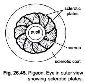
The eyeball is not spherical, the lens and cornea bulge forwards in front of the posterior chamber. This form is maintained by a ring of bony sclerotic plates. In most birds the whole eye is thus broader than it is deep. Eyeball is longer in those birds whose sight is very acute and in some eagles and crows it is tubular.
Eyelids:
The eyebrows or eyelashes are absent. There occur two inconspicuous eyelids, a slightly movable upper eyelid and a more movable and well developed lower eyelid which rises upwards to close the eye during the sleep. A semi-transparent third eyelid or nictitating membrane occurs as a fold at the anterior angle of the eye.
It can be drawn posteriorly over eye with great rapidity. It cleans the eyeball and also protects the eyes from wind and during flight and from water during swimming in aquatic birds. It also protects the eyes from glare of the sunlight during day in nocturnal birds.
Glands:
The nictitating membrane is lubricated by the oily secretion of a Harderian gland occurring in the inner angle of the eye. The tear gland or lacrymal glands are also well developed and lie below the outer angle of lower eyelid. Their watery secretion nourishes the non-vascular cornea, and also keeps it clean.
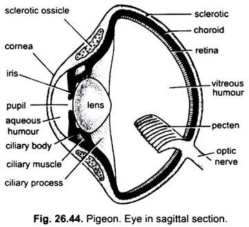
The wall of hollow eyeball consists of three usual layers, namely, an outer sclerotic, a middle choroid and an inner retina.
Sclerotic:
The external coat of the eyeball is sclerotic. In the posterior hidden part of the eye, it is opaque, white, dense and cartilaginous. In front in the exposed part of the eye, it bulges out to form a convex, transparent and horny cornea of connective tissue.
The cornea is externally covered by a thin, transparent, sensitive and vascular epithelial membrane, the conjunctiva, which is formed by modified epidermis and is continuous with the mucous lining of the eyelids. Anteriorly, at the junction of cornea and sclerotic coat, the latter is strengthened by a ring of 10-12 small overlapping bony, sclerotic plates or ossicles.
Choroid:
The sclerotic coat is followed by the middle layer, the choroid, which is thin, dark pigmented and highly vascular. The choroid closely lines the sclerotic, but it separates in front to form a circular, pigmented diaphragm, the iris, perforated by a rounded aperture, the pupil. The iris regulates the amount of the entering light. It contains intrinsic circular and radial smooth muscles, the circular muscles contract the pupil, while radial muscles dilate it.
Along the peripheral margin of the iris, the choroid forms a ring-like ciliary body which is a thickened fold containing smooth ciliary muscles. From ciliary body arises striated ciliary processes or suspensory ligaments and attached to the lens. The ciliary muscles are divided into anterior Crampton muscles and posterior Brucke muscles.
The Brucke muscles draws the lens forward into the anterior chamber so that since the shape of the eye is fixed by the sclerotic plates, the lens becomes more curved and, hence, accommodated for near vision. Contraction of the iris sphincter assists in this process. At the same time the Crampton muscles pull the cornea reducing its radius and further assist in accommodation. The circular and radial muscles of the iris and ciliary muscles are under the control of the autonomic nervous system receiving sympathetic and parasympathetic fibres.
Retina:
The innermost coat of the eyeball is a thin, light sensitive nervous layer called retina. It is transparent, devoid of blood vessels, thick and consists of nerve fibres, nerve cells and minute rods and cones. Pigeon, being a diurnal bird, has largely more cones than rods. The high resolving power and high powers of discrimination and of movement detection depend on the great density of the cones, as many as 1 million per square millimetre in the fovea of a hawk. Nocturnal bird’s retina is composed mainly or completely of rods.
Sensitive Spots:
The retina has two sensitive spots or fovea. The central fovea, which lies near the centre of retina as a slight depression, is more sensitive and used for lateral or monocular vision. The second fovea, the temporal fovea, lies more towards the outer side of the eye and is used for forward or binocular vision. The foveae have comparatively more cones and give more distinct vision. The cones of birds often contain carotenoid oil droplets, which are also found in frogs, turtles and marsupials.
In diurnal birds, they are red, yellow, orange or colourless, but in nocturnal ones pale yellow or colourless. Colour droplets may produce narrow-band sensitivity channels for the mediation of colour discrimination. The lower part of retina contains yellow droplets and upper part (dorso-posterior) contains red droplets. These droplets serve as fibers and increase the colour vision of birds up to great degree. The red may provide finer colour discrimination of objects in feeding.
Lens:
The lens is biconvex, soft, pliable, crystalline, colourless, transparent and is surrounded by a fibrous capsule. It remains suspended just behind the iris by suspensory ligaments. It divides the eye cavity into a small anterior aqueous chamber and a large posterior vitreous chamber.
The aqueous chamber is filled with a colourless watery fluid, the aqueous humour, while vitreous chamber contains a thick colourless, gelatinous vitreous humour. The two humours keep the eyeball tight or tense and also serve to focus the light rays on the retina.
Pecten:
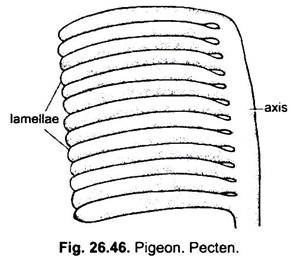
Pecten (Fig. 26.46) is a pleated, strongly pigmented vascular fold projecting into the cavity of the eye from the entrance of the optic nerve. It is large and much pleated in predatory birds, which detect minute movements at great distances, and is small and smooth in nocturnal birds. It is also well developed in diurnal birds (pigeon). Except kiwi, all birds have pecten.
Functions of Pectin:
There are many speculations about the function of pecten but none is known definitely. Probably its main function is to bring oxygen and nourishment to the retina, which in birds has no capillary circulation. It helps in accommodation, it is not likely that it actually assists in focussing, for instance, by pressing forward the lens, and no changes have been seen in it during accommodation. However, it might possibly assist by adjusting the intraocular pressure, which must be increased by the extensive changes in the lens during accommodation.
Vision:
In pigeon, because the cornea projects outwards and the posterior part is expanded, so the eye is broader than deep. Due to expansion of retina over the broad posterior portion, the distant objects are sharply focussed on it. Further, though the eyes are lateral in position, there is an overlapping of the two visual fields to some extent. This is called binocular vision. It is worth noting that there occurs little movement in pigeons’ eyes due to ill-development of extrinsic eyeball muscles and that is compensated by flexibility of neck which turns the neck very quickly.
