The birds have an efficient circulation, a high rate of metabolism and a high and constant temperature. The birds and mammals are the only vertebrates that have a complete separation of the respiratory and systemic circulations, making possible a high arteriolar pressure, which allows materials to reach the tissues rapidly.
The circulatory system of pigeon includes heart, arteries, veins, the blood and lymphatic system:
1. Heart:
The heart is four chambered 2 auricles and 2 ventricles. The sinus venosus is absent.
External Structure:
ADVERTISEMENTS:
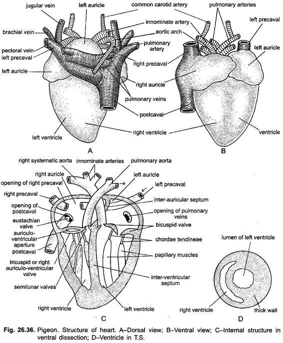
The heart of pigeon is large-sized, reddish in colour, triangular and compact. It lies midventrally in the thorax. It is enclosed in a thin, transparent membranous sac, the pericardium, which is a derivative of coelomic epithelium. The pericardial wall is made of an outer parietal layer and an inner visceral layer. The two layers enclose a narrow pericardial cavity, filled with a watery serous or pericardial fluid.
The pericardial fluid protects the heart from shocks and injuries and permits movements during beating. The auricles remain well marked from ventricles by an external deep, shallow, transverse groove, the coronary sulcus or the auriculo-ventricular groove which separates the two anterior, smaller, darker auricles from the posterior, larger, lighter in colour, the ventricles. The two auricles are also demarcated from each other by a faint, inter-auricular groove. The sinus venosus has disappeared being absorbed into the wall of the right auricle.
Internal Structure:
ADVERTISEMENTS:
Internally, the two thin-walled auricles are separated by a complete, thin, membranous partition, the inter-auricular septum. It has a small oval area in the middle, called the fossa ovalis, representing the foramen ovale in the embryo.
The two thick-walled muscular ventricles are also completely separated by a thick and muscular inter-ventricular septum. The left ventricle is larger with powerful muscular walls. It has a circular cavity. Its propulsive force has great power of pumping blood.
The right ventricle is smaller with a crescentic cavity in section. It partly encircles the left ventricle. The ventricles have bundles of muscle fibres arranged longitudinally to form columnae carneae.
The right auricle is larger than left auricle and it receives the three large veins, the right and left precavals and the postcaval, in its dorsal wall. The opening of the postcaval vein is gurarded by a muscular eustachian valve. The right auricle opens into the right ventricle by a crescentic aperture, the right auriculo-ventricular aperture.
ADVERTISEMENTS:
This aperture is guarded by a pair of muscular flaps, having no chordae tendinae, forming the right auriculo-ventricular valve. It represents the tricuspid valve of the mammals. The right ventricle gives off a single trunk, the pulmonary aorta, which bifurcates into two pulmonary arteries, each going to a lung. The opening of the pulmonary trunk is guarded by three semilunar valves.
The left auricle is smaller and receives four pulmonary veins from the lungs. It opens into the left ventricle by a circular left auriculo-ventricular aperture. It remains guarded by two membranous flaps forming the left auriculo-ventricular valve, which represents the bicuspid or mitral valve of mammals. The flaps are connected to the thick papillary muscles which arise from the wall of the left ventricle, by the chordae tendinae.
The left ventricle gives rise to the single right systemic or aortic arch which crosses over to the right side of the heart and continues as dorsal aorta. The left aortic arch, which is present in the frog and lizard, is absent in birds. The opening of right aortic arch is guarded by three semilunar valves. The cavities of both ventricles are traversed by bars of muscles, called trabeculae or columnae carneae.
Functioning of Heart:
The heart is a pumping station of circulatory system which pumps the blood to arteries. In pigeon, the venous and arterial blood never mixes in heart as in frog, but remains well separated like mammals since it is four chambered.
The right half receives and discharges only venous (un-oxygenated) blood, while the left half only arterial or oxygenated blood. Thus, birds possess a complete double circulation which includes a pulmonary circulation and a systemic circulation.
(i) Pulmonary Circulation:
The right ventricle pumps venous blood into the lungs through the pulmonary aorta and its two pulmonary arteries. The oxygenated blood from the lungs is returned into the left auricle through four pulmonary veins.
(ii) Systemic Circulation:
ADVERTISEMENTS:
The left ventricle sends blood into the right aortic arch, the dorsal aorta and through smaller arteries to the various organs and tissues of the body, where they break up into capillaries. These capillaries unite to form veins, which finally form three great veins, which return blood into the right auricle. From right auricle, the blood enters the right ventricle through right auriculo-ventricular aperture.
Heart Rate:
The heart gives a rapid heartbeat to ensure rapid blood circulation, high body temperature (102° to 112° F) and rapid metabolism, for production of energy. The size and rate of heartbeats vary with the size and activity of birds. The larger birds have in general relatively smaller heart and less heart beats. Rates increase with exercise and a maximum of 102 per minute has been recorded in a humming bird. The larger heart allows a greater stroke output and higher pressure than in mammals.
Heartbeats:
The heart of pigeon has two pacemakers, a sinu-auricular node in the wall of the right auricle, and an auriculo-ventricular node in the inter-auricular septum. The muscles of auricles and ventricles are not continuous (unlike amphibians and reptiles). A wave of excitation starts from the sinu-auricular node and goes to the auriculo-ventricular node from which fibres of the bundle of His convey the excitation wave to the ventricles.
The branches of the fibres break up into numerous Purkinje fibres, which spread as a network into the ventricle. When left auricle contracts, the oxygenated blood from the left auricle passes to the left ventricle. From the left ventricle the blood is taken by the right aortic or systemic arch and distributed to the body. Thus, systemic circulation completes. Likewise, when right auricle contracts, its unoxygenated or venous blood passes to right ventricle, the contraction of which forces the blood into pulmonary artery for oxygenation in the lungs.
2. Arterial System:
The heart pumps its unoxygenated blood to pulmonary aorta and oxygenated blood to aortic or systemic arch; both of which collectively constitute the arterial system of pigeon.
A. Pulmonary Aorta:
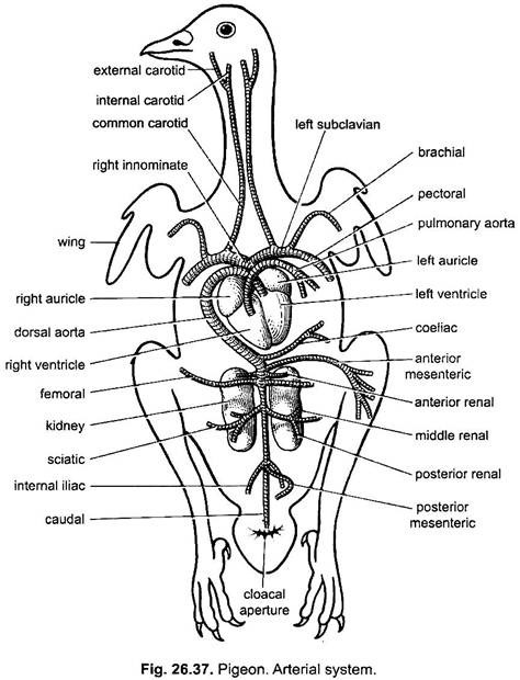
From right ventricle arises a single pulmonary aorta which passes ventral to aortic or systemic arch and bifurcates into pulmonary arteries, each of which enters into a lung.
B. Aortic Arch or Systemic Aorta:
The right aortic or systemic arch arises from the left ventricle, the left aortic arch being absent in adult birds. The right aortic arch gives out two small coronary arteries to the heart, just at the point of its origin. It then gives off a pair of short, stout and thick right and left in nominate arteries. Each in nominate artery divides into an anterior common carotid artery and a lateral subclavian artery.
The common carotid artery in turn divides into a verterbral to neck vertebrae, an internal carotid to the brain, an external carotid to the head and neck, and a comes nervi vagi, which joins the external carotid. The subclavian artery divides into a brachial artery going to the wing, and a very large pectoral artery which gives off a thin internal mammary artery to the ventral thoracic wall, then it forms three main branches going to the pectoral muscles of the flight.
Dorsal Aorta:
The right aortic arch now curves over dorsally toward the right side of heart and passes backwards mid-dorsally as dorsal aorta.
The dorsal aorta gives off following branches to different body organs:
(i) A single, median, strong coeliac artery which supplies branches to proventriculus, gizzard, duodenum, liver and spleen.
(ii) An unpaired anterior mesenteric artery to the greater part of small intestine.
(iii) Two anterior renal arteries to the first lobes of kidneys. In male, each renal artery gives off a branch to the testis; but in female, only the left renal artery gives off a branch to the ovary (the right ovary is absent in birds).
(iv) A pair of femoral arteries runs outwards dorsal to the anterior lobes of kidneys and supplies blood to pelvic muscles and outer muscles of the thighs.
(v) A pair of sciatic or ischiatic arteries passes outwards to supply blood to inner muscles of the thighs and the legs. Each sciatic artery gives off a middle renal artery to the middle lobe of the kidney and a posterior renal artery to the posterior lobe of the kidney.
(vi) A pair of iliac arteries to the hinder part of pelvic region.
(vii) An unpaired posterior mesenteric artery to the large intestine (rectum) and cloaca
(viii) At last the dorsal aorta continues into the tail as caudal artery.
3. Venous System:
The venous system of pigeon includes:
(i) The caval veins,
(ii) The pulmonary veins,
(iii) The hepatic portal system and
(iv) The renal portal system.
(i) Caval Veins:
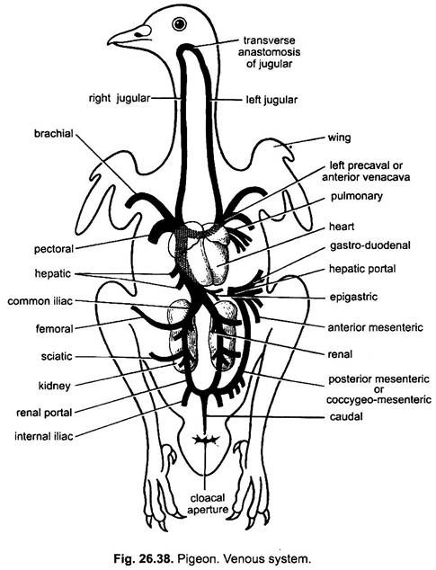
The non-oxygenated or venous blood of entire body of pigeon is collected and conveyed to the right auricle of the heart by three large veins, two precavals from the anterior part of the body and one postcaval from the posterior region of the body.
a. Precavals:
The precaval vein of each side (smaller right and larger left) is formed by the confluence of a large common jugular vein and a subclavian vein as in lizard. The common jugular vein is formed by the union of external and internal jugular veins from the head and a vertebral vein from the spinal cord and neck muscles.
The jugular veins of the two sides are joined just beneath the base of the skull by a transverse loop called jugular anastomosis. The jugular anastomosis prevents blocking of the flow of blood from the head when one vein is momentarily shut off by twisting the neck.
The subclavian vein receives a slender brachial vein from the wing and a large pectoral vein from the pectoral muscles. A small internal mammary from the ventral thoracic walls also joins the pectoral vein.
b. Postcaval:
The postcaval vein is formed by the fusion of all the veins from the posterior part of the body. A short, unpaired caudal vein from the tail after entering the abdomen bifurcates into right and left hypogastric or renal portal veins. From the bifurcation of the caudal vein arises a median coccygeo-mesenteric vein which is characteristic of birds. It receives the blood from cloaca and rectum, and runs forward to join the hepatic portal vein.
Each renal portal vein instead of breaking up into capillaries in the kidney, gives off only a few small branches, afferent renal veins, which carry blood to the kidney. The main renal portal vein passes forwards through the substance of the kidney and receives the femoral and sciatic veins from the leg to form the iliac vein. At the junction of iliac and renal veins is present a valve which probably regulates the flow of blood into the renal portal vein.
The femoral vein also receives the efferent renal veins from the kidney. As soon as it leaves the kidney, it is called a common iliac vein. The common iliac veins of the both sides unite to form a short postcaval vein, which runs forwards through the substance of right lobe of liver and receives three pairs of hepatic veins, and then enters the right auricle.
Thus, the main part of blood from the caudal and pelvic regions is taken directly to the heart, and not through the renal capillaries as in most fishes, and all amphibians and reptiles.
(ii) Pulmonary Veins:
From lungs two pulmonary veins return oxygenated blood into the left auricle. Both pulmonary veins before opening into auricle unite together and open by a single aperture in left auricle.
(iii) Hepatic Portal System:
The blood of various parts of alimentary canal is returned to liver by a hepatic portal vein which is formed by the union of gastro-duodenal, anterior mesenteric, posterior mesenteric, and coccygeo-mesenteric veins. Coccygeo-mesenteric vein arises from the point of bifurcation of the caudal vein, runs parallel with the rectum and joins with the hepatic portal vein. An epigastric vein returns blood from the fat-laden peritoneum (great omentum) and joins one of the hepatic veins.
The hepatic portal vein collects blood from rectum, ileum, duodenum and gizzard, and it divides into two branches, one entering each liver lobe into which they form capillary network. The blood of hepatic portal vein is filtered through the liver and then conveyed to the postcaval vein through the hepatic veins.
Because the coccygeo-mesenteric vein connects the two portal systems (i.e., renal portal and hepatic portal), it is considered homologous with the anterior abdominal vein of amphibians and reptiles. But this homology is not accepted by some authorities. They regard the epigastric vein to be the homologous of the abdominal vein of amphibians and reptiles.
(iv) Renal Portal System:
The renal portal system of pigeon is greatly reduced and includes two hypogastric or renal portal veins, each of which gives off only a few afferent renal veins to each kidney and does not form capillary system in the kidney. Thus, the blood from the posterior region of body reaches directly to the heart, and not through the renal capillaries, as in amphibians and reptiles.
4. Blood:
The blood of birds is rich in red blood corpuscles. They are oval and nucleated. They are large 10-15 µm x 5-8 µm and numerous 2.5-3.5 million per cmm. They carry a large amount of haemoglobin that gives up its oxygen suddenly at a relatively high oxygen tension. The red corpuscles are smaller in actively flying birds than in the larger flightless birds.
Haemopoietic tissue is widespread in the young, but is restricted mainly to the marrow in the adult although it may also be found in liver and spleen. The white blood corpuscles are more numerous than in mammals. Platelets are not found in pigeons, but it clots quickly. Thrombocytes are present. Every part of the body is nourished by the blood.
Bird’s temperature is considerably higher than that of mammals, usually about 42°C to nearly 45°C in some birds. In the absence of sweat glands, how it is kept constant is not known. Heat loss is minimised by the absence of vascularised extremities (feet). The information of wing from large avascular surfaces is the secret of the success of birds.
Air sacs may serve to conserve heat by providing an air cushion for the viscera, which might otherwise lose heat. There is a system of direct arterio-venous connections in the feet, and elsewhere. These anastomotic regions have powerful muscles, whose contraction close them and force the blood through the capillary system.
There must have been a system of nervous pathways for the control of upward and downward temperature regulation. To avoid heat loss one species (a nightjar) is known to hibernate. Certain humming birds and sunbirds have a serious heat loss problem so they become poikilothermic in night.
5. Lymphatic System:
A lymphatic system with a few lymphatic glands is associated with the blood vascular system. Lymph glands are found in some aquatic and wading birds. There is a pair of lymph hearts in the sacral region of the embryo and these may persist in the adult.
The lymph vessels unite to form two thoracic ducts (homologous with subvertebral trunk) opening into the precaval veins. Adult pigeons lack any lymph hearts. The spleen is a small, oval and reddish gland attached to the right side of the proventriculus by a peritoneal fold.
Spleen:
It is a small, ovoid red body attached by peritoneum to the right side of the proventriculus.


