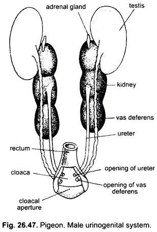
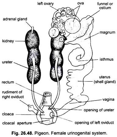
The excretory and reproductive organs of pigeon are very closely connected with each other structurally, so are studied together under the heading urinogenital system.
But, both systems, viz., excretory and reproductive, are physiologically independent, so should be studied separately as follows:
1. Urinary or Excretory Organs:
The excretory organs of pigeon are two kidneys and two ureters. The urinary bladder is absent in adults to reduce the body weight (flight adaptation). Among birds, ostrich only has the urinary bladder.
i. Kidneys:
ADVERTISEMENTS:
The main excretory organs of pigeon (birds) are two, dark brown, flattened, three-lobed metanephric kidneys lying posterio-dorsally in the body cavity and embedded in the hollow of the pelvic girdles. Their ventral surface is covered by peritoneum. They are provided with venous blood by the renal portal veins and arterial blood from the renal arteries.
The renal arteries supplying the glomeruli, while the tubules are supplied both by efferent glomerular arterioles and branches of the portal veins. A small yellowish, elongated and streak-like adrenal body lies attached ventrally to the anterior lobe of each kidney. It is an endocrine gland.
Each kidney consists of masses of numerous tightly packed, convoluted uriniferous tubules. Birds have comparatively more tubules than those of mammals. Each uriniferous tubule has small glomerulus and a specialised portion, called the loop of Henle. Loops of Henle are long. The glomerulus filters the blood and filtrate passes through the long loop, where much of its water is reabsorbed. Thus, much concentrated urine passes down the ureter.
ii. Ureter:
ADVERTISEMENTS:
Uriniferous tubules of each kidney unite to form a ureter or metanephric duct. The ureter is a narrow, straight tube, arising ventrally from the anterior lobe of the kidney and running backwards to open into the middle compartment of the cloaca, the urodaeum, through its dorsal wall.
Physiology of Excretion:
The excretory system is highly specialised for water-saving. The birds are urecotelic like reptiles, because in them the end product of urinary excretion is relatively insoluble uric acid which is synthesised in liver.
Semisolid viscous urine comes from the kidneys into the urodaeum from where it passes up into the coprodaeum and large intestine, where further water is resorbed and the mixed faeces and urinary products are then excreted as the characteristic semisolid white guano. Urine is hyperosmotic to the blood. Birds also excrete salt by nasal glands, especially well developed in marine forms.
2. Male Reproductive Organs:
ADVERTISEMENTS:
Sexes are separate and sexual dimorphism is absent in pigeon. The male reproductive organs of pigeon (Fig. 26.47) are testes and vasa deferentia and there occurs no penis like mammals. Among birds, a definite penis (and also clitoris) is found in ratites, anseriformes and a few other birds. Ostrich and ducks only have an erectile penis.
i. Testes:
Two oval and white testes, each attached to the anterior end of a kidney by mesorchium (fold of peritoneum). The right testis is slightly smaller than the left. The testes increase many times in size during the breeding season, usually under regulation by changes in illumination. The weight of testis is about 1000 times greater in the breeding season than it is in the non-breeding period, when it contains only spermatogonia.
Both testes have masses of coiled seminiferous tubules with groups of interstitial cells between them. The cells of lining epithelium of seminiferous tubules produce spermatozoa by undergoing spermatogenesis. The interstitial cells are also called Leydig cells and produce sex hormone testosterone. The seminiferous tubules join to form a long epididymis.
The testes are permanently retained within the body cavity. There is no temperature-regulating scrotum, as found in mammals. It is supposed that the abdominal air sacs may reduce testis temperature and be concerned indirectly with spermatogenesis, but this is not yet proved.
ii. Vasa Deferentia:
From the inner border of each testis arises a convoluted Wolffian or mesonephric duct, its anterior end is an epididymis and the rest is a vas deferens. The epididymis is connected with the seminiferous tubules of the testis by extremely fine tubules, the vasa efferentia. Vas deferens runs backwards along the outer side and parallel to the ureter of that side to open into the urodaeum on a very small erectile papilla posterior to the ureter.
It is the only copulatory organ of most birds. For temporary storage of spermatozoa, the hind end of each vas deferens becomes swollen, called the seminal vesicle. There is no copulatory organ in pigeon.
3. Female Reproductive Organs:
ADVERTISEMENTS:
The adult female has only the left ovary and left oviduct as their main reproductive organs. In the embryo there are two ovaries and two oviducts, but during development, the ovary and oviduct of right side become more or less completely atrophied.
i. Ovary:
A large-sized, irregular-shaped left ovary occurs at the ventral side of anterior lobe of left kidney. It remains attached to the kidney by mesovarium, a double-fold peritoneum. During breeding season, the size of the ovary increases considerably due to the influence of FSH and LH of anterior pituitary. The ovary secretes oestrogen which modifies the accessory sexual organs and behaviour.
The surface of ovary remains studded with numerous follicles or ovisacs of different sizes and each contains a single ovum or oocyte. The ova are at various stages of development. These follicles project from the surface of the ovary in birds.
Of the large number of oocytes only few ripen to make the large follicles. Each follicle produces a large yolky, polylecithal ovum which when mature escapes by the rupture of the follicle into the coelom. After each follicle has bursted it quickly regresses. There is no corpus luteum. The released ova in the coelom are caught by the enlarged ciliated and muscular funnel of the oviduct. Ovulation depends on the cyclic release of luteinizing hormone (LH).
ii. Oviduct:
The left oviduct (Mullerian duct) is a long, broad, thick-walled, convoluted tube passing backwards to the cloaca. It remains attached to the dorsal body wall by mesotubarium, a double fold of peritoneum. The oviduct anteriorly has an expanded muscular and ciliated coelomic funnel or ostium or infundibulum with fimbriated margin. It opens by a wide, slit-like aperture into the coelom near the ovary.
The oviduct has various parts. As an ovum enters the ostium and passes down, the walls of ostium secrete the thin chalaziferous layer of dense albumen around the egg. The succeeding part of the oviduct, called glandular part or magnum, has tubular glands which secrete the albumen or egg white. The magnum is followed by isthmus which secretes a parchment-like double shell membranes around the albumen.
The next portion is called uterus. Uterus is thin walled and is lined by nidamental glands which form an outer hard calcareous shell around the shell membranes. The last portion of oviduct is vagina which is muscular, thick-walled and contains mucus secreting unicellular glands which secrete pigment, external cuticular layer of the cell and mucus for expelling the egg. The oviduct opens into urodaeum.
As much as one-third of the weight of calcium in the wholes skeleton is needed by the pigeon for its two eggs. A reserve is collected as the ovarian follicles mature. The oestrogen they produce increases the uptake of calcium from the food and stimulates its deposition in the bones. After ovulation the oestrogen level falls, the calcium is then mobilised from the bones, probably by parathyroid hormone, and its concentration in the blood becomes very high, until used by the eggs.
In many birds the egg shells are coloured, pigments derived from bile by breaking down haemoglobin are deposited in the shells by nidamental glands of the oviduct. In pigeon, there occurs a rudimentary right oviduct close to the right side of cloaca and sometimes vestiges of right ovary.
Significance of One Ovary:
Loss of an ovary and oviduct in birds reduce weight as well as decrease the number of eggs produced, which is an advantage in flight. Further, the retention of a single ovary also helps in the safe manipulation of large eggs with breakable shells.
Copulation:
In copulation the proctodaea of male and female are everted and pressed together so that the sperm is ejaculated direct into the urodaeum of the female and travel up the oviduct. The fertilisation takes place in the upper part of the oviduct, before the coverings (albumen, shell membranes and calcareous shell) are formed around the egg.
The female pigeon being oviparous lays two eggs in a rough nest. The eggs are incubated by both parents for 14 days at a temperature of 38° to 40°C. At the end of incubation the youngs called squabs hatch by breaking the shell with a caruncle. The squabs are altricial being naked and unable to fend for themselves. They are fed for a time by both parents on pigeon milk formed by the epithelium of the crop. Crop gland activity is controlled by prolactin hormone.

