The muscular system of birds is efficient and well developed. It is well specialised to meet the different needs of the bird. The diaphragm of pigeon is rudimentary. The muscles of back are atrophied due to inactivity or immobility of the trunk vertebrae since few thoracic vertebrae are fused and also the lumbars, sacrals and few anterior caudals are fused.
However, the muscular system of neck, wings, tail, legs and ventral side of the body is well developed. Feathers are provided with non-striated muscles at the base, with an autonomic innervations serve to move the feathers. Striated subcutaneous muscles move groups of feathers. Control of feather position is important for regulation of heat loss, for flight and in sexual display, etc.
Feathers can be studied under following heads:
Appendicular Muscles:
ADVERTISEMENTS:
The muscles which provide movement to forelimbs, wings and hindlimbs are called appendicular muscles.
Both types of appendicular muscles (flight muscles and perching muscles) are well developed and can be studied separately as follows:
(A) Flight Muscles:
The muscles which operate the forelimbs (wings) during flight are called flight muscles. These are pectoral, accessory and tensor.
(i) Pectoral Muscles:
ADVERTISEMENTS:
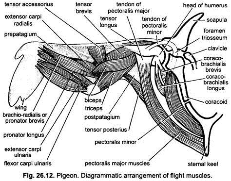
The most significant flight muscles of birds (pigeons) are pectoral muscles. These muscles remain attached to the keel of the sternum and to the wings, and provide up and down movements to the wings.
They are of following two types:
(a) Pectoralis Major:
ADVERTISEMENTS:
The pectoralis major is a very large, triangular and most powerful flight muscle which arises from the ventral side of the sternal keel and clavicle, one on each side, and forms the so-called ‘breast’. This muscle weighs about one-fifth as much as the entire body and has dark red colour due to rich blood supply.
In front each muscle is attached to the keel of sternum and the clavicle and anteriorly its fibres join the ventral side of the head of humerus by a broad and flat tendon. When the pectoralis major muscle contracts, the wing is pulled downwards and forwards, so that the body of pigeon is lifted up and propels itself through the air, because it causes the down stroke of wing, so also called depressor muscle.
(b) Pectoralis Minor:
Pectoralis minor, also called deep pectoralis, supracoracoideus or subclavius, is a small and elongated muscle which elevates the wing during flight. It lies deep to the pectoralis major. It arises from the anterior part of the sternum, dorsal to the pectoralis major. Its tendon passes through the foramen triosseum, an aperture occurring in between the junction of the clavicle, scapula and coracoid; and is inserted on the dorsal side of the head of humerus.
Pectoralis minor is an elevator muscle and it causes the upstroke of the wing. When it contracts, the foramen triosseum acts like a pulley for its tendon, pulling the humerus backwards and upwards, and, thus, raising the wing during flight. In pigeon, the pectoralis minor is especially developed and causes quick takeoff of the bird during flight.
(ii) Accessory Muscles:
Besides pectoral muscles, the accessory muscles also elevate or depress the wing during flight. A coraco-brachialis longus or coraco-humeral lies beneath the pectoral muscles. It arises from the coracoid and the costal process of sternum, and its tendon is attached to the posterior side of the head of humerus.
It lowers the hinder aspect of the wing. Another smaller and narrow muscle is coraco-brachialis brevis or scapulo-humeral lies in front of the longus. It also extends from girdle to humerus and raises the hinder edge of the wing. Both muscles rotate the wing in the glenoid cavity.
The biceps and triceps are the intrinsic muscles of the upper arm which operate the elbow and perform adjustments during flight. The extensor carpi radialis and the extensor carpi ulnaris muscles of the forearm help to stretch or fold the wing. The radius bone of the forearm is medially rotated by two brachioradialis muscles. The digital muscles of hand move the digits and feathers individually during flight. The policis muscles move the bastard wing or alula is attached to the first digit.
ADVERTISEMENTS:
(iii) Tensor Muscles:
Three muscles called tensor longus, tensor brevis, and tensor accessorius keep the prepatagium fully stretched when the wing is extended in flight. Similar tensor posterius alae keeps the postpatagium tensed during flight.
(B) Muscles of Hindlimbs and Perching Mechanism:
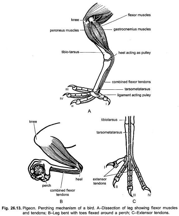
The musculature of hindlimb of pigeon and other birds resembles in every respect with that of mammals. But certain muscles in the legs and their tendons have a special arrangement so that when a bird sits on a perch (branch of a free, wire or rod), its toes are mechanically flexed and grasp the perch without any effort. Such muscles are called perching muscles and the arrangement of perching muscles in the leg is called perching mechanism.
The perching muscles of pigeon include following two sets of muscles, viz., flexor and extensor muscles.
1. Flexor Muscles:
There occur eight flexor muscles on the back of the tibiotarsus bone of hindlimb, all of them remain inserted up on the knee-joint. These flexor muscles perform the perching. Six of them pass their tendons to the phalanges of anterior toes, while the remaining two flexor muscles pass their tendons to the hind toe or hallux.
The important flexor muscles are following:
(i) Ambiens:
In some birds, an ambiens muscle arises from the ilium and passes along the inner surface of thigh. Its long tendon runs beneath the patella bone round to the outer side of the knee enclosed in a special sheath and passes to the outer side of the tibiotarsus to join the upper end of the flexor muscle of the second and third toes. This muscle has little role in perching.
(ii) Peroneus Medius:
This muscle occurs singularly on the anterior aspect of the shank, attached to the upper part of the tibiotarsus bone. Its tendon divides into three tendons, going to the three front digits.
(iii) Gastrocnemius:
It is a calf muscle mainly concerned with producing flexion of the toes in the act of perching and occurs on the back of tibiotarsus. Its tendon passes behind the ankle and trifurcate to supply the three anterior toes along with peroneus medius muscles. These tendons often act as a single unit.
(iv) Flexor Perforans:
This muscle is attached to the upper part of the tibiotarsus (above the knee). Its tendon going to the hallux and is joined by a slip with the peroneus medius. Thus, a pull up on one tendon flexes all the toes.
When a bird sits on its perch the legs are bent at the knee and the ankle, thus, the tendons are flexed and the digits are automatically closed around the perch. A pull on any tendon will close all four digits. Flexing of digits is due to a grip reflex which is inflated by a sensory receptor on the lower surface of the foot, the moment the foot touches the perch. When a bird sleeps with body relaxed, the weight of the body bends the ankles more and the tendons become tighter ensuring a firmer grip of the perch, so that a sleeping bird cannot fall. This is automatic perching mechanism.
A second perching mechanism is a locking device that holds the toes flexed. The lower surface of flexor tendon is ridged at the metatarso-phalangeal joint, where the weight of the body presses it against a branch. The upper surface of the tendon sheath is also ribbed and as the bird settles on its perch, the two sets of ridges are interlocked.
2. Extensor Muscles:
The toes are unlocked by raising of body and also by extensor muscles. In pigeon, several extensor muscles are found at the front of the tibio-tarsus. The tendon of the tibialis anterior muscle passes down in front of the intertarsal joint and then divides into three branches, to supply one branch to each anterior toe. These tendons of flexor muscles are attached to the upper surfaces of the phalanges. The contraction of tendons of extensor muscles serves to open the toes, when the bird raises its shank while taking off the perch.
3. Dermal Muscles:
The dermis of pigeon has some muscles which derive from parietal muscles and move individual feathers.

