In this article we will discuss about the external features of Indian frog with the help of suitable diagrams.
Shape and Size:
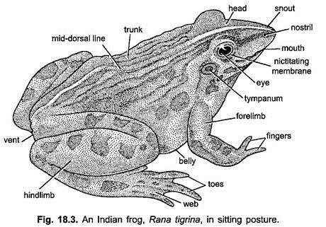
Besides aerial mode of life, frog also leads aquatic mode of life. Therefore, it has streamlined body which is the characteristic of the aquatic animals and assist in swimming in water. The two ends, the anterior and the posterior, of the body are pointed and the triangular flattened head, with its blunt apex directed forward, is broadly united to the trunk. Thus, there is no neck to connect the head and trunk together and no tail.
The size of the frog varies from species to species or even in the same species depending upon the age of the individual. The size may range from few centimetres to many centimetres.
ADVERTISEMENTS:
Colour:
The colour of the body at the dorsal side is green with black spots and streaks but ventrally it is paler. This type of colouration harmonises with that of surrounding environment.
Skin is smooth, thin, moist and slimy, and fits loosely on the body. Skin of back is folded or thickened longitudinally called dermal plicae.
ADVERTISEMENTS:
Division of Body:
The body of the frog is divided into two parts, the head and trunk, the true neck and tail of tadpole being absent. The head and trunk are broadly joined.
Head:
The head is almost triangular and somewhat flattened. Its anteriority directed blunt apex is known as snout which terminates into a large, transverse mouth. At the tip of the snout are two laterally placed nostrils or external nares communicating with the buccal cavity through internal nares, serving in respiration. The head dorsolateral bears two large prominent bulging eyes. This position enables the frog to see in all the directions and, thus, compensate the disadvantage on land due to the absence of the neck.
ADVERTISEMENTS:
The eyes are protected by two eyelids, the upper eyelid is thick, fleshy, opaque and almost immovable but the lower one is thin, transparent and movable, capable to cover the eye. Beneath it there is a transparent third eyelid or nictitating membrane which is merely an outgrowth of the lower eyelid that can cover the eyeball in water and also keep it moist in the air.
The frog can see through it. In the middle of the head, just in front of the eyes, there is a light coloured patch-the brow spot which represents the vestigial pineal eye. There are no external ears but behind and below each eye there is a nearly circular obliquely placed a tough transparent membrane-the tympanic membrane or ear drum. In the male frog under the head on either side are placed two bluish wrinkled patches of skin-the vocal sacs which are used to produce croaking sound to attract the females for copulation.
Trunk:
The head is broadly joined with short somewhat flattened ovoid trunk. At its dorsal side in the middle region in the resting stage there is a characteristic sacral hump which is due to the linking of the hip girdle to the vertebral column.
At the posterior end of trunk, in between the hindlimbs is present the cloacal opening or vent through which foecal matter, urine and reproductive bodies (sperms and ova) are discharged. Attached to the trunk are two pairs of limbs. The forelimbs are shorter, while the hindlimbs are larger.
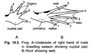
The forelimbs are meant to hold and support the front part of the body at the time of jumping but the hindlimbs assist in jumping and swimming as the webs are present in between the toes. Each forelimb comprises an upper arm (brachium), forearm (ante brachium), wrist and hand (manus) with four fingers (digits) and a vestigial “thumb” or pollex.
In male the base of the first (inner) finger is thickened especially in the breeding season, forming the nuptial pad for clasping the female at the time of amplexus. Each hindlimb comprises an upper thigh, shank or lower leg, ankle (tarsus) and long foot. The latter has a narrow sole and five slender toes connected by broad thin webs of skin which help in swimming.
Coelom and Viscera:
ADVERTISEMENTS:
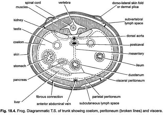
The coelom or body cavity is large and spacious in which are present viscera or internal organs. It lies ventral to the vertebral column or backbone. The walls of the body cavity and the visceral organs are covered by a thin, moist peritoneum. This membrane is perfectly continuous throughout and is simply reflected over the various organs.
The portion of the peritoneum surrounding the alimentary canal and its appendages is called the visceral layer and the part applied to the body wall is the parietal layer. The parietal layer on the dorsal side of the body is separated from the wall forming a large lymph space, the subvertebral lymph space. The kidneys lie in this space, hence, they are covered with peritoneum on the neutral side.
The alimentary canal and gonads are suspended from dorsal body wall by thin sheet of membrane called the mesentery. The coelom is filled with a transparent coelomic fluid which is like lymph. Near the anterior end of the body cavity lies the heart enclosed in a transparent sac, the pericardium.
Skin:
The skin is smooth, moist, slippery and lacking in the external protective scales or hairs. It is loosely attached by thin bands of connective tissue to the underlying musculature due to subcutaneous lymph spaces and, thus, these animals are easily skinned. It is considerably thicker on the dorsal side of the body than it is below.
At the dorsal side of the body it is thrown into a number of folds which extend from behind the eyes. The ridges, thus, formed by the thickening of the skin are known as dorsolateral dermal plicae. Its colour on the back and the limbs is dark green with dark coloured streaks and patches, while on the ventral side it is pale yellow.
Structure:
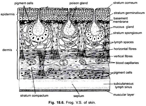
Structurally, like other vertebrates, the skin is composed of two layers, the epidermis and the dermis.
Epidermis:
The epidermis is an outer layer which is non-vascular, stratified and further composed of several layers of epithelial cells. It is usually shed and renewed at regular intervals by a process of moulting. The outermost layer is keratinized and made up of flattened, squamous epithelial cells. It is known as stratum corneum.
The innermost layer called stratum germinativum or stratum Malpighii is made up of active columnar epithelial cells which are capable in producing the new cells that pass towards the outer surface and become more and more flattened and ultimately lose their columnar shape as they reach the surface. It is due to the gradual change of protoplasm of these cells into a horny substance called keratin.
As the old cells are worn out due to friction, they are replaced by new ones formed by the cells of the layer stratum germinativum or stratum Malpighii. The stratum corneum is shed off from time to time and eaten by frog. The shedding of stratum corneum is due to the secretions of thyroid and pituitary glands. This activity is known as moulting.
Dermis:
The dermis forms a tough, flexible and somewhat elastic layer just underneath the epidermis. The dermis is separable into two layers, an outer comparatively loose layer (stratum spongiosum), which contains most of the glands, and an inner layer (stratum compactum) formed of dense connective tissue.
The stratum spongiosum consists of a loose network of fibrous connective tissue, richly supplied with lymph spaces and blood vessels. Just beneath the epidermis it forms a thin layer which contains numerous pigment cells. Each cell is irregular in shape with branched processes. They have black and yellow pigments and impart colouration to the skin. In the deeper portion are embedded the glands.
The stratum compactum is composed of a dense layer of connective tissue whose fibres run in a wavy course parallel to the surface of the skin. At intervals this layer is crossed by vertical strands, which often extend through the stratum spongiosum into the epidermis. In addition to fibrous connective tissue, these strands contain smooth muscle fibres, elastic fibres, nerves and bloodvessels.
The subcutaneous connective tissue forms a loose layer beneath the stratum compactum and a second very thin layer next to the muscles. The two layers are separated by large lymph spaces except in the septa, where they become continuous.
Glands:
In the skin of frog two types of glands are found—the mucous glands and the poison glands. Actually these glands are the derivatives of the epidermis but they lie in the stratum spongiosum of the dermis.
The mucous glands are somewhat smaller, flask-shaped found in abundance practically over the entire surface of the body. Their ducts are narrow and lined with a layer of small flattened epithelial cells. The body of the gland is also lined by a single layer of epithelial cells except near the opening of the neck, where there are two layers.
It is this epithelium which forms the mucus which is discharged into the lumen of the gland, and poured out through the neck over the surface of skin. The mucus is a colourless watery fluid which keeps the skin moist, glistening and sticky.
Outside of the epithelium of glands is a muscular coat of smooth muscle cells. The function of the muscle cells is the expulsion of the mucus of the glands.
They are more numerous on the dorsal side of the body and hindlegs, and they are especially abundant, and large in the dermal plicae.
These glands are lined with a single layer of epithelial cells and communicate with the exterior through their respective fine ducts which are narrow and lined with a layer of small flattened epithelial cells. Their epithelial cells are cylindrical nearly filled with granules.
Outside the epithelium like mucous glands, is a muscular coat and a connective tissue coat. The secretion of the poison glands is a whitish fluid with a burning taste. It protects the animal in some degree from the enemies.
Functions of the Skin:
The skin of frog performs the following functions:
1. It gives definite shape and texture to the body and also acts as a protective covering over the body.
2. It protects the body against the invasion of foreign bodies and fungal spores.
3. The mucous glands keep the skin moist, glistening and sticky. The mucus also prevents the invasion of the water and other harmful materials dissolved in water.
4. It forms a chief respiratory organ as its moist surface brings about an exchange of respiratory gases (O2 and CO2) in between the body of the animal and the environment.
5. Being devoid of sweat glands it acts as an excretory organ as the shedding of stratum corneum from time to time helps in removing the excretory wastes which are no longer needed for the body.
6. Due to presence of nerve endings it acts as an important sensory organ.
7. The frog never drinks the water through buccal cavity but absorbs through skin and, thus, compensates the loss of water from body. The skin is loosely attached to the body, and a considerable quantity of water may collect in the large subcutaneous lymph spaces.
8. The skin of frog larva produces hatching enzymes which dissolve the egg membrane so that hatching may occur.


