Frogs lay their eggs in water in early spring. In pseudocopulation or mating, the male frog firmly clasps the body of the female frog by his forelegs and enlarged thumb pads (nuptial pads). These nuptial pads help in clasping the body of female. This sexual embrace is called amplexus. As the eggs are extruded through the cloaca of female (oviposition), the male deposits sperm cells over them (insemination). Thus, fertilisation is external, taking place in water.
Spawning:
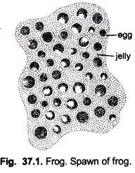
The mesolecithal eggs of frog enclosed in a protective gelatinous albumen are laid in water. The cluster or masses of eggs which remain stick together is called spawn. A spawn of Rana tigrina may have 3000 to 4000 ova. The spawn is laid during pseudocopulation or amplexus.
Fertilisation:
In frog, fertilisation is external and occurs at once in water outside the body of the oviparous female.
ADVERTISEMENTS:
The entire sperm penetrates the ovum anywhere around the animal hemisphere. If fertilisation is delayed, the albumen layers around the ovum become too thick for the sperm to pass through them and the ovum also starts to show degeneration. In the fertilisation process, vesicular sperm nucleus and vesicular female nucleus (or pronuclei) fuse together to form zygote nucleus.
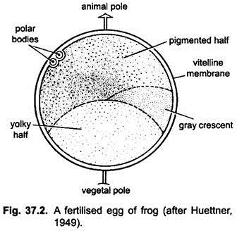
The fusion of both male and female pronuclei is called amphimixis. The fertilised egg or zygote is about 1.6 mm in diameter; it rotates within the vitelline membrane so that the animal pole becomes dorsal. The upper half of the zygote or animal hemisphere is pigmented black and it contains the cytoplasm and a nucleus, the lower vegetal hemisphere is white and full of yolk.
On one side between the black and white areas is a gray crescent region which marks the future dorsal side. At this region cortex becomes thin and this area is crescent-shaped.
The plane passing through the centre of grey crescent and the animal pole defines the median plane of bilateral symmetry. It coincides with the embryonic axis and is the only plane which separates the egg into two equivalent parts, each containing half the crescent material.
Cleavage and Blastulation:
ADVERTISEMENTS:
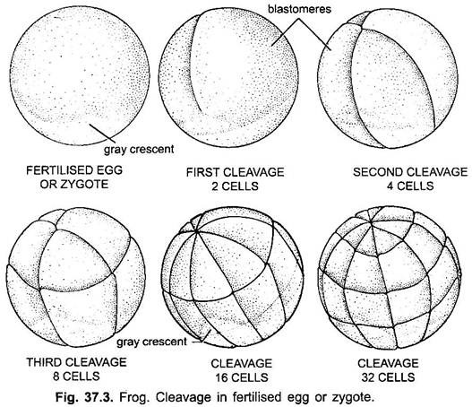
Cleavage or segmentation is holoblastic and unequal. A vertical furrow from the animal to the vegetal pole divides the zygote completely into two equal-sized cells. A second vertical furrow at right angles to the first divides the zygote into four cells.
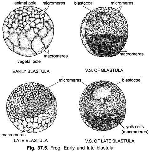
The third cleavage is horizontal and above the equator which segments the zygote into upper four smaller, black-coloured cells, and lower four larger, white-coloured cells. The cells formed by cleavage are blastomeres, the upper black blastomeres are called micromeres, and lower white ones are macromeres.
Further cleavages divide the micromeres more rapidly than the lower macromeres whose division is hindered by yolk. The blastomeres’ mutual pressure flattens their surfaces in contact with each other but free surfaces of each blastomere remain spherical. At this stage the whole embryo acquires a characteristic appearance reminiscent of a mulbery and so it is called morula.
ADVERTISEMENTS:
About fourth and fifth cleavage stages a small space, the blastocoel appears between the blastomeres of morula. In the beginning it is like narrow crevices between blastomeres of morula, which gradually increases as the cleavage goes on.
The blastocoel shifts more and more towards the animal pole due to more rapid multiplication of the micromeres, and infiltrated by water and albuminous fluid secreted by the surrounding cells. The blastocoel also enlarges due to uptake of more water. As cleavage proceeds, the blastomeres arrange themselves into a true epithelium called blastoderm.
It is two-cell thick towards animal pole of the egg and forms the roof of blastocoel, while the sides and floor of the blastocoel is occupied by multilayered blastoderm of large yolky blastomeres. Thus, the resulting embryo having fluid-filled blastocoel is called blastula.
Presumptive Areas:
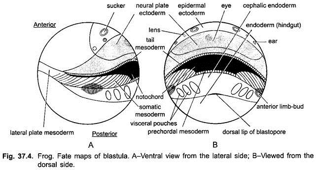
In the blastula, the blastomeres which have to form different germinal layers and different organs of the adult frog can be observed by artificial-vital staining methods of Vogt (1925) and prospective organ region maps or fate maps have been prepared.
According to the fate map studies, the whole surface of blastula can be divided into the following three areas:
(i) Prospective ectoderm area is present on and around the animal pole and it is pigmented black. The neural ectoderm occurs largely on the future dorsal side of blastula, while the epidermal ectoderm occupies the antero-ventral side of the blastula. Inside the neural ectoderm occurs a small sub-area that develops into the eye of the embryo. The sub-area of nose, sucker, ears and mouth are present inside the epidermal ectoderm.
(ii) Prospective notochord and mesoderm area is present behind the pigmented animal hemisphere. It is crescentic gray area, the marginal zone along the equator of blastula. It has blastomeres for the formation of notochord and mesoderm of the embryo. The large area of dorsal side of the gray crescent is occupied by notochordal cells.
Beneath the notochordal area, toward the vegetal pole lies a narrow strip of cells which form the pre-chordal plate of the embryo. On either sides of notochordal area, the part of grey crescent forms the segmental muscles (somites) and tail mesoderm is a narrow strip of cells on the dorsal side, toward animal hemisphere. Lateral and ventral parts of grey crescent give rise to ventro-lateral mesoderm.
ADVERTISEMENTS:
(iii) Prospective endodermal area is the entire non-pigmented area of the vegetal hemisphere, which give rise to endodermal lining of the mouth, gill region, pharynx, midgut and hindgut, and other organs such as liver, pancreas, urinary bladder and certain endocrine glands.
Gastrulation:
Gastrulation is a process of migration and re-arrangement of prospective organ forming cells already present in the blastula. It is brought about by several types of morphogenetic movements taking place at the same time.
i. Invagination of Endoderm:
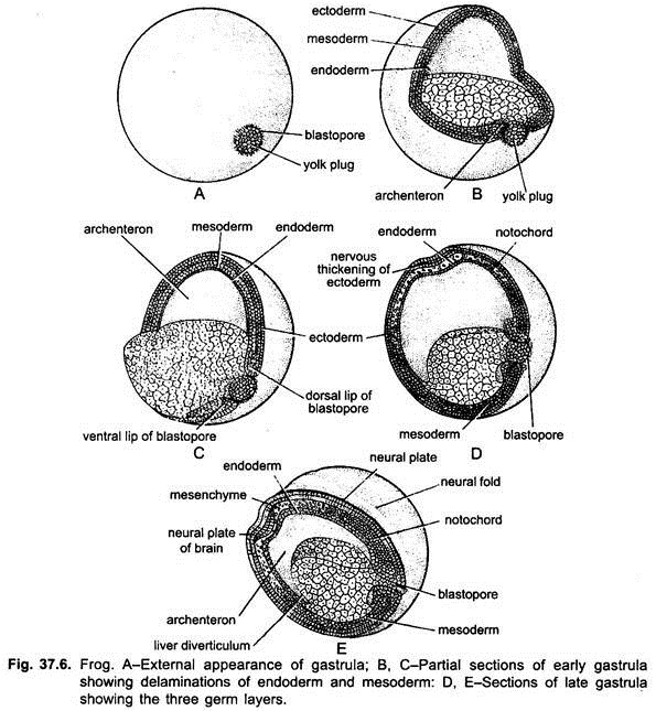
Certain prospective endodermal cells just beneath the mid- dorsal point of gray crescent of blastula assume the elongate shape of a bottle and move toward the interior of the blastula. Their steadily elongating necks remain attached to the surface of the blastula with the outermost cementing layer.
Thus, as the bulky-cell bodies move inward, a pull exerted along their attenuated necks and creates an indentation at the surface. With continued multiplication and attenuation of bottle cells, the invagination deepens, and expands internally to form the archenteron or gastrocoel and its outer opening (original indentation) is called the blastopore lying at the future posterior end. The area immediately above the blastopore is the dorsal lip of blastopore.
Gradually, the blastoporal invagination extends circulo-laterally, so the blastopore becomes crescentic, then horse-shoe-shaped and finally circular. Thus, lateral lips and ventral lip of blastopore are also formed and fused with each other along with dorsal lip, forming circular lip of blastopore.
The endoderm of foregut involute over the dorsal lip along chorda-mesoderm. The rest of prospective endoderm of vegetal region passes into the interior of the embryo passively and come to lie in the floor of gastrocoel.
ii. Involution of Pharyngeal Endoderm and Chorda-Mesoderm:
The endodermal cells bordering the dorsal lip of blastopore form the prospective pharyngeal endoderm, which is followed by pre-chordal plate, notochord and tail mesoderm. When dorsal lip is formed, the pharyngeal endoderm cells involute over the dorsal blastoporal lip. These cells move to the interior and their place take the converging prechordal plate cells and they also involute.
Behind these cells are present notochordal cells and tail mesoderm cells, which also involute and move to the interior. As these materials move inward around the dorsal lip they become considerably narrowed and elongated. The prospective pharyngeal endoderm in later stages of gastrulation forms the foregut whose lateral, ventral and anterior walls consist of a thin layer of endoderm. The dorsal wall of foregut consists of prechordal plate and anterior tip of notochord.
The notochord cells of the posterior region also involute and move anteriorly over the dorso-lateral lips of blastopore. Thus, the notochord forms the mid-dorsal wall of the archenteron, which is in the form of strip. The prechordal plate also forms the dorsal wall of the archenteron in front of notochord. The tail mesoderm remains near the blastopore, and marks the posterior end of the embryo.
The mesoderm (i.e., trunk somites and ventro-lateral mesoderm) rolls over the lateral and ventral lips of blastopore and then invaginates. After rolling inside the entire mesoderm (i.e., notochord, prechordal plate mesoderm, somites and ventro-lateral mesoderm) move from posterior side (blastoporal end) towards the anterior side as a single unit called chorda-mesodermal mantle in between outer ectoderm and inner endoderm. Thus, it occupies the entire space between ectoderm and endoderm except a small space at the anterior end of embryo where mouth will be formed in late stage.
iii. Epiboly of Ectoderm:
Throughout gastrulation the embryo retains its spherical shape and a uniform size. After involution of gastral endoderm and entire mesoderm, this space is taken up by the ectoderm (epidermal and neural). The expansion of ectoderm from animal hemisphere over the vegetal hemisphere is an active process. The presumptive epidermal ectoderm expands in all directions, but the presumptive neural ectoderm expands mainly in the longitudinal direction, i.e., from anterior end towards the blastopore and also contracts transversely.
Thus, the ectoderm expands up to the circular lip of blastopore through which unpigmented endodermal cells is visible, which form the yolk plug. Due to contraction of circular lip of blastopore, yolk plug slightly comes outside. Thus, in the end of gastrulation a new cavity gastrocoel is formed and the blastocoel is obliterated.
Due to accumulation of endodermal mass on the future ventral side, the gravity is shifted and embryo rotates within fertilisation membrane so as to bring its dorsal side uppermost. The protruding yolk plug gradually withdraws to the interior, and the blastopore steadily contracts to form a slit-like opening in the end of gastrulation.
iv. Gastrula:
Thus, gastrulation changes the radially symmetrical single layered blastula into a spherical, bilaterally symmetrical, triploblastic gastula having a head-to-tail axis. It is externally covered by ectoderm and endoderm, and mesoderm lies in the interior. Gastrocoel forms the lumen of the forming gut. Its lateral walls and floor is formed by the endoderm and its roof is formed of chorda-mesodermal cells.
Neurulation:
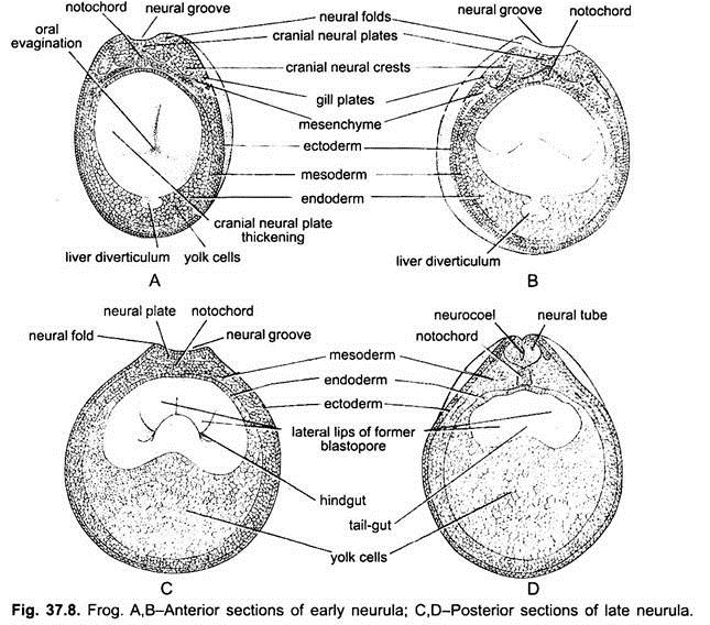
By the time gastrulation is being completed, the ectoderm along the mid-dorsal side of the embryo thickens to form a pear-shaped medullary or neural plate. The neural plate cells change in shape and become elongated and arranged themselves into a columnar epithelium.
The epidermal cells remain more or less flat and arranged as a stratified epithelium usually two cells thick. The edges of the neural plate become thickened and slightly raised above the general level as ridges called neural folds.
The neural plate narrows transversely especially in its posterior parts and the neural folds raised higher due to which a neural groove is formed along its length. The neural folds grow and fuse with each other in the mid-dorsal line to form a neural tube.
The lateral epidermal ectoderm of either side also meet and fuse at the mid-dorsal line above the neural tube, thus, enclosing it. The neural tube remains open in front for a time as a neuropore, posteriorly the neural folds cover and fuse over the blastopore so that the cavity of the neural tube communicates with the archenteron by a neurenteric canal which is the narrow canal-like opening of blastopore.
The anterior broad part of the neural tube forms the brain and the remaining narrow posterior part becomes the spinal cord. The neural tube also forms neuroglia cells of the nervous system.
Neural Crest Cells:
The cells from the neural folds that come to lie between the dorsal epidermis and the dorsal part of the neural tube are the neural crest cells. These lie along the dorso-lateral sides of the neural tube. The neural crests give rise to melanocytes, dorsal root ganglia of spinal nerves, parts of the autonomic nervous system and adrenal glands, and to some mesenchyme cells which form the visceral arches.
Notogenesis:
The notochord cells separates off from the prechordal plate of mesoderm as a narrow rod of cells. This notochordal rudiment also separates off from the rest of the chorda-mesodermal mantle and notochordal cells transform into colligocytes.
These cells secrete a collagenous sheath around them. Soon fluid-containing vacuoles appear in the notochordal cells which push the nucleus and cytoplasm toward the periphery. Thus, the notochord becomes round, turgid and elongated in antero-posterior axis.
Mesoderm Differentiation and Coelom:
The chorda-mesodermal mantle at the time of closure of blastopore, separates off from the endoderm and mesoderm, thus, lies in between ectoderm and endoderm. The notochordal cells also splits off from the prechordal plate at the anterior end.
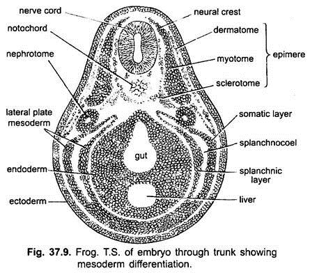
Simultaneously the tip of mesoderm at each side of notochord thickens and subdivides transversely, beginning at the anterior end, into a series of cell masses or somites. Each somite remains separated from its neighbours but remains joined to the lateral and ventral parts of the mesodermal mantle on each side by strands of cells.
These lateral and ventral mesodermal mantles are called lateral plates and these strands of cells in between somites and lateral plates are called mesomeres or nephrotomes, which later on forms the kidney tissue.
Each somite or epimere (dorsal part of mesoderm) on either side of notochord splits into three layers:
(i) Inner is the sclerotome or skeleton-forming tissue around the notochord,
(ii) Middle is the myotome whose cells differentiate to form the striated muscle fibres of the somatic muscles, and
(iii) Outermost narrow strip is the dermatome which forms the dermis of skin.
The hypomere or lateral plate of mesoderm of each side is divided by a split which passes downwards on each side to separate the hypomere into an outer somatic or parietal layer, and an inner splanchnic or visceral layer and the space between these two layers is a splanchnocoel or perivisceral coelom.
The inner visceral layer gives rise to smooth muscles of the intestine and to the blood and blood vessels, and outer somatic layer with the ectoderm forms the somatopleure. Anteriorly the coelom is restricted to the ventral side only as a pericardial cavity below the pharynx which gets separated from the splanchnocoel by a transverse septum.
Primitive Gut:
The nerve cord endoderm differentiates slowly and eventually forms the enteron or primitive gut. At the end of gastrulation it is an open trough. After the formation of notochord and separation of mesoderm, the free margins of endoderm fuse in the mid-dorsal line beneath the notochord to form the definitive gut or enteron. The floor of enteron persists as thick layer of large yolk-ladden cells.
At the antero- ventral end the enteron makes contact with the ectoderm, where later on mouth invagination occurs which communicates with the enteron. Later, the lungs, liver, pancreas develop as evaginations of the gut. The pharyngeal pouches develop as lateral out-pushings of the foregut, which later forms the gill-clefts.
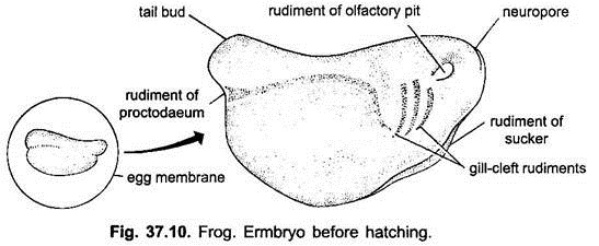
During neurulation, embryo elongates in antero-posterior axis, it also flattens laterally. The part of the embryo above the blastopore elongates beyond the blastopore forming the tail bud or rudiment of tail. The neurula at this stage is called tail bud embryo.
Post-Neural or Pre-Hatching Development:
The neurula of Rana tigrina develops into tail-bud embryo, having bulges of gill-plates, optic bulges, one on either side, stomodaeal groove at antero-ventral side of head and a pair of ectodermal adhesive organs, cement glands or oral suckers in the form of conical protuberances, which later on unite to form U or V-shaped adhesive sucker.
In trunk of embryo are present a pair of myotomes. Soon tail of embryo elongates and is provided with epidermal tail-fins. Trunk has about 12 pairs of myotomes and gill-plates has rudiments of three gill-clefts. Above the gills appear invaginations of auditory pits and at the front of head are present a pair of olfactory pits. The heart is simple S-shaped without chambers. At the base of tail is present a slit-like cloacal aperture.
Hatching:
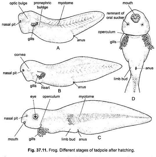
When the embryo is 4.5 mm long, it has a fully developed tail with tail-fins and myotomes extending up to the half-length of tail. A pair of external gills in the form of buds is erupted from each gill-plate. Stomodaeum is cup-shaped and V-shaped oral sucker becomes enlarged. The embryo hatches out by rupturing the egg membrane and is now called tadpole larva.
Tadpole Larva:
The newly hatched tadpole larva has no mouth, and is unable to feed. It is found attached with the leaves of aquatic plants, etc., with the help of its adhesive sucker. It gets its nourishment from the yolk present in the endodermal cells of the floor of midgut.
Its ectoderm is ciliated, nervous system and rudiments of sense organs are present. Gut is a straight tube with proctodaeum. Heart is S-shaped without chambers and blood vessels are being formed. The kidneys are pronephros with ducts opening into proctodaeum.
Post-Hatching Development of Tadpole:
A pair of folds of the skin called operculum grows backwards and downwards till the two meet on the ventral side, they have no skeletal support, and are not homologous with the operculum of bony fishes. Posteriorly each operculum covers the gill-clefts, external gills, and the area from which forelimbs will develop.
External gills begin to degenerate and the skin covering the third, fourth, and fifth pairs of visceral arches forms paired internal gills lying below the operculum. The internal gills are like the external gills, their filaments are covered with ectoderm, unlike the gills of fishes.
By now the external gills have disappeared and the tadpole breathes by its internal gills which are washed by a current of water entering through, the mouth and passing through the gill-clefts.
I. External Gill-Stage of Tadpole:
Tadpole at this stage is about 5.5 mm long and the following structures are visible in it:
1. Five pairs of branchial, pharyngeal or visceral pouches are formed in the gill-plate area due to out pushing of the endodermal lining of pharynx. The epidermal ectoderm also invaginates and fuses with the out pushing of pharynx and then a slit-like opening is formed which is called gill-slit or gill-cleft, by which pharynx communicates with the outside. In frog only four pairs of gill-slits are perforated.
2. Soon from the sides of head in the pharyngeal region three pairs of external gills are projected out, which are feathery extensions of the integument above the gill-slits. There are bathed by the surrounding water.
3. Stomodaeum develops in the form of pit, whose outer opening is the mouth. The mouth is surrounded by fringed lips and also acquires a pair of horny jaws. The fringed lips has two rows of tiny, needle-like horny teeth. A 10.5 mm long tadpole contains 3 rows of fringed teeth.
4. Adhesive suckers disappear.
5. The larval gut is differentiated into pharynx, oesophagus, stomach and intestine. Liver and gall bladder also develop. The intestine is very long and coiled like a watch spring due to herbivorous mode of feeding.
6. Myotomes extend up to the tip of tail.
7. Melanophores appear in the skin of dorso-lateral surface of head, trunk and tail.
8. The cornea becomes transparent and eye lens is visible.
9. Lateral line sensory system is visible on either lateral side of the tail.
10. A pair of giant neurons called Mauthner cells appear in the hindbrain. It is an interneuron that transmits impulses from the lateral line and auditory receptors to the motor output system of spinal cord.
11. A pair of pronephric kidneys becomes functional and excretes ammonia.
12. On either side of cloacal aperture, at the junction of trunk and tail, a pair of hindlimb buds appears.
The tadpole swims actively with the help of tail and feeds on algal and other aquatic vegetation.
II. Internal Gill-Stage of Tadpole:
1. The opercular folds grow backward from the hyoid arch of each side covering the external gills and gill-slits and finally fuse with each other ventrally and with the belly wall. Thus, an operculum or gill-cover is formed enclosing the external gills and gill-slits and open outside by a ventro-lateral opening, the spiracle located on the left side of the body.
2. External gills later fall off and four pairs of filamentous internal gills develop on the walls of gill-slits.
3. Intestine is still coiled and long.
4. Different parts of hindlimbs such as thigh, shank, ankle, foot and five toes become well formed in the tadpole of 40 mm long.
5. A pair of forelimb buds appear behind the head but remain hidden within operculum. As development proceeds, the left forelimb emerges through the spiracle. The right forelimb appears later.
6. In a mature tadpole, a pair of lungs develop from the pharynx. Now the larva breathes by both, the internal gills and lungs.
Thus, a fully formed tadpole larva is a fish-like creature. It has a well-developed locomotory tail with tail fin and muscles. It has a pair of eyes, nostrils, mouth, long spirally coiled intestine, cloacal opening and spiracle.
Adhesive glands have lost. It respires with the help of internal gills but in later stage lungs develop, so it breathes by both internal gills and lungs. Soon gill-slits close, internal gills absorbed and opercular cavity disappeared and, thus, it breathes only with the help of lungs.
It frequently comes to surface to gulp air. Lateral line system is well developed. Mesonephric kidneys are formed. Hindlimbs appear first and later the forelimbs, which are hidden within operculum in the beginning.
Metamorphosis:
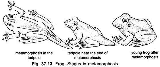
Metamorphosis (Gk., metamorphoun = to transform) is the abrupt transition from larval to adult form. It includes morphological, anatomical, physiological and behavioural, hormonally regulated changes in the larval form to transform it into the adult form.
In frog, metamorphosis is of progressive type. In frog, it is associated with a transition from an aquatic to a terrestrial mode of life and from a herbivorous to carnivorous mode of feeding. The metamorphosis involves numerous structural, biochemical and physiological changes.
These changes include the degeneration of existing structure, construction of new structures and modification of larval structures. Metamorphosis is controlled by hormones such as thyroxine of thyroid gland.
The following changes occur during metamorphosis:
1. The first sign of metamorphosis is that tadpoles frequently come to the water surface to take air into their lungs through the mouth because internal gills are resorbed, gill-slits are closed, opercular fold also falls off and lungs develop for aerial respiration.
2. The tadpoles stop feeding, they are nourished on the substance of the tail, the tail becomes shorter and resorbed. The ciliated larval skin along with the epidermal horny jaws with horny teeth and labial fringes are cast off.
3. The vascular system of larva having aortic arches for internal gills are reduced and becomes modified for aerial respiration.
4. The larval haemoglobin in RBCs having higher affinity for oxygen and independent from pH is replaced by adult haemoglobin which has lower affinity for oxygen and highly sensitive to acid.
5. The intestine shortens because of the change from a herbivorous to a carnivorous diet. The stomach and liver increase in size and start secretion of ceruloplasmin, a copper-binding serum protein.
6. Lateral line system and Mauthner cells of brain degenerate and disappear.
7. The limbs increase in size and differentiate.
8. The mouth widens, true jaws develop and tongue becomes long. Eyes become prominent on the dorsal surface of head with eyelids and nictitating membrane. Rhodopsin visual pigment appears.
9. Middle ear develops in connection with the pharyngeal pouch located between mandibular and hyoid arches. Tympanic membrane also develops.
10. Larval pronephros change into mesonephros of adult.
11. Erythropoesis occurs in spleen instead of liver.
12. Brain also becomes highly differentiated.
13. The synthesis of melanin and serotonin (a local vasoconstrictor) begins in the skin.
14. In stomach peptic activity starts for the digestion of animal tissue.
At cellular level, the cell modifications are evident in eye and eyelids, limbs, skin, operculum, tongue, liver, pancreas, intestine and lungs. Probably no cell or tissue or organ remain unaffected.
Endocrine function of pancreas also starts at metamorphosis to turn over the carbohydrates in liver. Thus, a young frog is formed, it still has a stump of the tail and it leaves water for some damp land, it feeds on insects and continues growing till it assumes the adult form and colour.
Hormonal Control of Metamorphosis:
The metamorphosis is under control of hormones secreted by hypothalamus, hypophysis and thyroid. Thyrotropin-releasing factor (TRF) and prolactin-releasing-inhibiting factor (PIH) of hypothalamus, and thyroid-stimulating hormone (TSH) and prolactin hormone (PH) of hypophysis regulate the secretion of thyroid hormone (TH) such as throxine from thyroid gland.
Thyroxine causes the degeneration and necrosis of some cells or tissues and organs in the larva, and stimulates the growth and differentiation of organs needed in the adult frog. Iodine is an essential component of thyroxine hormone and it has been found that iodine alone can cause metamorphosis in frogs’ larva.
Significance:
The study of embryology of frog is practically useful to us in a variety of ways:
1. It helps in interpretation of avian and mammalian development.
2. It explains the evolutionary transition of lower chordates into higher chordates.
3. It explains the evolution of lung-breathing animals from gill-breathing animals.
4. It also explains the evolution of various physiological requirements present in air-breathing and land-living animals.
