Development of Branchiostoma (Amphioxus) is indirect involving a larval stage. Early embryology of Branchiostoma is simple. Therefore, the transformation of egg, which having less yolk, into a complex and differentiated animal is far easier to follow than in any other vertebrate.
The early development of Branchiostoma (Amphioxus) is of great phylogenetic significance because it resembles with those of invertebrates like echinodermates on one hand and vertebrates on the other. Development of Amphioxus was described by many scientists such as Hatscheck (1882, 1888), Wilson (1883), Cerfontaine (1906), and Conklin (1932). The work of Conklin (1932) is the most recent and accepted one.
Gametes:
Spermatozoa:
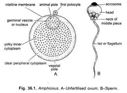
Spermatozoon of Amphioxus has a typical structure of flagellate spermatozoon, i.e., it contains a spherical head, a very short middle piece and a long tail.
ADVERTISEMENTS:
Ova:
The unfertilised ovum or secondary oocyte of Amphioxus is 0.10 mm to 0.12 mm in diameter. It is microlecithal and isolecithal. It is surrounded by a plasma membrane, which is further surrounded by a thin vitelline membrane of mucopolysaccharides. Its nucleus is eccentric lying at the animal pole.
The egg undergoes its first maturation division before it leaves the ovary. The animal pole is free from yolk and the cytoplasm towards the vegetal pole contains some yolk distributed evenly.
Fertilisation:
Fertilisation is external, taking place in the surrounding sea water where eggs and spermatozoa are shed. Before fertilisation, the ovum has outer thin vitelline membrane enclosing a peripheral cytoplasmic layer, central yolky cytoplasm mainly towards the vegetal pole and a fluid-filled germinal sac or nucleus towards the animal pole.
ADVERTISEMENTS:
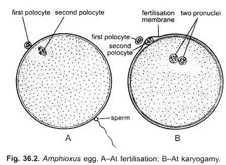
During fertilisation, the sperm enters the egg near the vegetal pole which provides a stimulus for the egg (secondary oocyte) to undergo its second maturation division, a polar body is extruded which comes to lie near the animal pole inside the fertilisation membrane, within perivitelline space.
It is a space formed by the elevation of fertilisation membrane from the plasma membrane. It marks the animal pole of the egg. The nuclei of sperm and egg migrate towards the equator of the egg and fuse to form zygote nucleus. The site of fusion is generally above the equator of the egg and slightly toward the side which will eventually be the posterior end of the embryo.
Presumptive Areas:
ADVERTISEMENTS:
Immediately after fertilisation the cytoplasm of the zygote gets so arranged as to foreshadow the future parts of the embryo. The clear cytoplasm occupies the animal hemisphere develops mainly into the ectoderm, the granular yolky cytoplasm at the vegetal pole gives rise to endoderm, a basophilic cytoplasmic crescent at the posterior end forms mesoderm, and another ill-defined cytoplasmic crescent around the anterior side, opposite to the mesodermal crescent is the dorsal crescent forms the notochord and nervous system.
Thus, it includes both notochordal and neural ectodermal material. This precocious differentiation of cytoplasm of the zygote is strikingly similar to that of Urochordata, and it shows a close relationship of two groups of Protochordata.
Early Embryonic Development:
Cleavage and Blastulation:
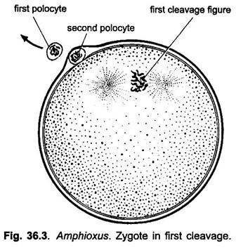
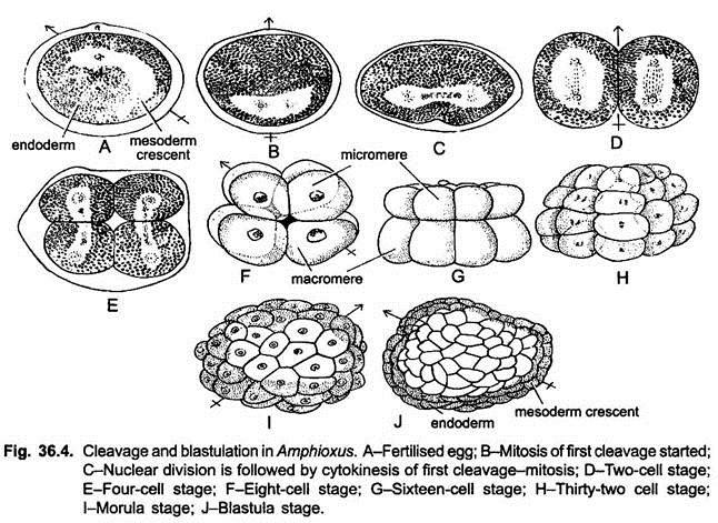
Cleavage is complete or holoblastic and almost of equal type. First cleavage is meridional which cuts through the egg along its median axis, resulting into two equal sized blastomeres, the right and left, which forms the right side and left side of the adult animal. The second cleavage is again meridional, but at the right angle to the first and divides the first two blastomeres into four equal-sized blastomeres.
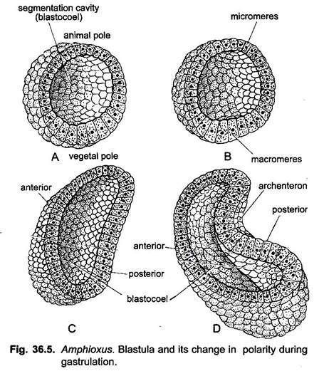
The third division is horizontal passing just above the equator forming four upper smaller micromeres near the polar body, and four lower large macromeres at the vegetal pole. Fourth cleavage is again meridional cutting all the eight blastomeres synchronously. Sixteen-cell stage is formed having eight micromeres and eight macromeres.
The fifth cleavage is latitudinal or horizontal and synchronous, resulting into thirty-two cells arranged in four tiers. The sixth cleavage is meridional and synchronous producing sixty four blastomeres. Later cleavages will not be synchronous up to this stage, the blastomeres remain loosely packed and form the morula. Meanwhile, a semifluid material accumulates in the centre of the morula.
ADVERTISEMENTS:
It pushes all the blastomeres outward during further cleavage of the morula, so that they become arranged in a single layer called blastoderm, enclosing a central fluid-filled cavity the blastocoel. This embryonic stage is called the blastula and a fully developed blastula of Amphioxus is called the coeloblastula.
The cells at the vegetal pole of blastula are larger than at the animal pole and the blastoderm is, therefore, thicker at the vegetal pole. The cells of ventral or mesodermal crescent are small sized, spherical amoebocytes.
Gastrulation:
In Amphioxus, gastrulation is started when there are about 800 cells in the embryo, i.e., between 9th and 10th cleavage.
It involves two basic types of morphogenetic movements of the embryonic cells:
1. Epiboly and
2. Emboly.
Both the processes go on side by side.
1. Epiboly:
It is the expansion of ectodermal cells of the embryo.
2. Emboly:
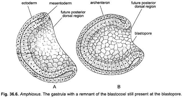
The blastoderm at the vegetal pole (endodermal plate) becomes flat and subsequently bends inwards (invagination). Thus, the embryo, instead of spherical becomes converted into a cup-shaped structure, having a large cavity, the archenteron, opening outside by a wide blastopore.
The cup has double walls, an external and internal epidermis, both of which are continuous with each other over the rim of the cup-shaped embryo. In this embryo, the prospective mesoderm lies in the ventral lip of blastopore and the prospective notochord lies in the dorsal lip of blastopore.
As the endodermal plate moves inward dorsal notochordal cells involute, move inward along with the endoderm due to which prospective notochordal cells come to lie beneath the prospective neural ectoderm. Similarly, the prospective mesoderm in the ventral lip of the blastopore also moves inward and side by side its lateral horns converge toward the dorsal side of the embryo, so that they come to lie on both sides of the presumptive notochord.
Now the embryo also elongates in antero-posterior direction and in this elongation all the prospective cells take part. The blastopore subsequently narrows due to contraction of the rim of blastopore. The blastopore leads into a newly formed cavity, the archenteron.
Thus, a two layered gastrula is formed, in which the outer layer of cells becomes ciliated but it is enclosed in the vitelline membrane. Growth continues and the gastrula becomes further elongated antero-posteriorly. Growth of the dorsal lip reduces the blastopore and shifts it from a nearly dorsal to a posterior position.
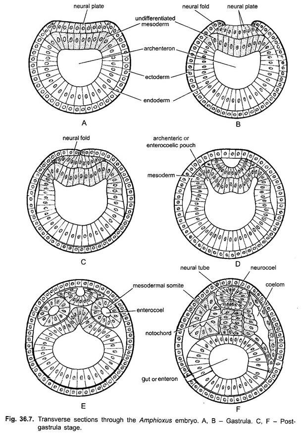
Gastrula has an outer layer of ciliated ectoderm cells, in which mid-dorsally are cells of neural plate and ventro-laterally are cells of epidermal ectoderm; the inner cells of the gastrula have a mid-dorsal strip of notochord cells, on the two sides of which are mesoderm cells, and the lateral and ventral inner cells are endoderm.
Fate of Germ Cells:
The ectoderm will give rise to epidermis, nervous system and receptors. Endoderm will form the alimentary canal and midgut diverticulum. Mesoderm will form muscles, connective tissue and germ cells.
Formation of Notochord:
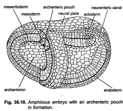
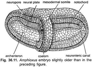
The cells which were invaginated from the dorsal lip of the blastopore lie in the mid-dorsal roof of the archenteron. They evaginate dorsally at the anterior end of embryo and become separated from the endoderm. This evagination of notochordal material also continues caudally and ultimately forms a solid, cylindrical cord of cells, which is called the notochord.
It lies below the neural tube and between the mesodermal somites. It extends in the entire length of the body. Its anterior extension into the rostrum takes place later. A notochordal sheath of fibrous connective tissue will eventually surround the notochord.
Formation of Neural Tube:
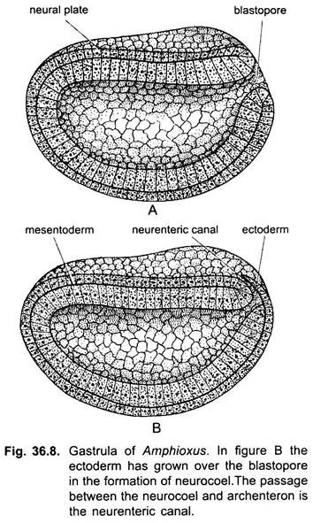
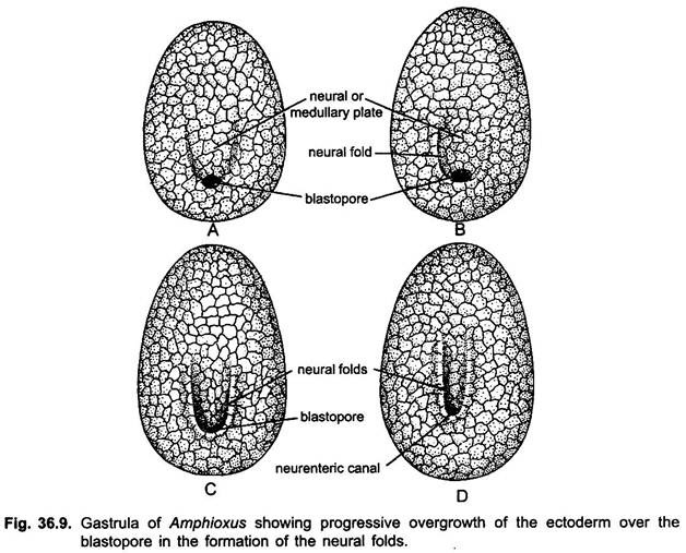
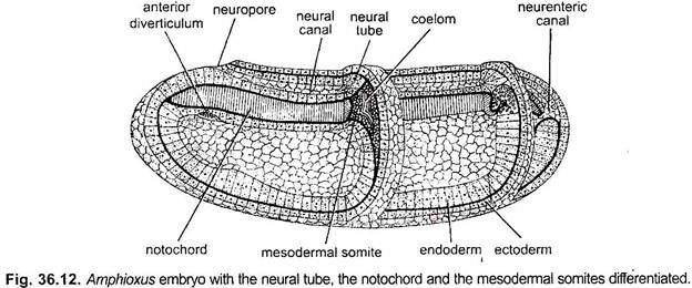
The prospective neural ectoderm cells along the mid-dorsal line become flat and thickened to form a neural plate which sinks inwards below the level of lateral epidermal ectoderm. The neural plate is then covered by the free edges of the epidermal ectoderm. At the same time the lateral edges of the neural plate grow upwards and meet in the mid-dorsal line to from a neural tube, enclosing a canal, the neural canal or neurocoel.
The neural tube opens in front by a neuropore which finally closes and its place is indicated by a Kolliker’s pit in the adult. The epidermal ectoderm spreads over the surface of the neural plate from the sides and from the area posterior to the neural plate.
Posteriorly the neural folds extend around the lateral lips of blastpore and when they meet above they close the blastopore, so that the neural tube communicates with the archenteron by a short neurenteric canal. The neurenteric canal persists for a short time until the cavity of the neural tube becomes completely separated from the cavity of archenteron, which in turn becomes the cavity of alimentary canal. The neural tube becomes spinal cord and its canal becomes the central canal of the central nervous system.
Development of Mesoderm and Coelom:
The cells in the dorso-lateral roof of the archenteron are presumptive mesodermal cells, they lie on either side of the notochord. Mesoderm cells form two lateral bands, which also get separated from the endoderm by dorsal evagination. In each mesodermal band a longitudinal groove appears. The groove deepens and opens widely into the archenteron.
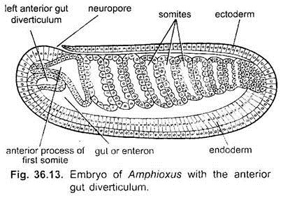
Beginning at the anterior end these longitudinal mesodermal grooves become transversely divided into distinct segments or somites, which lie on either side of notochord, one behind the other along the entire length of the body. The first two anterior somites retain a portion of enterocoel or archenteron and the rest are solid blocks of mesoderm. They ultimately become cut off from the endoderm.
Soon, each somite grows ventrally and becomes differentiated into following parts:
1. Some portion of each somite continues to grow around notochord and neural tube and becomes thickened to form the myotome.
2. The portion of each somite close to the epidermal ectoderm is called the somatic parietal mesoderm.
3. The portion of each somite close to the endoderm is called the visceral or splanchnic mesoderm.
4. The space found in between the somatic and visceral layer of mesoderm is called the enterocoelic coelom, because it is originally derived from archenteron.
Further Development of Coelom:
The spaces enclosed by somites are the forerunners of the coelom of adult Amphioxus. When each somite differentiates into dorsal myotome, the portion of coelom found in between somatic and splanchnic mesodermal layers is called ventro-lateral coelom or splanchnocoel.
As the myotomes enlarge, the splanchnocoelic space becomes more and more displaced ventrally and most of it comes to lie on either side of the enteron. Eventually, the splanchnocoels of each pair of somites grow ventrally to the lower portion of the enteron and ultimately fuse with each other.
Gradually, the splanchnocoels of all the segments fuse antero- posteriorly forming a continuous, antero-posterior, splanchnocoelic space below and around gut tube is formed. A horizontal septum, the intercoelomic membrane also appears, separating the myocoels above from the splanchnocoelic cavity below.
Thus, in Amphioxus the coelom is dual in origin – anterior most part of coelom is enterocoelic appeared in first two anterior most somites. The rest of coelom is schizocoelic in origin, i.e., solid mesoderm splits into two layers with an intervening coelom.
Differentiation of Myotome:
Each myotome has a median muscular portion and a laterally placed, thin-walled parietal part which surrounds the myocoel or coelomic space. The muscular portion of each myotome forms > -shaped myotome or muscle plate of adult Amphioxus.
The myocoelic portion in each segment gives rise:
(i) Lower sclerotomic diverticula which extend between myotome and notochord, and nerve cord. Its inner wall wraps around the notochord, and nerve cord and forms a skeletogenous sheath of connective tissues which ensheath these structures. Whereas its outer wall covers inner side of myotome with a connective tissue covering.
(ii) Ventral diverticulum extends between lateral wall of the splanchnocoel and epidermal layer of the body wall and separates the parietal mesoderm of the splanchnocoel from the epidermal wall. This ventral diverticula together with parietal wall of the myocoel above, forms the dermatome. Both the inner and outer layers of ventral diverticulum fuse to form the dermis of the skin.
(iii) The dorsal slerotomic diverticala forms the fin-ray cavities in dorsal fin.
Development of Head Cavities:
After about eight mesodermal somites have been separated, the dorsal portion of the anterior end of the gut cavity gives rise a dorsal or anterior gut diverticulum. This diverticulum splits to form a left and a right diverticula.
The right diverticulum extends into the anterior part of embryo in between ectoderm and notochord, in front of first pair of myotomes, and forms the head cavity or cavity of oral hood. The left diverticulum remains small and later fuses with an ectodermal invagination to form the Hatschek’s pit.
Development of Gut:
After separation of notochord and mesoderm from the endoderm, the edges of these endoderm cells grow towards each other and fuse below the notochord to form a tubular gut or mesenteron. Later the cells lining the gut become ciliated, and the splanchnic layer of mesoderm surrounding them gives rise to a layer of smooth muscles of the gut.
After separation of anterior gut diverticulum, cells of the floor of anterior part of gut become columnar and form an elongated groove, the hypopharyngeal groove or endostyle. Much later a midgut diverticulum or liver will grow out from the gut on the right side. The anterior part of gut also enlarges to form the pharynx.
Hatching of Larva:
Soon after gastrulation, when only two pairs of somites are formed, the ciliated embryo hatches from the vitelline membrane. The embryo is now called a larva. It swims actively on the surface of sea with the help of cilia. It does not feed since it has no mouth and anus.
Post-Hatching Development:
The ciliated larva swims actively and elongates gradually, becomes laterally compressed, the number of somites increases with elongation. Later on cilia disappear. Each larva undergoes following changes to become an adult.
1. Mouth:
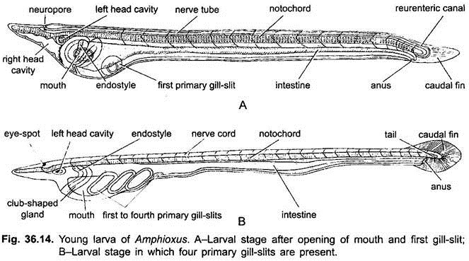
It develops when the larva acquires about 16 to 18 pairs of mesodermal somites. At about the level of second mesodermal somite, on the left side of the ventral surface of head, the ectoderm fuses with the endoderm and then there a perforation occurs forming the mouth opening. The mouth gets bordered with cilia.
2. Anus:
At the caudal end of the larva on the ventral side, a small area of endoderm fuses with the ectoderm and then a perforation occurs at this site to form the anus. Actually it lies slightly on the left side of the mid-ventral line. The larva begins to feed on plankton. Dorsal to the anus a tail grows out posteriorly.
3. Gill-Slits:
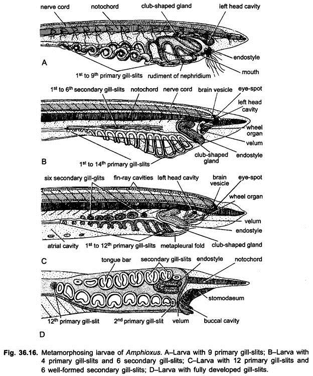
At the time of mouth formation, the endoderm opposite to the first pair of somites pushes ventrally and fuses with the ectoderm. This site perforates to form the first gill-slit. This gill-slit later on moves up on the right side of the body. Later fourteen more gill-slits also appear in a series and eventually shift to the right side of the larval body.
Out of these fourteen gill-slits, the first and the last five gill-slits degenerate or close up. These remaining eight gill- slits are called primary gill-slits. Soon another set of eight gill-slits called secondary gill-slits appear at the dorsal side of the primary gill-slits.
Thus, two rows of gill-slits are formed on the right side of the body. Eventually, first set of primary gill-slits is shifted to the left side of the pharynx. These gill-slits of both sides, later on, are subdivided by tongue bars and new gill-slits are added at the posterior end of both sets of gill-slits.
The first opening or cleft arises as a diverticulum from the floor, it lies on the right of the pharynx and is formed before the mouth, it becomes a tube opening at one end into the pharynx and at the other on the ventral side of the body, eventually it forms a coiled, glandular club- shaped gland which is a larval organ and disappears during metamorphosis.
4. Club-Shaped Gland:
From the anterior end of the gut floor grows out a coiled tube, which acquires an opening at its one end with the gut on the right side and at the other end with the exterior, slightly to the left side, just ventral to mouth. This is called club- shaped gland. Its cells are glandular. It begins to disappear when first set of eight gill-slits are formed.
5. Endostyle:
A V-shaped thickening arises from the wall of the gut, this is the rudiment of the endostyle. It is made of ciliated and glandular cells and is formed on the right side at the anterior end of the pharynx in front of club-shaped gland. The two limbs of the V fuse and grow back along the mid-ventral line to become the endostyle.
6. Myotomes:
Myotomes on either side of body increase in number.
7. Nephridia:
They also develop during larval life and are ectodermal in origin. Hatschek’s nephridium develops as a small tube behind the pre-oral pit.
8. Atrium:
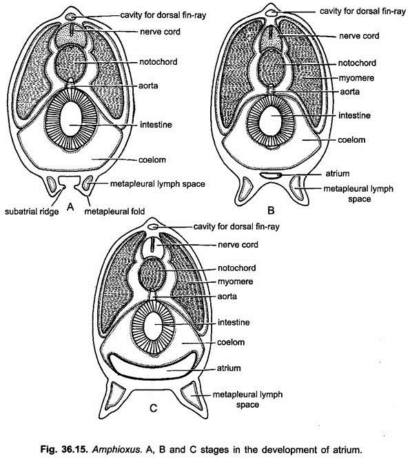
Shortly before the secondary gill-clefts of the right side are formed, the body wall forms two longitudinal metapleural folds on the ventral side. The atrial folds start appearing from posterior end where they are quite close to each other.
Anteriorly they grow and enclose the gill-slits in between them. From the inner side of each metapleural fold arises a shelf called the epipleur. The epipleurs grow towards each other and fuse together to enclose a space, the atrium.
The atrium is lined with ectoderm. It becomes closed anteriorly but remains open posteriorly by an atriopore. The atrium gradually enlarges displacing the body wall and reducing the splanchnocoel around the anterior part of the gut.
Metamorphosis:
For some time the larva continues to feed on plankton of the surface of the sea for about three months, and sinks to the floor of the sea where it grows and undergoes a slow metamorphosis to become an adult in several months.
Some very significant metamorphic changes are the following:
1. In this process ectodermal cilia and club-shaped gland are lost.
2. The mouth shifts from left to antero-ventral side of body. Its margins grow in to form a velum, folds of skin form an oral hood and oral cirri inside which a ciliated wheel organ is formed.
3. Larva acquires the habit of filter-feeding.
4. Gill-slits divide by the appearance of tongue bars and more gill-slits appear behind the first sets.
5. The development of atrium causes the reduction of coelomic cavity in the pharyngeal region, in the myocoel and in gonocoel.
6. Gonads develop from the anterior lower angles of myotomes and project into myocoelic spaces, which are now called gonocoels.
7. The tail region elongates and it assumes a considerable degree of bilateral symmetry to become an adult.
Significance:
The study of the development of Branchiostoma (Amphioxus) is significant from the following point of view:
1. The study of embryology (development) provides an insight into the evolutionary history of chordates.
2. The position and the way of the formation of blastopore in developing embryos divide bilaterally symmetrical animal into two groups, viz., Protostomia (where blastopore marks the area of mouth) and Deuterostomia (where blastopore marks the anus). The Branchiostoma falls in the later together with other chordates and echinodermates.
3. The way of formation of coelom in Branchiostoma (Amphioxus) brings it closer to echinodermates.
Based on the fate of blastopore, manner of mesoderm formation, muscle chemistry and similarity in sera proteins, it is believed that coelenterates onwards two stocks of invertebrates evolved. Branchiostoma (Amphioxus) is supposed to be a representative of primitive chordate and links chordates with invertebrates and shows the evolutionary steps followed by vertebrates.
