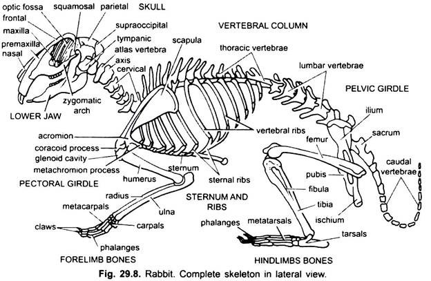
The endoskeleton of rabbit is chiefly formed of bone and cartilaginous part is very little.
Exactly like those of other vertebrates, the skeleton of rabbit can also be divided into two parts:
(i) The axial skeleton is present along the longitudinal axis of the body and consists of the bones of skull, the vertebral column, the ribs and the sternum;
(ii) The appendicular skeleton lies at right angle to the longitudinal axis of the body and consists of the bones of limbs and the girdles.
Axial Skeleton:
ADVERTISEMENTS:
Characteristics of Skull:
Some important characteristic points in the mammalian skull are as follows:
1. Since there is a general tendency to increase in the size of the brain, the skull has a short posterior cranial part for lodging the brain and the long anterior facial part comprising mainly the jaws. In higher mammals the facial part lies below the cranial part.
2. The number of bones in the skull is much reduced, many of them are fused intimately so that their separating boundaries are marked only by the sutures.
ADVERTISEMENTS:
3. Skull is dicondylic, i.e., 2 occipital condyles. Each exoccipital bears an occipital condyle.
4. Tropibasic skull-a vertical interorbital septum is present in between two orbits. Cranium does not extend into orbital region.
5. The food passage is well separated from the nasal passage due to the development of palate which is formed of premaxillae, maxillae and palatines.
6. A zygomatic arch on either side of the skull is formed by squamosal, jugal and maxillary bones.
ADVERTISEMENTS:
7. The auditory capsules are formed by the union of periotic and tympanic forming a swollen tympanic bulla.
8. The articular and quadrate of the jaws become separated and free, and form malleus and incus respectively (two ear-ossicles of the three). Stapes forms the columella.
9. Otic bones, prootic, epiotic and opisthotic, are fused to form a single periotic.
10. Turbinal bones are much folded and, thus, increases the olfactory surface of nasal chambers.
11. Only a single bone, the dentary, forms one half of the lower jaw.
12. Jaws suspensorium is craniostylic, i.e., dentary, articulates with the cranium (skull) by squamosal.
13. Prefrontal, postfrontal, parasphenoid and quadratojugal are lacking. Pterygoids scale-like.
14. Premaxillae, maxillae and dentaries bear the thecondont teeth (teeth embedded in sockets). Teeth are diphyodont (milk and permanent) and heterodont (different types). Canines are absent leaving a space, called diastema.
Dental formula is as follows:
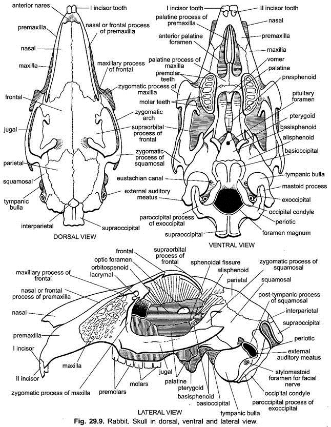
Parts of Skull of the Rabbit:
The skull of the rabbit can be divided into:
1. Cranium which encloses the brain,
2. Sense capsules (olfactory, auditory and orbital) closely attached with the cranium, and
3. Visceral skeleton includes the upper and lower jaws and hyoid apparatus.
1. Cranium:
The various bones constituting the cranium can be grouped into three segments, a posterior occipital segment, middle parietal segment and anterior frontal segment.
(i) Occipital Segment:
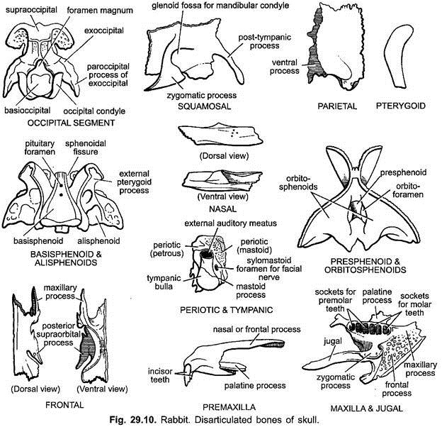
It is the hindermost part of the cranium. It consists of four cartilaginous bones completely fused together and encircling a large passage, the foramen magnum, through which spinal cord comes out. The bone making the dorsal boundary of the foramen magnum is flat shield-like square-shaped supraoccipital, the ventral boundary is marked by a flat bone, the basic occipital, and the lateral boundaries are marked by a pair of exoccipitals.
Each ex-occipital bears a prominent oval or a somewhat elongated occipital condyle and also gives off a downwardly directed paroccipital process which lies in close contact with postero-ventral surface of tympanic bulla.
Supraoccipital articulates in front with the parietals, and on lateral sides with the squamosals and periotics.
Basioccipital anteriorly articulates with basisphenoid and laterally with penotics.
The occipital condyles articulate with the facets of the first vertebra, the atlas.
(ii) Parietal Segment:
It is present in front of occipital segment. On lateral sides, both are separated from each other by auditory capsules and squamosals. It consists of five bones, a triangular basisphenoid on the mid-ventral side, alisphenoids on lateral sides and two parietals and interparietal on the dorsal side.
Basisphenoid and Alisphenoid:
Basisphenoid is a flat, median triangular cartilage bone. Its apex joins anteriorly with the presphenoid and its broad posterior end is connected with the basioccipital by a thin plate of cartilage.
Dorsal surface of basisphenoid has a depression, the sella turcica, in which pituitary gland is placed. Basisphenoid also bears a pituitary foramen at about its middle. Basisphenoid laterally articulates with the alisphenoids.The alisphenoids are broad wing- like and firmly united with the basisphenoid.
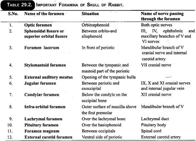
Each alisphenoid is produced below into a bilaminate process, the pterygoid process, connected with the palatine and bounded in front by a slit-like aperture, the sphenoidal fissure (foramen lacerum anterius) opening into the cranial cavity.
Interparietal:
It is a small, median wedge-like, triangular bone on the dorsal side in between parietals and supraoccipital.
Parietal:
The parietal bones are a pair of thin, slightly arched bones protecting the brain dorsolaterally. Both the parietals are firmly united with each other by a suture along the mid-dorsa line. They remain separated from alisphenoids by the squamosals. Each parietal gives off a ventral process from its posterior outer border which extends beneath the squamosal.
Squamosal:
It is roughly a rectangular bone situated ventral to parietal on either side. Outer surface of squamosal is produced into a strong zygomatic process which forms the posterior part of zygomatic arch. It unites anteriorly with jugal in the formation of a complete zygomatic arch. Below the root of the process is a hollow glenoid fossa for articulation with the condyle of mandible. Squamosal from its posterior side gives off a slender process, the post-tympanic process, which becomes applied to the outer surface of the periotic.
(iii) Frontal Segment:
This segment also consists of five bones, a presphenoid ventrally, two orbitosphenoids one on either side and two frontals dorsally.
Presphenoid and Orbitosphenoids:
Presphenoid forms the floor of the frontal region of cranium. It is a median, thin, small bone situated in front of the basisphenoid and connected with it by a cartilage. The presphenoid bone forms the lower and anterior boundary of the optic foramen. Orbitosphenoids are connected with the lateral sides of the presphenoids. These form the sides of the frontal region.
In rabbit orbitosphenoids are partially fused and form a thin, vertical median interorbital septum, which surrounds the optic foramen. These bones form the lateral wall of cranium and orbits. Orbitosphenoids articulate with palatine in front, with frontals above and behind with the squamosals and alisphenoids.
Frontals:
The frontals are large, and strong bones protecting the brain from the dorsal and lateral sides. Both the frontals are united with each other by a suture along the mid-dorsal line. Outer middle part of each frontal forms a prominent ridge over the orbit, known as the supra-orbital process. Frontals unite posteriorly with the parietals and anteriorly with the nasals.
A slender maxillary process arises from the anterior side of each frontal running in between the premaxilla and maxilla. On the ventral side each frontal articulates with the orbitosphenoid.
(iv) Ethmoidal Region:
It lies anterior to the cranial cavity and not well separated from olfactory capsules. It includes a single mesethmoid.
Mesethmoid:
It is a median vertical bone which extends forwards and downwards in front of presphenoid. It is called nasal septum which separates the two olfactory chambers from each other. Posterior part of mesethmoid forms a narrow plate of bone, the cribriform plate, which forms the anterior boundary of the brain case. It is perforated by small foramina through which the fibres of the olfactory nerves enter the cranial cavity.
2. Sensory Capsules:
Sensory capsules are olfactory lodging the organs of smell, auditory containing organs of hearing and orbits for eyes. These are closely fused with the cranium.
(i) Olfactory Capsules:
Nasal:
Roof of olfactory chambers is formed by two flat bones, the nasals which unite with each other in the mid-dorsal line. Each nasal has on its inner surface a very thin, hollow process the nasoturbinal. Anterior end of each nasal forms the upper boundary of external nostril. Each nasal articulates laterally with the premaxilla.
Vomer:
Two vomers are long slender fused bones forming the floor of olfactory capsules. These dorsally articulate with ventral (inferior) edge of the mesethmoid (nasal septum). Vomers possess delicate lateral wings.
Turbinals:
Each nasal or olfactory chamber or cavity separated from other cavity by a median septum, partly cartilaginous, partly bony by the mesethmoid, contains the turbinals or turbinate bones of its side. These are ethmoturbinals, maxilloturbinal and nasoturbinal.
(ii) Auditory Capsules:
The auditory capsules are found closely fused with the postero-lateral region of the cranial region of the skull. Each capsule is formed by the fusion of two bones, the periotic and tympanic, and encloses the internal ear.
Periotic:
Each periotic is an irregular cartilage bone formed by the fusion of prootic, epiotic and opisthotic bones. It is found in between squamosal and occipital segment and externally visible forming a bulge and perforated in the adults.
The periotic consists of two parts:
(i) An internal, hard, bony petrous part and
(ii) An outer posterior porous mastoid part which is produced downwards into a mastoid process which is closely applied to the posterior side of tympanic.
Petrous part bears two apertures-an anterior fenestra ovalis and another posterior fenestra rotunda. It also bears a ventral rounded swelling called the promontory for lodging the cochlea.
Tympanic:
It is a flask-shaped membrane bone attached to the outer surface of the periotic. Its lower, flask-shaped swollen part is known as tympanic bulla. It encloses the tympanic cavity of middle ear containing three ear ossicles arranged linearly-the malleus, incus and stapes. The neck of the flask constitutes the external auditory meatus closed at the base by the tympanic membrane in life. Tympanic on its posterior side bears a stylomastoid foramen for the facial nerve.
(iii) Orbits:
The orbits are situated on the sides of the frontal segment of the cranial region. A median bony inter-orbital septum separates the two orbits. Each orbit has a median wall formed of dorsal frontal which project above the orbit as supra-orbital ridge, median ventral orbitosphenoid, alisphenoid and ethmoid bones. The outer side of the orbit is protected by the zygomatic arch and the lower side of the orbit is unossified.
3. Visceral Skeleton:
It includes the upper and lower jaws and hyoid apparatus.
(i) Upper Jaw:
Bones of the upper jaw are premaxillae, maxillae, jugals, pterygoids and palatines. These are closely associated with the cranium and olfactory capsules.
Premaxillae:
These are large bones and form the anterior most part of the upper jaw. Both the premaxillae unite in front in the median line. Each premaxilla ventrally bears two sockets for incisor teeth. Each presmaxilla also bears two processes-a nasal process extending backward up to frontal in between nasal and maxilla; a palatine process (ventral) which meets with its fellow of other side in the mid-ventral line and both extend up to palatine processes of maxillae.
Maxilla:
Maxilla is a large, irregular-shaped bone forming the greater part of the upper jaw and side of the face. The ventral surface of maxillae bears 6 sockets for the cheek teeth. Each maxilla gives off a number of processes. A median flat palatine process is given out from the ventral surface of each maxilla. Both the palatine processes are united by a suture in the middle line forming the anterior part of hard palate.
A flat stout process from the outer surface of each maxilla, called the zygomatic process, passes backwards and joins with a similar forwardly directed process of the squamosal, thus, forming the zygomatic arch. Each maxilla anteriorly gives off a maxillary process which joins with the premaxilla.
Palatine:
The palatines are thin, more or less vertically situated bones in the mid-ventral side. Both palatines form an inner process in front, which meet together to form the posterior part of hard palate. Each palatine posteriorly unites with the alisphenoid and pterygoid and encloses a pterygoid fossa between it and basisphenoid.
Pterygoid:
The pterygoids are small, irregular, bony plates situated behind the palatines and joined with the pterygoid process of alisphenoids behind.
Jugal:
Molar or jugal is a laterally compressed bone which extends in between zygomatic processes of maxilla and squanosal. It forms the greater part of the zygomatic arch.
(ii) Lower Jaw:
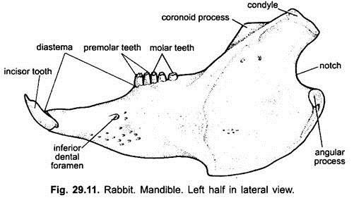
The lower jaw consists of two halves or rami which are united in front by the mandibular symphysis. Each ramus is formed of a single bone, called dentary or mandible. Each dentary has a horizontal part which bears sockets for the attachment of teeth like incisors, premolars and molars, and a gap without teeth, called the diastema.
The posterior vertical or ascending part of dentary carries the articular surface or condyle for articulation with the glenoid cavity of squamosal. In front of the condyle is the compressed coronoid process. On the postero-lateral surface of the mandible is present a slight depression, called messenteric fossa, in which are attached large masseter muscle.
Temporal muscle is attached with the medial side of coronoid process. The posterior, lower border of the dentary gives off a rounded and inwardly directed angular process. On the outer anterior surface of dentary has a inferior dental or mental foramen and on the inner side is present the mandibular foramen.
(iii) Hyoid:
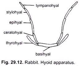
The hyoid is bony in rabbit and consists of a small stout thick body, called basihyal, placed transversely between the two halves of mandible and bears two pairs of cornua or horns. The small anterior cornua or ceratohyals are situated below the tongue and consists of a series of small bones such as ceratohyal, epihyal, stylohyal and tympanohyal.
The last one is fused with the tympanic and periotic bones. The long and posteriorly directed posterior cornua consist of a single thyrohyals.
Important Foramina in the Skull of Rabbit:
The important foramina in the skull of rabbit can be easily explained with the help of the following table:
Vertebral Column:
The vertebral column of rabbit, like that of birds and lizards, is differentiated into the following five regions. The total number of vertebrae in rabbit is about 45-47.
1. Cervical
2. Thoracic
3. Lumbar
4. Sacral
5. Coccygeal or caudal
Vertebral formula of rabbit: C7, T12-13, L6-7, S4, Cd15 where C, T, L, S, Cd stand for cervical, thoracic, lumbar, sacral and caudal respectively.
Vertebral column of mammals is distinguished from other vertebrates in the following respects.
Important Characteristics of Mammalian Vertebrae:
1. The centra are more a less flattened on both the surfaces, i.e., amphiplatyan type. The centra on either side, is provided with small bony plate, epiphysis.
2 A thin epiphysial cartilage separates the centrum and epiphysis in the embryonic condition, which later become fused with the centrum. Thus, in adults no epiphysial cartilage is found.
3. In between the adjacent centra are present intervertebral discs of central portion of the intervertebral disc is called nucleus pulpous, which represents the remnant notochord. The discs are shock-absorbing cushions and probably represent the hypocentrum.
A. Cervical Vertebrae:
Out of the seven cervical vertebrae, first and the second are highly modified, known as atlas and axis respectively. Remaining 3rd to 7th are more or less alike and can be called typical cervicals.
1. Atlas Vertebra:
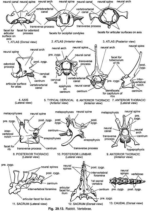
It is ring-like without any solid centrum and zygapophyses. It consists of a large neural arch but a reduced neural spine (The centrum is, however, present in the embryonic condition which later fuses with the axis vertebra and known as odontoid process). The neural canal is large and divided into two parts in living condition.
The upper part is the neural canal for the passage of spinal cord and lower part is occupied by the odontoid process of the axis. Two large concave occipital facets are present in the anterior face of the altas for articulation with the occipital condyles of the skull.
This atlanto-occipital joint allows movements of the head in sagittal plain, as in nodding the head. The transverse processes arising from the sides are broad, long and wing like for the attachment of muscles that hold and rotate the head and neck.
These are not transverse processes but are enlarged flattened cervical ribs, perforated basally by the vertebrarterial canals. A pair of lateral and a mid-ventral articular facet is present on the posterior face of the atlas for the odontoid process of axis.
2. Axis Vertebra:
It is the second cervical vertebra. Its neural spine is high, ridge-like, laterally compressed and elongated antero-posteriorly. Its centrum bears a peg-like odontoid process in the anterior face which is developmentally the centrum of atlas. This process forms the atlanto-axial joint, which allows the rotation of the skull and atlas on the axis.
This movement is facilitated by a pair of smooth articular surfaces on the anterior face of the axis one on either side of the odontoid process. The transverse processes are short and perforated basally by vertebrarterial canals for vertebral artery. A pair of post-zygapophyses are found but pre-zygapophyses are absent.
3. Typical Cervical Vertebra:
A typical cervical vertebra is broad and has a small flattened centrum, a large neural arch and a small neural spine. Pre-and post-zygapophyses are well developed. Transverse processes are bifurcated into dorsal and ventral lamellae perforated basally by a foramen transversaria or vertebrarterial canal. The cervical ribs are much reduced and more or less incorporated in the vertebra. The transverse processes and reduced ribs provide surface for the attachment of neck muscles.
The cervical vertebrae from 3rd to 6th are similar in structure. But the 7th cervical differs from others in having a more elongated neural spine, in having its transverse processes simple and imperforate and in the presence of a small concave semilunar facet at the posterior edge of the centrum for the articulation of thoracic ribs.
B. Thoracic Vertebrae:
The thoracic vertebrae are 12-13 and each vertebra is provided with a well-developed centrum, a neural arch, long neural spine and pre- and post-zygapophyses. The transverse processes are short and stout. Each bears near its extremity a small smooth articular surface or tubercular facet for the tubercle of a rib.
On the anterior and posterior borders of each vertebra is a little semilunar facet, the capitular facet, situated at the junction of the centrum and neural arch. The neural spines of the anterior thoracic vertebrae are more or less straight and directed backwards. Posterior 4 or 5 thoracic vertebrae are little different from the anterior thoracic vertebrae.
They possess longer and stout centra, short neural spines, reduced transverse processes with no tubercular facets, and zygapophyses are distinct. Capitular facets are present near the anterior of centrum. Neural spine is directed upward. Metapophyses and anapophyses are present on the anterior end of neural arch and the posterior end of neural arch below post-zygapophyses respectively.
Ribs:
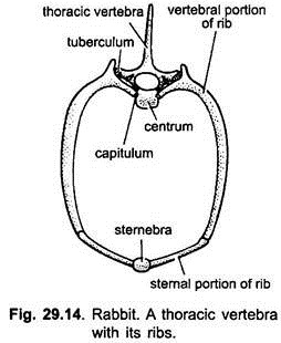
Thoracic ribs are 12 or 13 pairs and found articulating with the thoracic vertebrae. Each rib is a bony curved rod divided into a dorsal longer bony vertebral rib and a ventral smaller cartilaginous sternal rib. Vertebral part is biramus (double headed), the heads are known as tuberculum and capitulum which articulate with the transverse process and with the demi-facets on the centra of two adjacent vertebra respectively.
The sternal part of the rib articulates with the sternum. The sternal parts of all the thoracic ribs, except the last five, meet the sternum below and are called true ribs. The last three or four ribs have no sternal parts and they along with one anterior to them are not connected with the sternum and, hence, known as floating ribs.
C. Lumbar Vertebrae:
They are 7 and out of which the first two are called anterior lumbars. Each anterior lumbar has a large, stout, strongly built centrum, a neural arch enclosing a broad neural canal and well developed forwardly directed neural spine. The transverse processes are large, distally expanded beside pre- and post-zygapophyses and directed downwards and forwards.
There are two pairs of bony processes, called mammillary processes, i.e., metapophysis is present at the anterior end of neural arch and it is sloping forward. Beneath it lies the median pre-zygapophysis; anapophysis is present at the posterior end of neural arch beneath the post-zygapophysis and it is a small backwardly directed process. Each anterior lumbar also has a median ventral process beneath the centrum, called hypapophysis.
Posterior Lumbar:
From third to seventh lumbars are called posterior lumbars. These resemble the anterior lumbars in all details but the hypapophysis is absent, only a small ridge is present in its place.
D. Sacral Vertebrae (Sacrum):
There are four vertebrae in the sacral region of rabbit which fuse together to form a compound bone, the sacrum but only the first articulates with ilium of the pelvic girdle. It is believed that out of these four vertebrae constituting sacrum, only first vertebra is the sacral vertebra and the remaining three vertebrae are the anterior caudals.
Each vertebra of the sacrum is provided with a long neural spine, tubercle-like projections on the upper side representing zygapophyses, and intervertebral foramina. The first vertebra is the largest and has large, flattened, stout transverse processes, probably fused sacral ribs.
These articulate with the ilia of pelvic girdle. This joint is called sacro-iliac joint. Hypapophyses and anapophyses are absent and the metapophyses are relatively small. The sacro-iliac joint provides strength to the pelvic girdle and the vertebral column at the time of throwing the body forward when the hindlimbs are straightened.
E. Caudal Vertebrae:
The caudal vertebrae are sixteen in rabbit. Only the anterior caudal vertebrae are provided with well-developed neural spines, neural arches and zygapophyses. These processes being gradually reduced until the terminal vertebrae near the end of the tail is only left in the form of cylindrical centrum. The transverse processes are absent in caudal vertebrae. The muscles attached to the anterior caudal vertebrae provide movements of the tail in many planes.
Sternum:
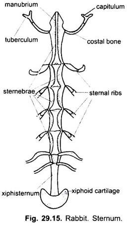
The thorax of rabbit is bounded mid-ventrally by the sternum which consists of five elongated bony pieces, known as sternebrae. Thus, the sternebrae together constitute the main body of the sternum, called mesosternum. The first anterior most sternebra is the longest and called manubrium or presternum.
It is ventrally produced into a keel. The first pair of sternal ribs articulate with it in the middle. Sixth sternebra is the smallest of all and the last one is long and slender. Except first rib, all the sternal ribs called xiphisternum terminating into an expanded xiphoid cartilage are attached at the intersternebral junctions.
Appendicular Skeleton:
1. Pectoral Girdle:
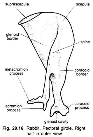
It consists of two separate halves placed dorsal to the anterior thoracic ribs in between the forelimbs, it supports forelimbs and protects the soft parts of the body from the ventral side. Each half of the pectoral girdle is known as innominate. Thus one half of the pectoral girdle is formed of a broad, more or less triangular bony plate, called scapulo-coracoid and a small clavicle bone.
Scapulo-Coraciod:
It is mainly formed of scapula which is a thin, flat, and more or less triangular bony structure. Its outer surface bears a prominent ridge, called the spine which divides its surface into antero-dorsal and postero-dorsal portions to which are attached muscles. The spine terminates ventrally into an expanded knob-like structure, the acromion process which posteriorly bears backwardly directed metacromion process.
The apex of scapulo-coracoid is directed downwards and forwards terminating below into a concave glenoid cavity for the head of humerus. Above this cavity is present a small hook-like coracoid process, the rudimentary coracoid. The suprascapula is in the form of a thin strip of cartilage situated along the dorsal or vertebral border of the scapula.
Clavicle is a thin, slightly curved bone extending between the acromion processes and manubrium of the sternum.
2. Pelvic Girdle:
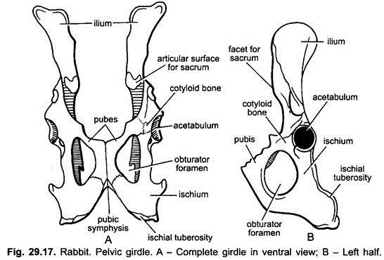
The pelvic girdle is also formed of two equal halves or os-innominate. Both the halves are joined together mid-ventrally by pubic symphysis to form a stout and strong girdle situated in the pelvic region between the two hindlimbs. Each half or os-innominatum is formed of three bones, the ilium, ischium, and pubis. All these bones are fused together to form a single hip bone.
The ilium is the antero-dorsal longest bone, which bears a rough flat articular surface roughly at about the middle of its length for sacrum. The anterior and dorsal edge of the ilium is raised into iliac-crest. The ilium extends posteriorly up to the acetabulum. The postero-dorsal part of the os-innominate is formed by the ischium.
The posteriormost part of ischium is broad and projects outwards into a prominent ischial tuberosity. The pubis is a narrow bone and forms the ventro- median portion of innominate. Both the pubes unite with each other on the mid-ventral line to form a pubic-symphysis.
The pubis does not take part directly in the formation of acetabulum, because a cotyloid bone is present in between the acetabulum and pubis. A big obturator foramen is present between the ischium and pubis which is always covered by the obturator membrane and muscles in the lifetime. Acetabulum is only formed by the ilium and ischiam on both sides of the girdle and into it articulates the head of humerus.
3. Forelimbs Bones:
Humerus:
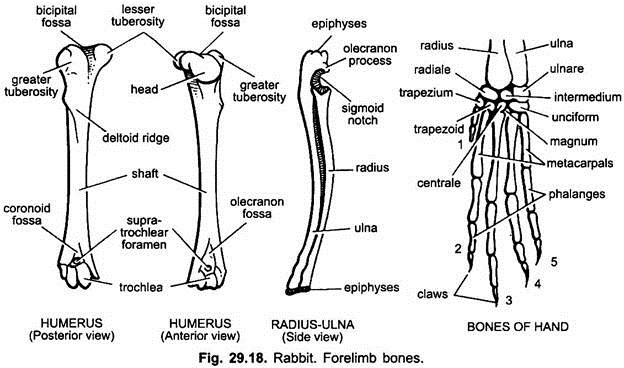
The bone of the upper-arm is the humerus, which is a long bone having a proximal large rounded head for the articulation in the glenoid cavity of scapula. The proximal end of the humerus near the head is provided with a bicipital groove in between the two tuberosities (greater and lesser) for the attachment of muscles.
The anterior surface of the humerus below the head has a projection called deltoid ridge for the attachment of muscles. The distal end of the humerus is provided with a pulley-like trochlea for articulation with ulna. Just above trochlea are present two fossae (depressions)-anterior smaller is coracoid fossa and posterior larger is olecranon fossa. Both the fossae communicate with each other through a supra-trochlear foramen.
Radius and Ulna:
The bones of the forearm are radius and ulna which are closely held together at the two ends, so that they cannot move over each other. The ulna is the long bone and proximally bears a prominent projection called the olecranon process. It articulates with the olecranon fossa of humerus.
Olecranon process is basally notched called the sigmoid notch into which fits the trochlea of humerus.The radius is the smaller bone than ulna and situated towards the inner side. The radius and ulna are distally provided with epiphysis and articulate with the wrist bones.
Bones of Hand:
The wrist bones or carpals are 9 small bones arranged in two rows. Proximal row has three carpals, called radiale, ulnare and intermedium. The radiale is situated below the radius, ulnare below the ulna, while the intermedium is situated between them.
The distal row consists of trapezium and trapezoid situated below the radiale, centrale and magnum situated below the intermedium and unciform situated below the ulnare. The unciform is actually fused two carpals. Besides these, a sesamoid pisciform is present on the ventral side of carpus.
The bones of the palm or manus are five long metacarpals, which support the five digits having 2, 3, 3, 3, 3 phalanges respectively. The terminal phalanx of each digit bears a horny claw.
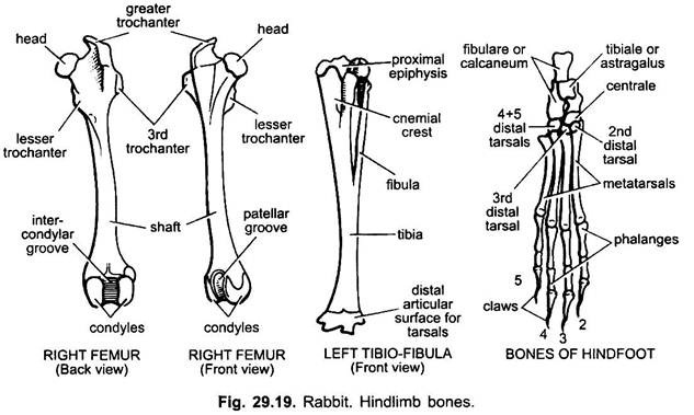
4. Hindlimb Bones:
Femur:
The bone of the thigh is the femur which is a long bone with a flattened proximal end. Its flattened proximal end bears a rounded smooth head towards the inner side for the articulation with the acetabulum. The proximal end of the femur also bears three trochanters for the attachment of muscles.
The first or greater trochanter is situated above the head, the second or lesser trochanter is situated below the head, while the third trochanter is situated below the greater trochanter. The deep groove below the head and greater trochanter is the digital fossa.
The main body of the femur or shaft terminates distally into a pair of expanded condyles enclosing the intercondylar groove for the articulation with tibio-fibula. On the anterior side of this groove is called the patellar groove into which moves the patella bone. These condyles are provided with articular facets for articulating with the tibia.
Tibio-Fibula:
The shank of the hindlimb is provided with long stout and straight tibia and a small slender fibula bones. The fibula is a reduced slender bone which is fused with the tibia distally, while proximally it is free. The proximal end of these bones is provided with proximal epiphysis which articulates with the condyles of the femur. Tibio-fibula articulate distally with the bones of the ankle or tarsus. A cnemial crest is present on the proximal dorsal end of tibia whose two depressions articulate with two condyles of femur.
Bones of Foot:
The ankle is formed of six tarsus arranged in three rows. The first proximal row is formed of two tarsals, a tibiale and intermedium, both fused to form the astragalus located on the preaxial side.
The other largest is the calcaneum which is produced into a process behind its articulation with the tibio-fibula. The astragalus bears a pulley-like surface for articulation with tibia. The middle row has a single bone, the centrale, just in front of astragalus. The distal row has three tarsals which are mesocuneiform, ectocuneiform and cuboid.
The bones of sole or foot are 4 long metatarsals (the first is absent). Four digits or toes are only present, each formed of three phalanges. The last phalanx of each digit is provided with a claw. Hallux (first toe) is absent.
