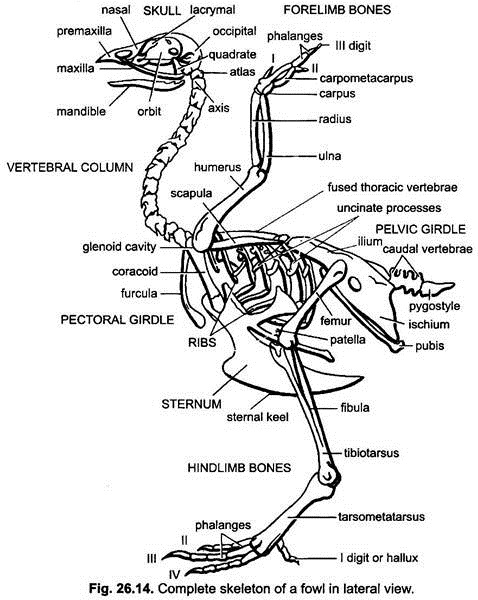
The endoskeleton of birds (fowl or Gallus) is quite light and completely ossified and contains no marrow. They are filled with air and are pneumatic. The endoskeleton comprises the usual two divisions, viz., the axial skeleton and the appendicular skeleton.
The axial skeleton forms the thin axis of the body and includes the main skull, vertebral column, sternum and ribs. The appendicular skeleton includes the bones of the limbs and girdles by means of which they are connected to the axial skeleton.
In pigeon, the bones of the forearm and hand, and of the leg, are non-pneumatic. In very good fliers (such as some birds of prey and albatross), wing and leg bones are pneumatic. The long bones have no epiphysis at the ends.
The skeleton of backbone and limb girdles allows to carry the weight of the body on the wings or on the legs. For this purpose there are two plate-like girders, the sternum and synsacrum, curved in opposite directions.
Axial Skeleton:
ADVERTISEMENTS:
Characteristics of Skull:
1. The skull is large but very light due to spongy bones.
2. There has been a complete fusion of bones so that it has no sutures, but it is not deviated from the reptilian type of skull.
3. Number of bones is reduced.
ADVERTISEMENTS:
4. Jaw bones form a toothless beak.
5. Monocondylic, i.e., having a single occipital condyle.
6. Cranium is rounded and large for accommodating the well-developed brain.
7. Cranium does not extend forward into orbital region (tropibasic).
ADVERTISEMENTS:
8. Large orbits separated by a thin membranous interorbital septum.
9. Autostylic jaw suspensorium.
10. In fowl, palate is schizognathous (short vomers and palatines meet together).
Regions of Skull:
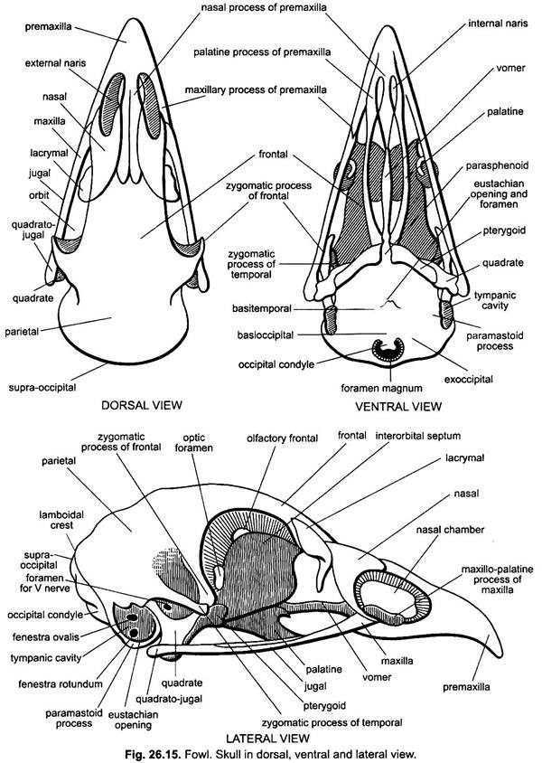
The skull of fowl consists of cranium, sense capsules and visceral skeleton including the upper and lower jaws and hyoid apparatus.
1. Cranium (Brain Case):
It encloses the brain. For the study of the cranium it can be further divided into the following regions:
(i) Occipital Region:
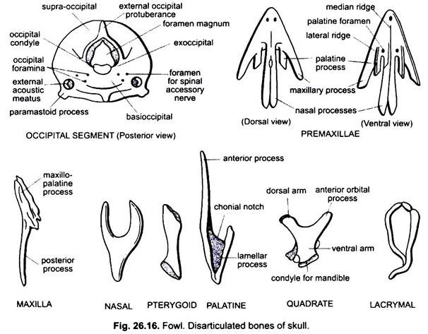
It is the back part of the skull. It is formed by the usual bones, the basioccipital (basal), the two occipitals (lateral sides) and the supraoccipital (above), all of which enclose a large rounded opening, the foramen magnum. Immediately below the foramen is a single rounded occipital condyle, mostly formed by the basioccipital. The supraoccipital anteriorly joins the parietals.
(ii) Parietal Region:
It lies in front of the occipital region. Its roof is formed by two fused square-shaped parietals. The lateral sides of parietal region are formed by the squamosals and alisphenoids and the floor by a large basisphenoid.
Ventrally the basisphenoid is covered over by a paired broad membrane bone, the basitemporal or parasphenoid. The basisphenoid continues forwards as in lizards, by a slender parasphenoid rostrum which represents the anterior portion of parasphenoid. Each squamosal is produced outward and forwards into a zygomatic process. The squamosal bounds the tympanic cavity and firmly united to other cranial bones.
(iii) Frontal Region:
It is anterior to parietal region. The roof of the frontal region consists of a pair of large frontal bones. The lateral posterior end of each frontal is drawn out into a strong zygomatic or post-orbital process which joins the zygomatic process of the squamosal. The alisphenoids are continued forward into the orbitosphenoids forming the sides of the frontal region, while its base is formed by a poorly developed presphenoid, lying above the parasphenoid rostrum.
2. Sense Capsules:
These are closely united with the cranium and lodge the various sense organs.
The sense capsules include auditory, optic and olfactory capsules:
(i) Auditory Capsules:
A pair of auditory capsules, enclosing the organs of hearing, is attached behind one on either lateral side of the occipital region of the skull. Each capsule is mainly formed by the prootic bone. (The small opisthotic of early embryo unites with the exoccipital and the epiotic with the supraoccipital.) A large cup-like tympanic cavity of the middle ear is present on each posterior lateral side of the skull bounded by the squamosal above and the basitemporal below.
The tympanic cavity contains an tympanic membrane attached just within its prominent outer edge, and a slender rod-like columella made of bone and cartilage. Its inner portion is like a slender rod-like bone, called columella auris, and its outer part is called extra-columella which is a triradiate cartilage. Three rays of extra columella are the supra- stapedial, extra-stapedial and infra-stapedial. Within the tympanic cavity are several openings; two of these lie about the middle of the cavity. The upper one is fenestra ovalis and the lower one is fenestra rotunda.
The inner end of columella auris fits into the fenestra ovalis, while its outer end touches the tympanic membrane. The tympanic cavity communicates with the mouth through an eustachian canal, the posterior funnel-shaped opening of which lies in the anterior ventral part of the tympanic cavity. The eustachian canal runs forwards and inwards between the basitemporal and the basisphenoid and joins with its fellow of the opposite side to open by a common median aperture in the roof of the posterior part of the mouth cavity.
(ii) Optic Capsules:
The two orbits are very large to accommodate the relatively massive eyes. They are separated from one another by a narrow vertical partition, the inter-orbital septum, which is formed by a mesoethmoid together with presphenoid, parasphenoid rostrum and orbitosphenoids. Each orbit is bounded anteriorly by the frontal and large lacrymal, dorsally by the frontal and posteriorly by the post-orbital process of frontal and alisphenoid.
The lacrymal is produced below in a curved pointed process hanging freely. Ventrally the orbit is incomplete. The interorbital septum is perforated by many openings. A large median optic foramen lies in the posterior part of the orbit. It actually perforates the orbito-sphenoid and communicates the two orbits with one another.
(iii) Olfactory Capsules:
The olfactory capsules are cartilaginous and greatly reduced in size in accordance with the poor sense of smell. They are present in front of cranium. The ecto- ethmoid or turbinals are comparatively poorly developed. The nasals are a pair of thin, triangular plate-like bones forming the roof and sides of the nasal chambers.
Each nasal sends out three processes, a posterior process uniting with the frontal and also with lacrymal. Other two anterior processes furnish the posterior boundary of the external nare and uniting with the process of premaxilla. The two vomers fuse into a small, median, slender bone, lying in continuation with the parasphenoid rostrum at the base of nasal chambers, and separating the two nasal chambers at the base. In pigeon vomers are absent.
Visceral Skeleton:
The visceral skeleton includes the upper jaw, lower jaw and hyoid apparatus. Jaws are not toothed and form boundaries of the mouth gape. Hyoid apparatus supports the tongue.
(i) Upper Jaw:
The upper jaw on each side forms two bony arcades- the outer arcade or infra-orbital arcade or sub-orbital bar consisting of premaxilla, maxilla, jugal and quadrato- jugal and the inner arcade or palato-pterygo-quadrate bar is formed by pterygoid, palatine and quadrate.
Premaxillae:
Premaxillae are united in front into a large triradiate bone which forms practically the whole of the upper beak.
Posteriorly it is produced into three paired processes:
(i) The nasal processes are the longest, run backwards closely along the inner side of nasals to unite the mesethmoid and frontals forming the upper margin of the external nares.
(ii) The maxillary processes run backwards and slightly outwards forming anterior part of the upper jaw.
(iii) The inner palatine processes are the smallest, run horizontally backwards below maxillae and join ultimately with palatines. They form the anterior part of roof of buccal cavity (palate).
Maxilla:
Maxilla is a slender rod-shaped bone. Its anterior end articulates with maxillary process of maxilla and produced inwards into spongy maxillo-palatine process. The slender posterior end of maxilla is continued backwards by a slender jugal and quadrato-jugal to the quadrate. The posterior zygomatic process of the maxilla forms the anterior part of the outer arcade or infraorbital arcade or suborbital bar or zygomatic arch.
Jugal:
The jugal is a very slender rod which forms the middle part of the sub-orbital bar lying dorsally upon the other two components of the bar, i.e., maxilla and quadrato-jugal.
Quadrato-Jugal:
The quadrato-jugal is also a rod-like slender bone and forms the posterior part of the sub-orbital bar. Its hind end is thickened and articulates with quadrate.
Quadrate:
The quadrate is a stout, triradiate bone articulating by two facets on its otic process with the roof of the tympanic cavity. Its dorsal arm articulates with the squamosal slightly above the anterior margin of the tympanic cavity. Its ventral arm forms a condyle for articulation with the mandible. Condyle also articulates on the outer side with the quadrato-jugal and on the inner side with the pterygoid. Its anterior arm (orbital process) runs anteriorly parallel to the pterygoid and ends blindly.
Pterygoid:
It is a short and stout, rod-shaped bone. It lies obliquely behind the palatine between the presphenoid rostrum and quadrate of its own side. It articulates behind with the quadrate and in front with the parasphenoid rostrum and palatines.
Palatine:
It is slender bony bar lying in front of parasphenoid rostrum. Anteriorly it is attached with the processes of premaxilla and maxilla. Its posterior end articulates with the pterygoid. Posteriorly it is also expanded inwards into a broad lamella, the sides of which articulate with the rostrum.
Lower Jaw:
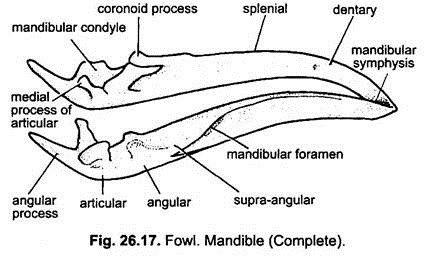
The V-shaped lower jaw consists of two long laterally compressed rami which are firmly united in front. Each ramus is composed of five bones, i.e., one replacing bone, the articular, and four investing bones, the angular, supra-angular, dentary and splenial, developing around a cartilaginous bar, Meckel’s cartilage. All these bones are fused together.
Articular forms the posterior expanded part of each ramus. On the dorsal surface it bears an elongated mandibular condyle for the articulation with the quadrate. Posteriorly it is produced into a medial and angular process. Angular is a slender bone and lies beneath the articular along the lower inner posterior third border of each ramus.
Supra-angular forms the dorsal part of about one-third of the posterior upper part of ramus. It bears a coronoid process. Dentary is the largest bone and forms about the complete distal half of the ramus. Anteriorly it is united with its counterpart of the other side forming the mandibular symphysis. It is devoid of teeth. Splenial is a thin splint-like bone lying on the inner surface of the middle part of ramus.
Hyoid Apparatus:
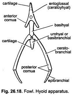
Hyoid apparatus lies in the floor of the mouth and supports the tongue. It is composed of three bones- an anterior entoglossal or glossohyal or ceratohyal, a median bony basihyal and a posterior urohyal. Entoglossal bears a cartilaginous process in front. Entoglossal gives off a pair of backwardly directed short anterior cornua which represents the free ends of ceratohyals.
From the junction of basihyal and urohyal arise a pair of long backwardly directed posterior cornua or thyrohyals. Each thyrohyal consists of two rod-like pieces, the proximal ceratobranchial and distal epibranchial.
Vertebral Column:
The vertebral column of birds is peculiar in having a large number of cervical vertebrae due to long mobile neck; thoracic, lumbar and sacral vertebrae are fused giving rigidity which is advantageous in flight. Caudal vertebrae are fused forming the pygostyle which supports the tail feathers. Epiphysis is lacking in long bones. Vertebral column of Gallus is divided into the following regions, i.e., cervical, thoracic, sacral and caudal.
1. Cervical Vertebrae:
These are 14 to 16 in number. Their articulation with each other allows greater movement of the long neck and head.
(а) Atlas Vertebra:
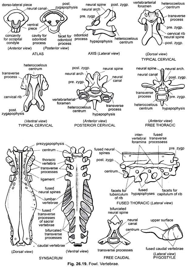
Atlas is the first vertebra of the cervical region. It is small ring-like in appearance. Centrum, neural spine, transverse processes, ribs and prezygapophyses are absent. It is formed of three pieces, a ventral and two dorso-lateral uniting mid-dorsally to form the neural arch. Its ventral piece is thick like the centrum and notched above to receive the odontoid process of the axis. Its anterior side bears a cup-like cavity to provide articulation to the single occipital condyle of the skull.
In life, neural canal is divided by a transverse ligament into an upper spinal canal and a lower odontoid canal for odontoid process of axis. The posterior border of the neural arch is produced into a pair of postzygapophyses for the articulation with the prezygapophyses of axis vertebra.
(b) Axis Vertebra:
Axis is the second vertebra of the cervical region. Transverse processes, ribs and vertebrarterial canal are absent. Centrum is produced in front into a peg-like odontoid process by which it articulates within notch present on the dorsal side of the ventral piece of the atlas vertebra. Neural spine is blunt. Pre and post zygapophyses are present. A small hypapophysis is present below the posterior face of centrum. Head and atlas get mobility over the axis.
(c) Typical Cervical Vertebra:
The typical cervical vertebra (3rd to 10th) has a long body or centrum. The centrum is heterocoelous or saddle-shaped. Anterior surface is concave from side to side and convex from above downwards, while the posterior surface is convex from side to side and concave from above downwards. The neural arch is short and notched both anteriorly and posteriorly.
The neural spine is rudimentary. Transverse processes are short and irregular, and arise laterally from the anterior end. These are fused with the rudimentary posteriorly directed spine-like cervical ribs. The bases of cervical ribs have vertebrarterial canal for vertebral artery. Prezygapophyses are flat articular facets on the anterior side of transverse processes facing upwards and inwards and postzygaphyses are projecting backwards from the posterior side of the neural arch and facing downwards. Posterior carvical vertebrae (i.e., behind Xth) are shorter.
Their centrum bears a ventral spine-like process, the hypapophysis. Neural spine is also distinct. Cervical ribs are no longer visible and vertebrarterial canal is also open groove-like, not closed.
2. Thoracic Vertebrae:
These are seven in number. First and sixth are free, while 2nd to 5th are fused and seventh is fused with the synsacrum.
(a) Fused Thoracic Vertebrae:
Fused thoracic vertebrae are formed by the fusion of 2nd to 5th thoracic vertebrae. The centrum is heteroceleous. Their neural spines, transverse processes and hypapophyses are fused with each other forming a continuous plate-like ridges perforated by intervertebral gaps. Fused hypapophyses form a ventral bridge. Fused transverse processes and centra of vertebrae possess on either side the tubercular and capitular facets for the articulation of thoracic ribs. Pre- and post-zygapophyses are present on the anterior and posterior side of the fused thoracic mass respectively.
(b) Free Thoracic Vertebra:
First and sixth free thoracic vertebra are slightly smaller than the typical cervical. The centrum is heterocoelous and neural spine is elongated, pointed and well-developed. Transverse processes are also well-developed and outwardly directed. Pre- and post-zygapophyses are present on the anterior and posterior surfaces respectively. Hypapophysis is prominent beneath the centrum. It also carries capitular and tubercular facets for the articulation of the thoracic ribs.
3. Synsacrum:
Synsacrum is a long compound bone formed by the fusion of 14 to 16 vertebrae, supporting the long ilium of the pelvic girdle on both sides. The fused vertebrae of synsacrum are last thoracic having a pair of free thoracic ribs and have no hypapophysis, six lumbars, two sacrals and about seven caudal vertebrae. All the fused vertebrae of lumbar region have free well-developed transverse processess which is applied against long ilia.
The neural spines of these vertebrae are also fused to form a vertical crest which is continuous in front with that of the last thoracic and attached with the dorsal margins of the ilia. Two sacral vertebrae lie behind the lumbars and fused with them. Their transverse processes are also united to form bony plates which support the ilia along their outer margins. Sacral vertebrae are fused with their transverse processes.
The hindermost part of the synsacrum is formed by the remaining seven anterior caudal vertebrae. The centra of few anterior caudal vertebrae are laterally compressed, while those of two or three posterior vertebrae are dorso-ventrally compressed. The transverse processes in each except the last one are divided into dorsal and ventral portions.
The dorsal portions of these fuse to form bony plates continuous with those of the vertebrae in front. The ventral portion is rod-shaped in the anterior four or five vertebrae and is regarded as ribs but in the remaining are smaller The components of synsacrum are so intimately fused that it is difficult to distinguish them separately.
4. Caudal Vertebrae:
Tail is short and has a few free vertebrae and pygostyle (fused caudal vertebrae).
(a) Free Caudal Vertebra:
Four or five free caudal vertebra are small. Their centra are heterocoelous, and neural spines are small and bifid. Transverse processes are somewhat cylindrical and directed downwards and outwards. Pre and post zygapophyses are absent. Movement of tail feathers is due to these vertebrae.
(b) Pygostyle:
Pygostyle is the last fused vertebra of the caudal region. It is commonly known as ploughshare. It is formed by the fusion of four or more of hindermost caudal vertebrae. It looks like a vertically triangular and laterally compressed plate. Centrum, neural spine, pre and post zygapophyses are absent. It supports the rectrices (tail feathers) and provides attachment to caudal muscles.
Sternum:
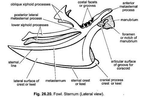
Sternum (Fig. 26.20) is a broad bone lying in the breast region, therefore, it is also known as breast bone.
It is a boat-shaped bone present in the ventral thoracic region and consists of the following parts:
1. Metasternum:
It is a broad plate-like bone forming the body proper. It is concave dorsally and convex ventrally. It is broader towards the anterior and posterior sides and narrow in the middle.
2. Keel:
Ventral convex surface of metasternum is produced ventrally into a median, triangular ridge called the sternal crest or carina or keel. The great pectoral muscles are attached with the keel.
3. Manubrium:
Manubrium is a small plate-like structure arising vertically from the anterior end of the ventral surface of the metasternum and called the manubrium or presternum. It also bears a large foramen at its base called the notch of manubrium.
4. Metasternal Processes:
There are a pair of large processes on either side of the metasternum, a large posterior and a small anterior metasternal processes. Anterior processes are also known as costal processes and posterior processes are also called xiphoid processes.
Each posterior metasternal process is divided again just at its origin in two large laterally compressed processes, the upper is oblique lateral xiphoid processes and posterior lateral xiphoid processess. The upper oblique processes terminate in a broad end just behind the last sternal rib. Lower larger xiphoid processes run downward and backward.
5. Costal Surfaces:
These are four cup-like depressions in the dorso-lateral margins of the anterior part of the metasternum. These depressions serve for the attachment of sternal ribs.
6. Coracoid Grooves:
There is a deep transverse groove on either lateral side of metasternum, at the base of manubrium which provide articular surface for the coracoid bones. Coracoid grooves communicate with each other through the foramen of manubrium.
Ribs:
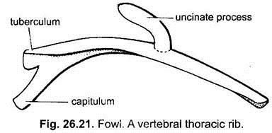
There are seven pairs of thoracic ribs in fowl. One pair of ribs articulates with each thoracic vertebra. Each thoracic rib consists of a dorsal vertebral and a ventral sternal portions meeting at an angle. By the sternal portion the ribs are united with sternum. First, second and sometimes the seventh ribs do not have sternal parts and do not reach the sternum.
The vertebral portion of the ribs leaving first and last pairs bear backwardly directed uncinate processes which overlap the next rib behind. The proximal end of vertebral portion is bifurcated into a dorsal tuberculum and ventral capitulum. The tuberculum is attached to the transverse process, while the capitulum is attached to the centrum of the corresponding vertebra.
Appendicular Skeleton:
Pectoral Girdle:
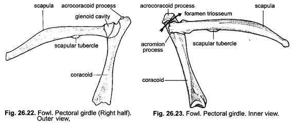
The pectoral girdle (Fig. 26.22 & 26.23) is very stout bony structure connected with the sternum on either side to support the wings. On either side it consists of a scapula, coracoid and a clavicle.
Scapula:
Scapula is long, flattened, slightly curved, sabre-shaped bone lying above the thoracic ribs and parallel to the vertebral column. The expanded anterior end of scapula is firmly united with the coracoid by ligament. Its anterior end bears a shallow depression forming a part of glenoid cavity. The glenoid end of scapula is produced into an acromian process to provide articular surface to the clavicle and to form a part of the foramen triosseum. Suprascapula is wanting.
Coracoid:
Coracoid is a stout, straight rod-like bone directed downwards and articulates with the coracoid groove on the antero-lateral edge of the sternum at the base of manubrium. The upper end of coracoid on its inner side articulates with the scapula and on its outer side it bears a deep cup-shaped depression which forms the greater part of the glenoid cavity. The upper end of coracoid is also produced into a hook-like acrocoracoid process which articulates with the clavicle.
Clavicles:
Clavicles are a pair of slender, curved delicate rod-like bones connected by their expanded upper ends with the scapula and acrocoracoid process of coracoid to enclose a circular foramen triosseum.
Furcula:
Ventrally the two clavicles are fused with a small interclavicle to form a laterally compressed disc or hypocleidium. It is connected with the rostrum of the sternum by a ligament. The V-shaped bone, thus, formed is known as furcula or merry thought or wish bone. It works as a spring-like connection between the two halves of shoulder girdle.
Pelvic Girdle:
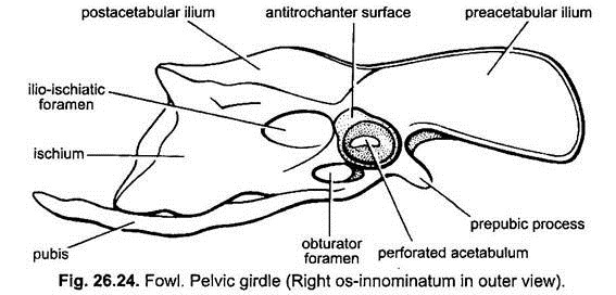
The pelvic girdle (Fig. 26.24) consists of two separate halves lying on either side of the synsacrum. Each half is known as os-innominatum. Each os-innominatum is composed of ilium, ischium and pubis. At the junction of three bones is present a concavity, the acetabulum, which provides surface for the articulation of the head of femur.
Ilium:
Ilium is an elongated and remarkably expanded bone extending both anterior and posterior to the acetabulum and is called preacetabular part and postacetabular part of ilium respectively. The inner margin of ilium is fused with the transverse processes and neural spines of the synsacrum. The outer surface of its anterior part is concave and posterior part is fused with the ischium. Ilium forms the dorsal part of acetabulum. On the outer surface above the acetabulum is a projection, the antitrochanter, which articulates with the great trochanter of femur.
Ischium:
Ischium is also dorso-ventrally flattened bone projecting backwards behind the acetabulum and parallel to the posterior part of the ilium. Ischium is fused posteriorly with the ilium but separated anteriorly from it by a large, oval ilio-ischiatic foramen.
Pubis:
Pubis is a long, thin, curved, slender bone directed backward parallel to the ventral margin of ischium with which it is usually fused. It forms ventral part of acetabulum. Behind the acetabulum the pubis and ischium are separated by an oval opening, the obturator foramen. Just in front and outside the acetabulum the pubis gives off a short and blunt process, the prepubic or preacetabular process.
Bones of Forelimb:
Forelimbs of birds are modified as wings and correspond to the forelimbs of a mammal.
1. Humerus:
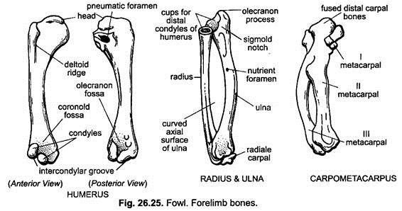
Humerus (Fig. 26.25) is the bone of upper arm of forelimb. It is an elongated stout bone expanded at both the ends. The proximal end is expanded into smooth convex surface the head which fits into the glenoid cavity of pectoral girdle.
The head is bordered by two tuberosities, preaxial and postaxial. From the smaller preaxial tubersity extends in front a short crest or deltoid ridge for the insertion of great pectoral and deltoid muscles. The postaxial tuberosity is larger and has a pneumatic foramen on the dorsal side of the proximal end communicating with its air cavity. The distal end possesses trochlea or condyles for the articulation with the radius and ulna.
2. Radius and Ulna:
Radius and ulna are the bones of the forearm.
Radius (Fig. 26.25) is shorter, slender and straight bone. Proximally it has a cup-shaped articular surface for the outer trochlea of humerus. Distally it bears a knob which articulates with the radiale carpel.
Ulna (Fig. 26.25) is stouter and larger than the radius and also slightly curved. Proximally it bears a small projection, the olecranon process, and a large articular facet for the inner condyle of the humerus. Distally it articulates with the ulnare carpal and radius.
3. Carpals:
Carpals are two irregular bones of wrist. Preaxial radiale is smaller and articulates with the radius and the postaxial ulnare is larger and articulates with the ulna.
4. Carpometacarpus:
Carpometacarpus (Fig. 26.25) is the bone of manus or palm. It is a compound bone formed by the fusion of three metacarpals with the distal row of carpals. The first metacarpal is very short, stumpy projection at the proximal end. The second metacarpal is strong and somewhat straight bone, the third metacarpal is thinner, slightly curved outwardly on the postaxial side and fused at both ends with the second metacarpal.
5. Phalanges:
Three metacarpals bear three clawless digits (fingers) which possess small rod-like bones called phalanges. First digit or pollex is preaxial and consists of a single phalanx, second digit (index) has two phalanges and third postaxial digit has a single phalanx.
Bones of Hindlimb:
In birds hindlimbs are modified for bipedal locomotion. Tarsus is absent.
1. Femur:
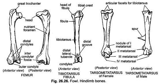
Femur (Fig. 26.26) is the bone of thigh of hindlimb. It is short and powerful bone flattened at both the ends. Proximally on the inner side it bears a rounded head which fits into acetabulum. On the outer side of the head is an irregular prominent process the great trochanter. Between the great trochanter and head is the articular surface for the antitrochanter of ilium Distally it has a deep groove or intercondylar fossa bounded laterally by two condyles for the articulation with the tibiotarsus. Outer condyle receives the upper end of fibula.
Patella is a sesamoid bone formed by the ossification of tendons. It is found in the tendon in front of femur-tibiotarsal joint.
2. Tibiotarsus and Fibula:
Tibio-tarsus and fibula (Fig. 26.26) are the bones of shank of hindlimb. Tibiotarsus is a long and stout nearly straight bone found on the inner side of shank. It is formed by the fusion with the proximal row of tarsals, i.e., astragalus and calcaneum. The proximal end of tibiotarsus is expanded and has on its anterior face a median cnemial or tibial crest and two articular surfaces for the condyles of the femur. Distally it articulates with the tarso-metatarsus by a pulley-like articular surface, i.e., a groove bounded by two condyles.
Fibula (Fig. 26.26) is a smaller, slender bone closely applied to the outer surface of the tibiotarsus. Proximally it articulates with the outer condyle of femur. Its distal end does not reach up to the lower end of tibiotarsus and terminates in a sharp point.
3. Tarsals:
In adult birds, the proximal row of tarsals is fused with tibia and distal row with metatarsus. In between the two rows is present the intertarsal ankle joint.
4. Tarsometatarsus:
Tarsometatarsus (Fig. 26.26) is a compound bone of foot or pes and formed by the fusion of distal row of tarsals with the second, third and fourth metatarsals It is a stout, long bone and proximally it bears two cup-like facets for the articulation with the tibiotarsus. First metatarsal is a small nodule-like bone attached to the inner and posterior surface of tarso-metatarsus near its distal end.
At its distal end, the three metacarpals become free each has a pulley-like articular surface for the corresponding digit. In male, the tarsometatarsus often bears a stout pointed spinous fighting spur on its medial side.
5. Phalanges:
These are small, slender bones. Metatarsus carries four toes- first or hallux is directed backward and has two phalanges, and the remaining three are directed forward. Second, third and fourth toes bear 3, 4 and 5 phalanges respectively. Terminal phalanx of each toe has a horny claw.
