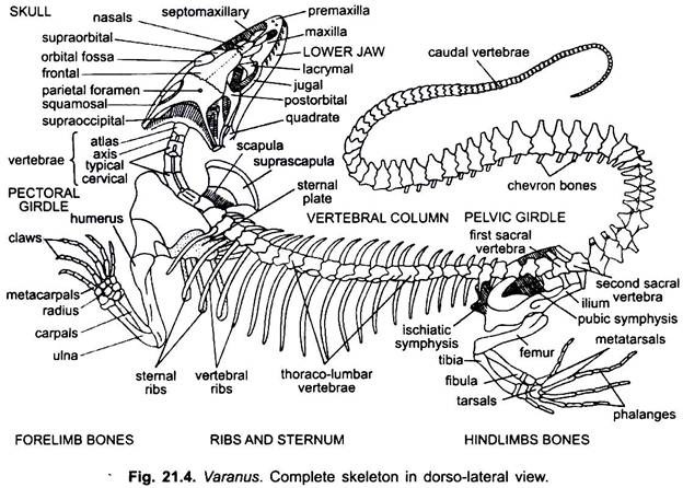
The skeleton of Varanus is divisible into two usual types, i.e., the axial skeleton and the appendicular skeleton. Axial skeleton consists of skull, vertebral column, sternum and ribs, while the appendicular skeleton consists of pectoral girdle, pelvic girdle and the bones of fore- and hindlimbs.
A. Characteristics of Skull:
i. It is monocondylic, i.e., single occipital condyle formed by the basioccipital.
ii. It is tropibasic, i.e., presence of interorbital septum separating the two orbits.
iii. Alisphenoids, orbitosphenoids and presphenoid are absent.
ADVERTISEMENTS:
iv. Prefrontals, supraorbitals and postorbitals are present.
v. A pair of parietals are fused and perforated by a median parietal foramen.
vi. Both the premaxillae are also fused to form a single bone.
vii. Presence of three vacuities (temporal fossae).
ADVERTISEMENTS:
viii. Presence of a single ear ossicle or columella in the tympanic cavity.
ix. Teeth polyphyodont, homodont and pleurodont.
x. Autostylic suspensorium, i.e., lower jaw articulates with the quadrate.
1. Skull:
ADVERTISEMENTS:
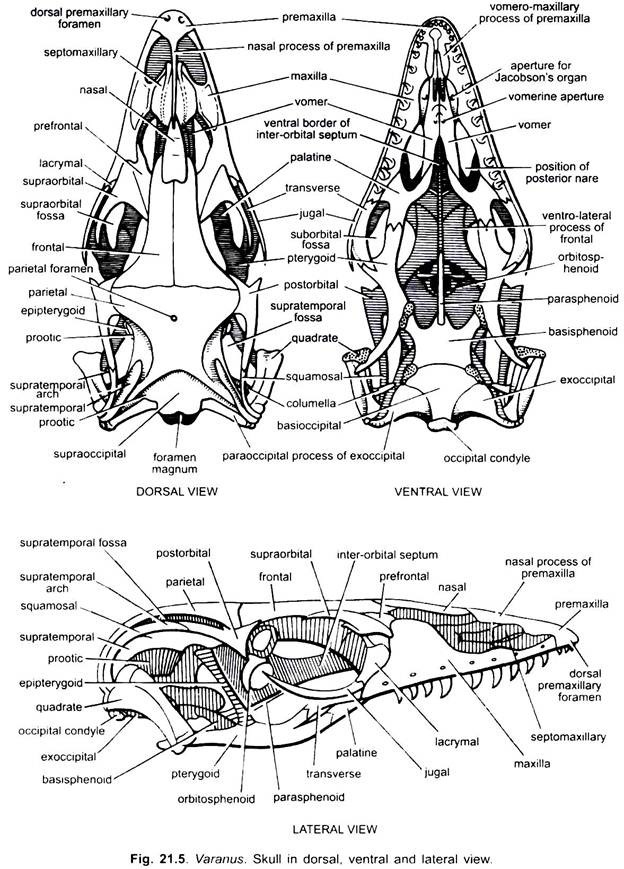
The skull of Varanus may be divided into four main regions, viz., the cranium, sense capsules (auditory, optic and olfactory) and the visceral skeleton comprising the jaws and hyoid apparatus.
Cranium:
The cranium proper forms the posterior part of the skull and lodges the brain. The cranium comprises three segments, i.e., the posterior occipital segment, the middle parietal segment and the anterior frontal segment.
(a) Occipital Segment:
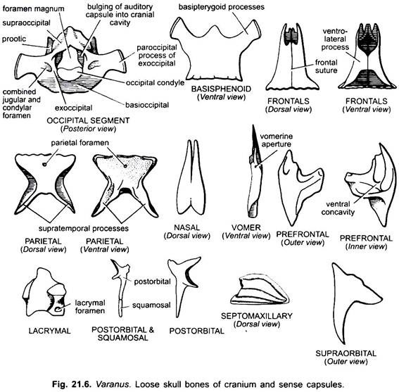
The occipital segment is the posteriormost part of the cranium. It encloses a large aperture foramen magnum in its centre. It consists of four bones, i.e., the dorsal supraoccipital, the lower basioccipital and paired lateral exoccipitals. Supraoccipital is more or less rectangular forming the roof of the hindermost part of the cranium.
It articulates anteriorly with the parietals and laterally with the prootics forming the post-temporal fossa. Basioccipital forms the floor of the hindermost portion of the cranium and posteriorly bears a single occipital condyle. It articulates anteriorly with the basisphenoid, laterally with the exoccipitals and prootics.
Exoccipitals surround the foramen magnum from its lateral sides. Each exoccipital is produced on its outer side into a paroccipital process articulating with the supratemporal, squamosal, parietal and quadrate. Each exoccipital has a combined juglar and condylar foramen for the exit of X, XI and XII cranial nerves and artery and vein.
Each exoccipital articulates with the supraoccipital, prootic and the basioccipital. Occipital segment articulates posteriorly with the atlas vertebra by the occipital condyle of the basioccipital. All the bones of this region are fused forming a ring-like structure.
(b) Parietal Region:
ADVERTISEMENTS:
The parietal segment comprises only two bones, parietals and a basisphenoid. Parietal. The parietal is a large flat bone on the postero-dorsal side of the cranium. It forms the roof of the cranium in the parietal region or segment. The two parietals are fused to form a single bone.
It is broad in front and narrow behind. It has a parietal foramen in its centre. Its postero-lateral angles are produced into supratemporal processes, each of which articulates with the quadrate, squamosal, supratemporal and paroccipital process of the exoccipital of its side. The supratemporal processes form the outer margins of the post-temporal fossae.
The parietals articulate anteriorly with the frontals, posteriorly with the supraoccipital, on the sides with the post-orbitals and below with the epipterygoid. Basisphenoid. The basisphenoid forms the floor of the cranium in the parietal region. It is a broad, flat and somewhat rectangular bone.
On its antero-lateral sides it gives off a pair of basipterygoid processes which unite with the pterygoids at their free ends. At the anterior margin it bears a median knob-like process which receives the hinder end of parasphenoid. Basisphenoid articulates posteriorly with the anterior margin of the basioccipital.
(c) Frontal Region:
The frontal segment is composed of only three bones, a pair of frontals and parasphenoid. Frontals. The frontals are large triangular bones lying on the dorsal side with their apex forwards. They meet one another along the median line by a frontal suture and with the parietal behind by a coronal suture. The frontals form the roof of the cranium in the frontal segment.
Frontals articulate in front with nasals, with prefrontals, palatines and postorbitals on the outer sides, with the parietals behind and below with the parasphenoid. The parasphenoid is a long, narrow, rod-like bone in the mid-ventral line.
2. Sense Capsules:
The sense capsules protect and lodge the organs of special sense. There are three pairs of sense capsules- auditory, optic and olfactory.
(a) Auditory Capsules:
The auditory capsules enclose the internal ears, the organs of hearing. They are situated on the lateral sides of the posterior end of cranium. Each capsule comprises a single small irregular vertical bone, the prootic lying just outside the supraoccipital and epiotic and opisthotic are indistinguishable. The epiotic is fused with the supraoccipital, while the opisthotic is fused with the exoccipital.
(b) Optic Capsules:
The optic capsules or orbits are a pair of large excavations on the lateral sides of the cranium. They enclose the eyes. The two orbits are separated from one another by a thin, longitudinal vertical partition, the interorbital septum. Each orbit is bounded by five bones- prefrontal, supra-orbital, lacrymal, post-frontal or post-orbital and jugal.
(i) Prefrontal:
The prefrontal is a small, more or less triangular bone bearing a deep cup-like concavity on its ventral side. It lies obliquely between the frontal and the maxilla. It forms the anterior boundary of the orbit and articulates with the suprarorbital posteriorly.
(ii) Supraorbital:
Supraorbital is somewhat a triangular bone overhanging the anterior portion of the orbit. Its broad base articulates in front with the prefrontal, while its free pointed apex is directed posteriorly towards the postorbital.
(iii) Lacrymal:
Lacrymal is a square-shaped bone forming the anterior boundary of the orbit. It is perforated by a lacrymal duct.
(iv) Postfrontal:
Postfrontal or postobital is an irregular bone producing four processes. In between the two inner processes is present a deep notch which receives the parietal and frontal. The posterior longest process articulates with the squamosal, whereas the antero-lateral process remains free. Postfrontal name is given because it is present behind the frontal, and postorbital name is given because it forms postero-dorsal boundary of orbit. It forms the postenor boundary of the orbital capsule.
(v) Jugal:
Jugal is a slender, curved, rod-like bone forming the ventral border of the orbit.
(c) Olfactory Capsules:
The olfactory capsules lie side by side in front of the cranium and contain the organs of smell. Each capsule is composed of dorsal nasal, septomaxillary and ventral vomer.
Nasals:
Nasals form the roof of the olfactory capsules. Two nasals fused in the mid-line into a single compound bone. Each nasal is a flat triangular bone, narrow anteriorly and broad behind. Nasals articulate in front with the nasal process of premaxiila and two septomaxillanes, and behind with the frontals.
Vomer:
The vomer is an elongated rod-shaped bone lying on the floor of the olfactory capsule. It meets with its counterpart in the mid-line in the anterior half and their posterior half remains free. It forms the inner margin of the posterior nare and is perforated by a vomerine aperture in its centre. It articulates with the vomero-maxillary process of premaxilla in front, with the palatine behind and Jacobson’s organ outside in the anterior region.
3. Visceral Skeleton:
It comprises the jaws, suspensorium and hyoid apparatus.
Jaws:
The jaws support the borders of the mouth. There are two jaws, i.e., upper jaw and lower jaw.
(a) Upper Jaw:
The upper jaw consists of two similar halves or rami which are fused together anteriorly in the middle line but diverge posteriorly. It is intimately fused with the cranium. Each half or ramus consists of nine bones divisible into two sets- the outer and inner. The outer set includes four bones- premaxilla, maxilla, jugal, septomaxillary, and quadrate, while the inner set includes five bones- pterygoid, palatine, transverse or transpalatine or ectopterygoid, epipterygoid and squamosal.
Premaxillae:
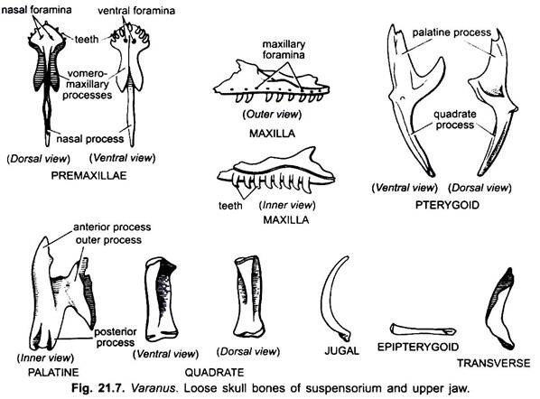
Two premaxillae are fused in the mid-line into a single bone and form the anterior limit of the snout. On its ventral surface it possesses 6-8 small conical teeth along its margins. Posteriorly it is produced into a long, narrow dorsal nasal process which is lodged into the groove present on the anterior part of the nasals. A pair of wing-like ventral vomero-maxillary processes articulating with the vomers behind. Premaxillae articulate on either lateral sides with the maxillae.
Maxilla:
The maxilla forms the major portion of the upper jaw on either side. It is a long irregular bone lying on either side behind the premaxillae. Outer surface of each maxilla is perforated by a number of maxillary foramina. The main body of each maxilla is known as alveolar part which bears a row of 8-10 teeth along its outer margin. The teeth are small, conical and pointed backward and pleurodont type.
The palatine process present on the inner side is poorly developed due to which a gap is present between maxilla and palatine. Maxilla articulate in front with the premaxillae, septomaxillary and vomer. Its upper process articulates with the prefrontal, supraobital, nasal and lacrymal, and behind with palatine, jugal and transverse bones.
Jugal:
Jugal is a slender curved, rod-like bone behind the maxilla. Besides forming a part of the upper jaw, it also bounds the orbit ventrally. It articulates anteriorly with the maxilla and lacrymal and on the inner side with the transverse, but free posteriorly.
Palatine:
Palatine is the bone of the roof of the buccal cavity. It is small, flat and more or less irregular bone. It is produced into three processes. The anterior long process joins with the vomer, the posterior small process with the pterygoid and the outer broad process with the maxilla and transverse. Palatines form the posterior boundary of inner nares.
Pterygoid:
Pterygoid is a large elongated and irregular bone. It has an anterior palatine process and a posterior quadrate process. It articulates anteriorly with the palatine on the inner side and transverse on the outer side, posteriorly with the basipterygoid process of basisphenoid on the inner side and with the quadrate on the outer side. Pterygoids are present on the roof of mouth.
Transverse:
The transverse is a small curved bone connecting the pterygoid with the jugal and the maxilla. It forms the floor of the orbit. It is also called the transpalatine or ectopterygoid.
Epipterygoid:
Epipterygoid is very slender rod-like bone extending almost vertically from the upper surface of the pterygoid to the prootic. It is also called columella crani.
Squamosal:
The squamosal is a curved rod-like bone attached to the posterior end of the arcade. Its anterior end meets a backward process of the post-frontal to form the supra-temporal arch, and its posterior end bends downwards to articulate with quadrate, supra-temporal, parietal and exoccipital.
Septomaxillary:
A pair of small, irregular flat bones is present on the dorsal side of vomers and one on either side of nasal process of premaxillae in the nasal region. These articulate with maxillae and nasals.
Suspensorium:
It includes quadrate and squamosal bones which suspend the lower jaw with the cranium and upper jaw.
Quadrate:
The quadrate is a small, thick, rod-like bone of the suspensorium. It lies obliquely in the postero-lateral side of the hind region of the cranium. Posteriorly it articulates with the squamosal, supra-temporal, supra-temporal process of the parietal and paroccipital process of exoccipital. Anteriorly it articulates with the quadrate process of pterygoid and lower jaw.
(b) Lower Jaw:
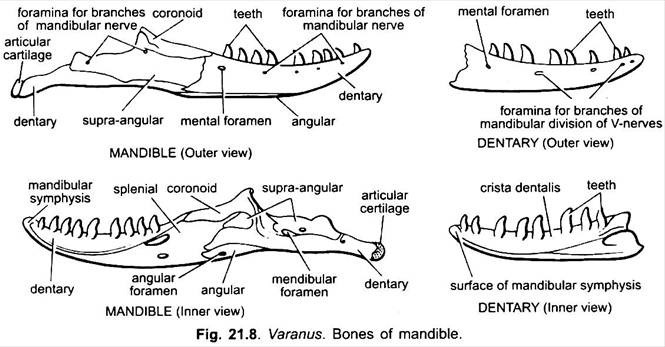
The lower jaw or mandible consists of two rami meeting anteriorly. Each ramus is composed of six bones, i.e., articular, angular, supra-angular, coronoid, splenial and dentary around an axial Meckel’s cartilage.
Articular:
The articular is the posteriormost bone of the ramus. On its dorsal surface it bears an articular surface for the quadrate and extends posteriorly terminating into an articular cartilage.
Angular:
Angular is a small splint-like bone fitted in between the dentary and articular and perforated by an angular foramen.
Supra-Angular:
Supra-angular is a flat, elongated and more or less rectangular bone lying in the middle of the ramus. It carries a pair of mandibular foramina for the mandibular nerves. It is present over the upper half of the outer surface of articular.
Coronoid:
Coronoid is a small more or less conical bone present above the supra-angular. It forms the dorsal side of the middle portion of ramus. It is produced into an upwardly directed coronoid process just behind the last tooth.
Splenial:
Splenial is a flat, irregular membranous bone lying on the inner side of the dentary.
Dentary:
Dentary is the largest bone forming the distal half portion of the ramus. It bears a row of 8 to 10 small, conical mandibular teeth which are of pleurodont type. Its outer surface also carries numerous foramina for the mandibular nerves. Both the dentaries unite suturally at their anterior ends.
Hyoid Apparatus:
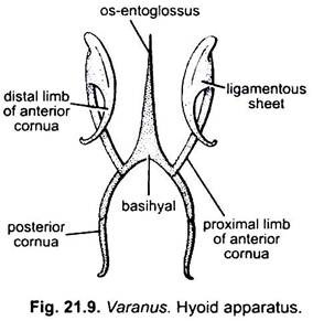
The hyoid apparatus lies embedded in the floor of the buccal cavity. It is cartilaginous structure which supports the tongue. It consists of a basihyal (main body) and two pairs of cornua arising from basihyal.
Basihyal:
Basihyal is in the form of an elongated, median, rod-like structure forming the body proper of hyoid apparatus. Its tapers anteriorly into os-entoglossus or lingual process.
Anterior Cornua:
The anterior cornua are elogated cartilaginous rods connected ventrally with the basihyal and running upwards curving round the gullet and ending on the ventral surface of the auditory capsule of their side. Each anterior cornua consists of a proximal and a distal piece, both joined together by ligaments.
Posterior Cornua:
The posterior cornua are also cartilaginous two segmental rods arising from the posterior end of the basihyal and running towards the hind end. Posterior cornua arise from branchial arches, while anterior cornua arise from hyoid arch.
B. Vertebral Column:
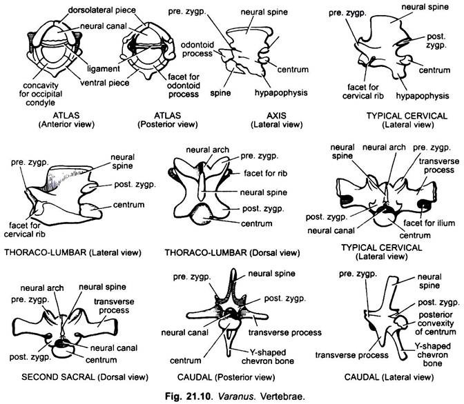
The vertebral column of Varanus consists of a large number (83-85) of vertebrae. Epiphyses are absent and centra of vertebrae are strongly procoelous. The vertebral column is divisible into four regions: the cervical containing 8 vertebrae, the thoraco-lumbar including 22 vertebrae, the sacral having only 2 vertebrae, and the caudal with about 51-53 vertebrae.
(a) Cervical Vertebrae:
A typical cervical vertebra consists of a stout and elongated centrum which is concave in front and convex behind, i.e., procoelous. Neural spine is crest-like. Prezygapophyses present at the anterior surface of neural arch are directed upwards and outwards, while postzygapophyses present on the posterior surface of neural arch are directed downwards and inwards. On the ventral surface, the posterior region of the centrum carries a backwardly directed hypapophysis. On the outer anterior surface the centrum carries articular facets for cervical ribs. Articular facets are present behind the third cervical vertebra.
(b) Atlas Vertebra:
Atlas vertebra is the first vertebra of cervical region. It is small and ring like in shape. It has no distinct centrum. It is composed of three distinct bony pieces of which two are dorso-lateral and one ventral around the neural canal. The neural canal in living animals is divided by a ligament into the dorsal and ventral portion which is downwardly projected into a small spine. Ventral piece occupies the position of centrum.
The dorso-lateral pieces have a space filled by a membrane at the point of their union in the mid-dorsal line. Along the anterior face of the ventral piece is present a cavity for the articulation of occipital condyle of the basioccipital of skull. On the posterior side of the ventral piece is also present an articular facet for the odontoid process of the axis vertebra. Pre- and postzygapophyses are absent. Transverse processes are also entirely absent.
(c) Axis Vertebra:
Axis vertebra is the second vertebra of the cervical region. It resembles the typical cervical vertebra in structure but it is slightly larger and bears a posteriorly directed hook-like spine below the odontoid process and arising from the centrum. The neural spine is vertical crest-like. It is characterised by the presence of an odontoid process at the anterior face of centrum.
Below the odontoid process, from the anterior end of centrum arises a backwardly directed hook-like spine. It represents the intercentrum or hypapophysis of atlas vertebra. A hypapophysis is also present on the ventral surface of the centrum in the posterior region. Prezygapophyses are rudimentary represented by simple notches but postzygapophyses are well developed.
(d) Thoraco-Lumbar Vertebrae:
Thoraco-lumbar vertebrae occur in the thoracic and abdominal regions. Thoraco-lumbar vertebrae are similar to cervical vertebrae but they are slightly larger in size and lack hypapophysis. Centrum is procoelous. Neural spine is vertical crest-like. Pre- and postzygapophyses are present. On either side at the junction of the neural arch and centrum is present a capitular facet for articulation with the single-headed thoracic rib.
(e) Sacral Vertebrae:
Both the sacral vertebrae are firmly united. These are stout and strong giving support to the pelvic girdle. Anterior or first sacral vertebra is more stout than the second. Its centrum is short and strongly procoelous. The neural spine is small crest-like. Pre- and postzygapophyses are well developed. Hypapophysis is absent. Transverse processes are strong and expanded at the tips which are also notched to receive a part of the ilia bones of the pelvic girdle. Posterior or second sacral has also short, stout and strongly procoelous centrum, neural spine is low and crest-like, and zygapophyses well developed. Unlike first sacral, transverse processes though expanded but not notched at the tips.
(f) Caudal Vertebrae:
The caudal vertebrae progessively become smaller towards the posterior end, finally getting reduced to mere bony rod-like centrum. Anterior caudal vertebrae. In the anterior caudal vertebrae the centrum is longer and strongly procoelous. The neural spine is long, pointed and backwardly directed. Pre- and postzygapophyses are well developed. Transverse processes are long, slender, outwardly and downwardly directed.
The most characteristic feature of the vertebra is the presence of Y-shaped chevron bone attached to the ventral surface of the centrum which bears two nodules for this purpose. Y-shaped chevron bone has two haemal arches attached to the ventral surface of centrum and a long downwardly directed haemal spine.
It encloses the haemal canal through which passes the artery and vein. Posterior caudal vertebrae. Posterior caudal vertebrae resemble the anterior caudal vertebrae in structure except the absence of chevron bone. The various processes also become gradually reduced.
3. Sternum:
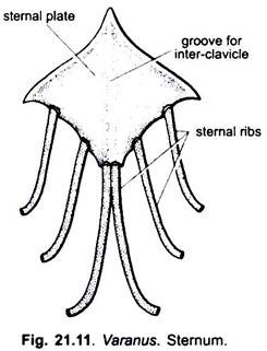
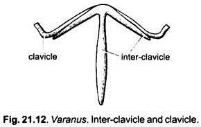
The sternum or breast bone is in the form of a rhomboidal plate of calcified cartilage lying embedded in the ventral thoracic wall. Its antero-lateral borders articulate with the coracoids and epicoracoids of the pectoral girdle. Along its each postero-lateral border it bears two small facets for the articulation with the 2 sternal ribs. At its posterior end it also bears two sternal ribs. The posterior elongated stem of T-shaped interclavicle is lodged in a groove on the mid-ventral surface of sternal plate. A pair of slender curved rods, the clavicles, are applied to the anterior cross-piece of the interclavicle.
4. Ribs:
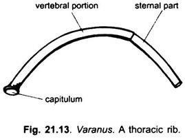
Ribs are unicephalous, i.e., single-headed. Their upper ends articulate with the capitular facets of corresponding vertebrae. Tubercular facets are lacking. Ribs are slender and curved formed of bone or bone and cartilage. Except first three cervicals, all the cervicals, thoracic and lumbars bear ribs.
Thoracic Ribs:
These are larger and differentiated into a dorsal bony vertebral portion articulating with the vertebra and a ventral cartilaginous sternal portion. First three thoracic ribs articulate with the sternum by their cartilaginous sternal parts. Rest thoracic ribs do not articulate with the sternum and, thus, remain free.
Cervical Ribs:
Leaving first three cervicals, all the remaining ones have cervical ribs. These are shorter and remain free ventrally and have no cartilaginous sternal portion.
C. Appendicular Skeleton:
Pectoral Girdle:
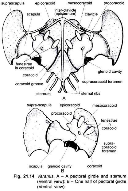
The pectoral or shoulder girdle (Fig. 21.14) is situated at the anterior end of the trunk. It protects the heart and lungs along with the sternum and ribs and also provides articulation to the bones of forelimbs. The pectoral girdle is composed of two similar halves one lying on either side of the T-shaped interclavicle and sternum. Each half of the pectoral girdle consists of scapula, suprascapula, coracoid and epicoracoid.
Scapula:
Scapula is bony, oblong and flat plate which is narrow in the middle. Its outer broader end articulates with the suprascapula, while its inner narrow end unites with the coracoid. Its lower posterior end forms a part of the glenoid cavity and its dorsal anterior end gives out an ossified process, the mesoscapula.
Suprascapula:
Suprascapula is more or less rectangular thin plate of calcified cartilage with a free distal border. It articulates proximally with the scapula and its distal border is free.
Coracoid:
Coracoid is large, flat and fenestrated bone. It is partly ossified and partly cartilaginous. Two large fenestrae divide the coracoid into outer procoracoid, middle mesocoracoid and inner coracoid proper. Inner anterior part of coracoid is cartilaginous forming the epicoracoid. It lies above the two fenestrae. Epicoracoid meet with the posterior long arm of interclavicle and sternum.
Interclavicle or Episternum:
It is a T-shaped bone located in between the two halves of pectoral girdle. Its lateral curved limbs lie anterior to the scapulae and the posterior long limb is closely set to the mid-ventral surface of sternum.
Clavicle:
It is a small curved narrow bone attached to the anterior side of lateral limb of interclavicle. Its outer distal end articulates with the union of scapula and suprascapula, while its inner limb does not reach up to the middle of interclavicle. At the joint of scapula and coracoid along the ventral side is present a glenoid cavity for the articulation of head of humerus.
Pelvic Girdle:
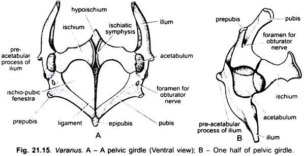
The pelvic or hip girdle (Fig. 21.15) is situated at the hind end of the trunk. It provides articulation to the bones of the hindlimbs. Pelvic girdle also consists of two similar halves meeting in the mid-line by a vertical ligament. Each half is known as os-innominatum and composed of ilium, pubis and ischium. All these three bones are not fused with each other. At their meeting points on the outer surface is a concave acetabulum for the head of humerus.
Ilium:
Ilium is strong, compressed, rod-like bone directed upwards and backwards to articulate with the sacral vertebrae (within the groove of transverse process of I sacral). On the outer side it is produced into a pre-acetabular process in front of the acetabulum and also contributes in the formation of about one-third part of acetabulum.
Pubis:
Pubis is flat and slightly curved bone, passes downwards to meet in front with its fellow of the other side in the middle line at the pubic symphysis. Between the anterior ends of two pubes is present a backwardly directed nodule of calcified cartilage, the epipubis. On the anterior end near its fusion with the ilium and ischium it has an oval foramen for the obturator nerve. Just external and slightly posterior to the foramen is a small rod-like process, the prepubis, which is directed outwards. Pubis also contributes to about one-third of the acetabulum.
Ischium:
Ischium is a flat and slightly curved bone, runs inward to meet its fellow of the other side at the ischiatic symphysis. It articulates on the outer side with the pubis and ilium of its side. A small rhomboidal piece of calcified cartilage, hypoischium, is present in between the two ischia at the anterior face of ischiatic symphysis and gives support to the ventral wall of cloaca. Ischium also forms about one-third of the acetabulum. The acetabulum is a cup-shaped cavity situated on the outer side at the point where all the three bones of each half of pelvic girdle join with each other. It provides articular surface for the head of femur.
A wide space is present between the pubes and ischia of both sides which is divided into two lateral ischio-pubic fenestrae by a median ligament. The ligament is often lost in the dried girdle.
Bones of Forelimb:
The forelimb contains humerus, radius and ulna, and the bones of forefoot.
Humerus:
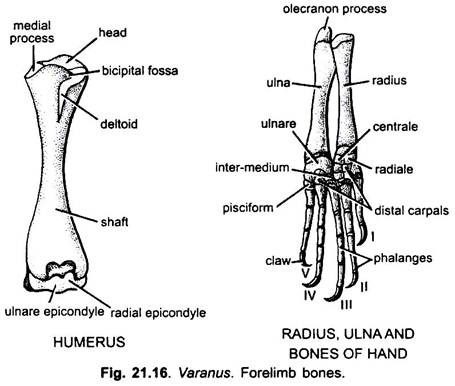
Humerus (Fig. 21.16) is the bone of the upper arm of the forelimb. It has an elongated shaft, both the ends of which are expanded and covered by epiphyses of calcified cartilage. The proximal end bears the rounded head which fits into the glenoid cavity of the pectoral girdle. A prominent crest-like deltoid ridge is present below the head. A bicipital fossa is enclosed between the medial process of proximal end and head. The distal end has a pulley-like articular surface, the trochlea, divisible into radial condyle and ulnare condyle by which it articulates with the radius and ulna.
Radius and Ulna:
Radius and ulna (Fig. 21.16) are the bones of forearm of the forelimb. Radius is slender and consists of a shaft and two epiphyses. At the distal end of radius is present a concave articular facet for the carpus and a preaxial styloid process. Ulna is somewhat stouter lying on the outer side of the radius. The proximal end of ulna is produced into an upwardly directed olecranon process, whereas distally it bears a concave articular surface for the carpus.
Forefoot:
Carpus or wrist consists of ten bony polyhedral carpals arranged in three rows. The proximal row contains three carpals- radiale, ulnare and intermedium. The central row has only one carpal, the centrale. The distal row has five small carpals. Besides the above, a pisciform is attached to the distal epiphysis of the ulna on its post-axial side.
Manus:
The digits are five each consisting of a metacarpal and varying number of phalanges, i.e., 2, 3, 4, 5 and 3. The terminal phalanx has a horny claw.
Bones of Hindlimb:
The hindlimb (Fig. 21.17) contains femur, tibia and fibula, and the bones of hindfoot.
Femur:
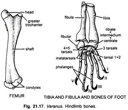
Femur is the bone of thigh of hindlimb. It is a stout bone having an elongated shaft and two epiphyses. The proximal end has a rounded head which fits into the acetabulum of pelvic girdle. A bit posterior to the head, on the pre-axial side is the prominent lesser trochanter and the greater trochanter lying on the post-axial side is greatly reduced. The distal epiphysis is pulley-shaped having two condyles for the articulation of tibia and fibula and a tuberosity in between the two.
Tibia and Fibula:
Tibia and fibula are the bones of the shank of hindlimb. Tibia is a stout bone and bears a longitudinal ridge, the enemial crest, along the anterior or dorsal surface. The proximal end of tibia bears two concave facets for the articulation with the condyles of femur. Fibula is relatively slender bone proximally articulating with the distal tuberosity of the femur. The distal end of both tibia and fibula provide surface for the articulation with the tarsals.
Foot Bones:
Tarsus contains only five tarsal bones, two in the proximal row and three in the distal row. Two tarsals of the proximal row are suturally fused and articulate with tibia and fibula. The distal row consists of three cuboid tarsals articulating with the five metatarsals of the foot. The number of phalanges in each toe is variable- the first toe has two, second toe has three, third toe has four, fourth toe has five and fifth toe has three phalanges. The terminal phalanx of each toe has a horny claw.
