In this article we will discuss about the Circulatory System in Earthworm:- 1. Introduction to Circulatory System in Earthworm 2. Circulatory System in First Thirteen Segments of Earthworm 3. After Thirteenth Segment in Earthworm.
Introduction to Circulatory System in Earthworm:
Earthworms possess a closed circulatory system in which blood always flows in the vessels and never comes in direct contact with tissues. The system consists of several tubes or vessels, blood glands and blood. The arrangement of different vessels is very complicated and that in the first thirteen segments differs from that of the rest body segments.
In general, there are few longitudinal vessels acting as collecting or distributing vessels which are connected with one another by some transverse vessels acting as heart. In this text, for the sake of convenience, the circulatory system of earthworm is described according to the segmental arrangement.
Circulatory System in First Thirteen Segments of Earthworm:
In the first thirteen segments following vessels are present:
ADVERTISEMENTS:
(a) Dorsal Vessel:
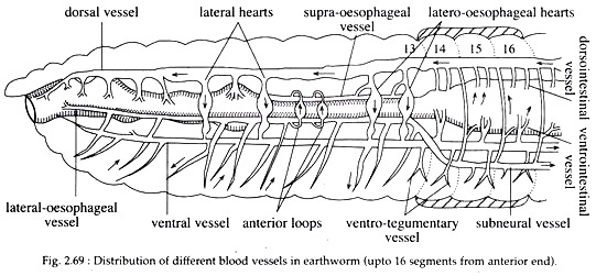
In the first thirteen segments, the dorsal vessel acts as a distributing vessel and extends upto the cerebral ganglion (Fig. 2.69). It gives off branches that supply the blood to the anterior regions of the body.
(b) Ventral Vessel:
ADVERTISEMENTS:
It extends anteriorly up to the 2nd segment (Fig. 2.69). It supplies the blood to ventral body wall, septal nephridia and reproductive organs. It gives a pair of ventro-tegumentary vessel in each segment.
(c) Supra-oesophageal Vessel:
It is a very short vessel extending between segments 9th and 13th. It collects blood from gizzard and stomach.
(d) Lateral-oesophageal Vessel:
ADVERTISEMENTS:
These vessels originate from the bifurcation of the sub-neural vessel at the 14th segment. They continue anteriorly along the lateral sides of the oesophagus. They collect blood from lateral regions of gut, body wall, septa and seminal vesicle.
(e) Ring Vessels:
The wall of the stomach between segments 10th and 13th bears dozens of circular vessels in each segment. These vessels carry blood from lateral oesophageals to the supra-oesophageals.
Circulatory System after Thirteenth Segment in Earthworm:
In this region, three large longitudinal vessels run parallel to each other and are as follows:
(a) Dorsal Vessel:
It runs along the mid-dorsal line of the body and just above alimentary canal (Fig. 2.69).
It acts as a collecting vessel and in each segment it receives:
(i) A pair of dorsointestinal vessels that bring blood from the intestine and
(ii) A commissural vessel running along the posterior border of each septum, collecting blood from skin and nephridia. The wall of dorsal vessel is muscular and its lumen is provided with a pair of valves in each segment which are directed forward and inward to prevent back flow of blood.
ADVERTISEMENTS:
(b) Ventral Vessel:
This long vessel runs along the mid-ventral line beneath the intestine (Fig. 2.69).
It distributes the blood through the following:
(i) A pair of ventro- tegumentary branches one on each side of each septum. These vessels pierce the septum, run upward and supply the blood to inner body wall and integumentary nephridium. Each vessel also gives a septonephridial branch which runs on the anterior face of the septum supplying blood to septal nephridia.
(ii) A median ventro-intestinal branch that supply the blood to the floor of intestine. In the lumen of ventral vessel valves are absent.
(c) Sub-neural Vessel:
It is a long, slender vessel extending from the posterior end to the 14th segment, and running along the mid-ventral line beneath the nerve cord (Fig. 2.69). It is a collecting vessel and in each segment it collects the blood by a pair of ventral branches from the ventral part of the skin.
The sub-neural vessel is linked to the dorsal vessel by a pair of commissural vessels in each segment. The vessel bifurcates in the 14th segment into two lateral oesophageal vessels.
Blood Plexus of the Intestine:
In earthworm, the intestinal wall is traversed by an internal and external network of capillaries. These capillaries are connected with the ventro-intestinal and dorsointestinal vessels.
Heart:
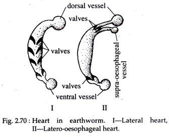
In earthworms, the dorsal and ventral vessels are connected to each other in segments 7th, 9th, 12th and 13th by means of transverse vessels which are commonly known as hearts (Fig. 2.70).
However, the anterior pair situated in the 7th and 9th segments are called lateral hearts while the vessels present in the 12th and 13th segments are called laterooesophageal hearts as they communicate dorsally both with dorsal and supra-oesophageal vessels (Fig. 2.70).
The hearts are provided with valves that open in single direction. Under electron microscope, four layers viz. epithelial tissue, connective tissue, circular and longitudinal muscle layers and vascular intima are observed in the wall of heart in earthworm.
Anterior Loop:
In the 10th and 11th segments, a pair of thin-walled, non-pulsatile, non-muscular loop like broad vessels without valves are present. These anterior loops convey blood from the lateral – oesophageals to supra-oesophageals.
Blood:
The blood of earthworm is red in colour and made up of plasma in which haemoglobin is dissolved and colourless nucleated corpuscles are suspended. In the European earthworm, Lumbricus, the blood cells are oval with a delicate cell membrane and finely granular cytoplasm. These cells bear resemblance with the coelomocytes and are excretory in function.
In Pheretima, five types of haemocytes have been recognized which are small amoebocytes, large amoebocytes, fibrocytes, granulocytes and illiocytes. Oxygen, carbon dioxide, dissolved food substances, excretory materials are distributed about the body by the blood and reach the tissues by diffusion through the walls of tiny capillaries.
Blood Forming Gland:
On the dorsal side of the oesophagus, in segments 5th and 6th blood glands are present. They are enclosed within the hinder part of the pharyngeal mass and consists of small spherical follicles of about 0.1 mm in diameter. The blood glands are red in colour as they contain blood.
Each follicle consists of a delicate fibrous capsule, within which lie a cup-shaped syncytial layer of nucleated protoplasm, separated from the capsule by a blood sinus. The function of these glands includes production of blood cells and haemoglobin. In addition, these may perform excretory function in some species.
Circulation of Blood:
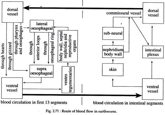
The circulation of blood through different vessels is carried out by the peristaltic contraction of heart. In front of each septum, there is a pair of ring valves which direct the blood forward or backward but prevent reverse flow. The route of blood circulation through different vessels is elaborated in Fig. 2.71.


