
In this article we will discuss about:- 1. External Features of Hirudinaria 2. Transverse Section of Hirudinaria 3. Locomotion 4. Digestive System 5. Feeding 6. Digestion 7. Blood Vascular System 8. Excretory System 9. Nervous System 10. Reproductive System.
Contents:
- External Features of Hirudinaria
- Transverse Section of Hirudinaria
- Locomotion in Hirudinaria
- Digestive System of Hirudinaria
- Feeding in Hirudinaria
- Digestion in Hirudinaria
- Blood Vascular System of Hirudinaria
- Excretory System of Hirudinaria
- Nervous System of Hirudinaria
- Reproductive System of Hirudinaria
1. External Features of Hirudinaria:
1. The body of the leech (Hirudinaria) is elongated, dorsoventrally flat, with a convex dorsal and a flat ventral surface (Fig. 24.25).

2. The body is broader at the middle and narrower towards both the ends.
3. A full grown leech varies in length from 300 to 350 mm with an olive green dorsal and an orange red ventral surface.
4. Each lateral line of the body bears an orange or yellow stripe, and ventral to each stripe is a broad black stripe.
5. Two suckers are present, one at the anterior and the other at the posterior end of the body:
ADVERTISEMENTS:
i. The anterior or cephalic sucker is oval shape and placed on the ventral surface at the anterior end of the body. It is formed by the fusion of a few anterior body segments and the prostomium. It bears a depression at the centre.
ii. The posterior or anal sucker is circular in outline, large in size and is a highly muscular disc at the posterior end of the body.
6. The body is divided into 33 segments, the last 7 fused to form the posterior sucker:
a. Each segment is subdivided into several, usually 5 rings or annuli by a series of close set transverse grooves.
ADVERTISEMENTS:
b. The number of segments is determined by counting the segmental receptor organs or sensillae. These are dot-like elevations on the skin of both the dorsal and ventral surfaces and restricted to the first annulus of each somite.
7. 5 pairs of sensillae (eyes), appearing as black dots on the dorsal surface of 2 to 6 segments, are called eyes.
8. The Mouth is a narrow triradiate aperture at the centre of the depression of the anterior sucker.
9. The anus is a small aperture on the dorsal surface at the junction of the body and the posterior sucker.
10. 17 pairs of nephridiopores are present on the ventral surface, a pair in the last annulus of each of the segments 6 to 22.
11. The leech is hermaphrodite, i.e. both the male and female reproductive organs are present in the same individual.
12. The gonophores are situated on the ventral surface of the body:
i. The male gonopore is single and median and lies in the groove between the second and third annuli of the 10 segment.
ii. The female gonopore is also single and median. It lies in the groove between the second and third annuli of the 11 segment.
ADVERTISEMENTS:
ADVERTISEMENTS:
2. Transverse Section of Hirudinaria:
1. The outermost layer is a thin, porous cuticle secreted by the underlying epidermis (Fig. 24.26, 24.27).


2. The epidermis is a single layer of cells which are wider at the outer and narrower at the inner curved ends. Some are modified as unicellular glands and others act as sense organs. It is not a continuous layer but broken at intervals.
3. Below the epidermis, there is a matrix of fibrous connective jelly containing connective tissue cells, pigment cells, fat cells, short muscle fibres and portions of various glands. This layer is often described as dermis.
4. Two layers of muscles are present. The outer circular muscular layer is a continuous one. The inner longitudinal muscular layer is also continuous but not uniformly thick and consists of several strata.
5. Certain muscles run across the body wall obliquely and dorso-ventral muscles run from dorsal to ventral surfaces.
6. Parietal layer absent.
7. Coelom absent. The space is filled up by spongy connective tissue, embedded in which are two lateral blood vessels, one dorsal sinus and one ventral sinus, enclosing the nerve cord. Botryoidal tissue is present just below the longitudinal muscular layer.
8. Parapodia and setae absent.
9. The gut wall is made up of an enteric epithelium, consists of a single layer of cells, some of which are glandular and others are absorptive. The epithelium is thrown into folds.
3. Locomotion in Hirudinaria:
Change of place is effected by swimming and looping movements.
1. Swimming:
The dorsoventrally flattened body moves in water by lateral undulations. The movement is produced by the contraction of the dorsoventral muscles and relaxation of the longitudinal muscles and this is repeated.
2. Looping movement:
Leeches move on a substratum by looping movement. Both the anterior and posterior suckers, circular and longitudinal muscles help in it.

It has two stages (Fig 24.28):
1. With the attachment of the posterior sucker to a surface, a simultaneous contraction of circular and relaxation of longitudinal muscles occur, resulting in lengthening of the body.
2. The anterior sucker is fixed on the substratum at a forward position, the posterior sucker released; longitudinal muscles contract, circular muscles relax; the posterior part is drawn forward and the body assumes the form of an inverted “U”. The posterior sucker is fixed behind the anterior sucker, and the process is repeated.
4. Digestive System of Hirudinaria:
The alimentary canal is a straight tube, beginning in the anterior ventral mouth and ending in a posterior dorsomedian anus (Fig. 24.29).

Mouth:
A triradiate opening at the base of the preoral chamber in the anterior sucker and guarded by the velum. Buccal cavity. A small chamber behind the mouth, opening in the pharynx. Three jaws—one dorsomedian and two ventrolaterals—are present on the walls of the buccal cavity.
a. A jaw (Fig 24.30) is a compressed muscular cushion, with a sharp, evenly curved free edge, covered with chitin, produced into numerous striations or teeth. Each jaw can be moved forward and backward through a certain arc, and the three acting together produce the typical triradiate wound in the skin of the prey.

b. The jaws bear papillae on both sides, bearing the openings of the unicellular salivary glands.
Pharynx:
A small, muscular chamber between the buccal cavity and the oesophagus. Numerous unicellular salivary glands are lodged in the space between the pharyngeal wall and body wall. Their secretion is discharged close to the mouth. Radial muscles run from the pharyngeal wall to the body wall.
Oesophagus:
Small and joins the pharynx with the crop.
Crop:
It is the largest part of the alimentary canal. The crop is thin-walled and extends from 8 to 18 segments and produced into eleven pairs of lateral, blind pouches or caeca. The first ten pairs are laterally directed, corresponding to each segment, while the last pair runs backwards as far as the 24 segments.
a. The lumen of the crop is divided between the pairs of pouches, corresponding to each segment by a transverse septum with an opening, controlled by sphincter.
b. The crop is capable of great dilatation and communicates with the stomach by a minute aperture.
Stomach:
A small, thin-walled chamber in the 19 segment, broad anteriorly and narrowing posteriorly, ending in the intestine. The wall bears transverse folds. Intestine. It is a continuation of the stomach, located in 20 to 22 segments. The intestine is narrow, thin-walled and bears longitudinal and transverse folds. It narrows posteriorly and opens in the rectum.
Rectum:
Thin-walled, extends from 22 to 26 segments and opens through the anus on the dorsal surface of the 26 segment in the last annulus.
5. Feeding in Hirudinaria:
The leech is a sanguinivorous animal, feeding on the blood of vertebrates. A crop- full of blood provides nourishment for several months.
i. The leech attaches itself to the victim’s body with the posterior sucker.
ii. The cup-shaped anterior sucker is pressed against victim’s skin; the jaws are protruded and the serrated margins of the jaw come in contact with the skin.
iii. With the operation of the muscles, the jaws move to and fro; a triradiate wound is
made in the skin and blood starts flowing out.
iv. Alternate contraction and expansion of pharynx due to the action of the radiating pharyngeal muscles, create a suction force and blood is sucked into the pharynx.
v. Secretion of the salivary glands containing hirudin, an anticoagulant, mixes with the blood, coagulation is prevented and the steady outflow of blood continues.
vi. Blood is stored in crop and its caeca; haemolysed and transformed into a jelly-like mass.
6. Digestion in Hirudinaria:
1. The sphincter between the last crop and the stomach controls blood flow and a small amount passes at a time from the crop to the stomach. The colour of the blood turns green from red.
2. The proteolytic enzymes secreted in the stomach digest blood.
3. Absorption takes place in the intestine and the residue is egested through the anus.
7. Blood Vascular System of Hirudinaria:
In leech, the blood containing spaces are of two types — blood vessels proper, with muscular wall and blood sinuses, without a definite wall and valves.
Two principal vessels, lateral in position and two principal sinuses — one dorsal and the other ventral, with branches and a number of spaces constitute the blood vascular system (Fig. 24.31). All the four principal channels are in communication at the posterior end of the body (Fig. 24.32). The longitudinal channels run from 6 to 22 segment.


Dorsal Sinus:
1. Mid-dorsal in position, runs beneath the body wall and dorsal to the alimentary canal; gives out two pairs of dorsal channels in each segment, ending in the dorsal and dorsolateral body wall and in the alimentary canal.
2. In the 6 segment, the dorsal sinus terminates in capillaries, forming plexus in the anterior five segments.
3. Posteriorly, the dorsal sinus bifurcates in the 22 segment. Running round the rectum, the branches join the posterior dilatation of the ventral sinus.
4. Haemolymph flows from posterior to anterior end.
Ventral Sinus:
1. It encloses the nerve cord and runs ventral to the alimentary canal. It is largest in diameter and encloses cerebral ganglia, peripharyngeal connectives and sub-pharyngeal ganglia anteriorly and terminal gangalia posteriorly.
2. At the level of nerve ganglia in each segment arise a pair of branches, each dividing into two, the ventral branch ending in the ventrolateral wall and the anterodorsal branch in the dorsolateral body wall.
3. In segments between 6 to 22, ventral sinus gives out a pair of nephridial branches in each segment.
4. Haemolymph flows from anterior to posterior end.
Lateral vessels:
One on each side of the alimentary canal.
1. In the 6 to 22 segments, a latero-ventral branch arises from each lateral vessel in each segment. The branch divides into two anterior and posterior, and join with the corresponding branch from other lateral vessel, in the mid-ventral line, below ventral sinus and form ventral commissure of the laterals. Three inter-segmental and longitudinal commissural branches connect the commissures of the successive segments.
2. ‘Valves are absent at the origin of the lateroventrals but present at the origin of its branches.
3. Each lateral vessel receives slender vessels from each segment:
a. A laterolateral from the lateral body wall and nephridium.
b. A laterodorsal from the lateral and dorsal body wall, gut and nephridium.
4. The laterodorsals of both sides meet above the dorsal sinus and form a dorsal commissure of the lateral vessels in the segments 6 to 22.
5. The laterodorsals and laterolaterals are joined by a lateral commissure in each segment.
6. The laterodorsals, laterolaterals are collecting vessels and their junctions with the lateral vessels are guarded by valves. The haemolymph flow is from posterior to anterior. Anteriorly, the lateral vessels branch into capillaries in the five first segments. Posteriorly, they end in a dilatation of the ventral sinus and a direct communication of ail the four channels established.
Course of Circulation:
Presence of haemoglobin in the fluid gives red colour to the haemolymph. Colourless corpuscles are less in number.
1. The dorsal and ventral sinuses are distributory, the lateral vessels are both collecting and distributory in function.
2. The dorsal, dorsolateral body wall and the gut receive haemolymph from the dorsal sinus.
3. Haemolymph from the dorsal and dorsolateral body wall passes to the lateral vessels through dorsolaterals and from the gut through the laterointestinals via dorsal commissures of the laterals.
4. The ventral, ventrolateral body wall and the nephridia receive supplies from ventral sinus and the haemolymph from there goes to lateral vessels through laterolateral and laterodorsal branches.
5. In each segment, the ventral body wall, gut, nephridia and genital organs receive supply from two lateral vessels through lateroventrals and the haemolymph returns to lateral vessels through laterodorsals and laterolaterals.
8. Excretory System of Hirudinaria:
1. The nephridia are one type, seventeen pairs, and one pair occur in each segment from 6 to 22 segments (Fig. 24. 33A). They are, however, absent in the first five and last four segments of the body and also in the segments forming the posterior sucker.

The nephridia may be placed under two categories:
Testicular Nephridia:
Elevon pairs, situated in 12 to 22 segments.
1. Body of the nephridum is attached to the ventral body wall and consists of a horseshoe-shaped main lobe and an inner lobe in the concavity of the main lobe (Fig. 24.33B).
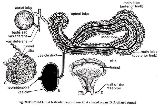
2. The body consists of a much coiled ciliated tubule richly supplied with blood.
3. The nephrostome is absent. The posterior limb of the main lobe runs forward as a stout apical lobe. The inner lobe runs up to the half of the apical lobe. The initial lobe is a cord of cells and coiling the apical lobe joins the main lobe posteriorly. Anteriorly, it runs towards the testis sac and ends in the ciliated organ (Fig. 24.33C) corresponding to the nephrostome.
4. The anterior limb of the main lobe turns backward and ends in a narrow vesicle duct. The duct opens in a vesicle, which in turn opens to the exterior by the nephridiopore placed on the ventral surface.
5. Waste products are temporarily stored in the vesicle and discharged to the exterior.
Pretesticular Nephridia:
The six pairs of nephridia situated in the segments 6-11, where the testes sacs are absent, are considered as pretesticular nephridia. Structurally, they resemble the testicular nephridia, but instead of ending in the ciliated funnel, the initial lobe ends loosely in the connective tissue on either side of the nerve cord.
In the adult Hirudinaria, the ciliated organ is not communicated with the body of the nephridium, although it is distinct in the embryonic stages. The ciliated organ loses its nephridial function and manufactures coelomic corpuscles, which are driven to the haemocoelomic stream.
Some workers are inclined to believe that the botryoidal tissue has got certain excretory function.
9. Nervous System of Hirudinaria:
The well-developed nervous system has three, components—the central, the peripheral and the sympathetic nervous system (Fig. 24.34).

Central Nervous System of Hirudinaria:
The cerebral ganglia or the brain, peripharyhgeal connectives, sub-pharyngeal ganglia and ventral nerve cord constitute central nervous system.
1. Nerve ring:
Situated in the fifth segment, the nerve ring is small, somewhat elongated and oriented vertically. The pharynx passes through it.
a. Cerebral ganglia or brain:
The two ganglia are fused to form the dorsal part of the ring or the brain.
b. Peripharyngeal connectives:
Two, constitutes the lateral sides of the ring around the pharynx and connect the brain with the sub-pharyngeal ganglia.
c. Sub-pharyngeal ganglia:
Large, triangular in shape/formed by the fusion of four pairs of embryonic ganglia and form the ventral part of the nerve ring.
2. Ventral nerve cord:
It is solid, consists of two nerves, right and left, enclosed in a sheath. It runs backward from the sub-pharyngeal ganglia ventral to the gut, being enclosed in the ventral sinus.
A ganglion in the ventral nerve cord is formed by the fusion of two ganglia and twenty-one such ganglia are situated, one in each, from 6 to 26 segments, in the first, annulus. The terminal ganglionic mass is large and formed by the fusion of seven pairs of embryonic ganglia.
Peripheral Nervous System of Hirudinaria:
Nerves emanating directly from the brain and ventral nerve cord are included in the peripheral nervous system:
1. Nerves from the brain innervate the first pair at eyes, prostrimium and walls of the buccal chamber.
2. The sub-pharyngeal ganglia send four pairs of nerves to 2 to 5 pairs of eyes and innervate buccal floor, body wall and segmental receptors of the first five segments.
3. Two pairs of nerves, anterodorsal and posterolateral, arise from each ganglion of the ventral nerve cord:
a. The anterolaterals supply the body wall, nephridia, reproductive organs and receptors.
b. The posterodorsals innervate body wall, viscera, muscles and testis sacs.
Sympathetic Nervous System:
Nerve plexus below the epidermis, in muscle layers of the body wall and in the gut wall, connected with the peripharyngeal nerve ring, constitute the sympathetic nervous system.
Receptors and Sense Organs in Hirudinaria:
These are of four types (24.35):

1. Eye:
Five pairs, one pair in each of the five segments (2 to 5), at right angle to the body surface.
a. Cylindrical in shape, the cells of the walls of the cylinder are pigmented and the structure is called pigment cup (Fig. 24.35A).
b. A convex, transparent cuticle and epidermal layer form the broad convex surface of the eye.
c. Large, clear, refractile cells bearing a crescentic hyaline substance, the lens or optic organelle at the centre, arranged in longitudinal rows, are present in the cylinder. The cells are photoreceptors and possibly can distinguish between light and darkness.
d. An optic nerve enters the cylinder near its base and runs along the middle line.
2. Segmental Receptor:
a. The receptors are small, white areas, segmentally arranged on elevated, elliptical papillae and located in the first annulus of each segment (Fig. 24.35B).
b. Each segment bears four pairs of the receptors on the dorsal and three pairs on the ventral surface.
c. Two types of cells, tactile or tango-receptor and photoreceptor, are present in a receptor.
d. The tactile cells are slender, with hair-like processes at the free ends. The optic organelles or the lens is present in the photoreceptor.
3. Annular Receptor:
These are thirty six in number, eighteen on the dorsal and eighteen on the ventral surface, arranged in a line across the middle annulus of each segment (Fig. 24.35C).
a. A receptor is formed of a group of flattened, overlapping cells, projecting beyond the integument.
b. The receptors are tactile in function.
4. Free Nerve Endings:
The free nerve endings are present all over the body between epidermal cells. The ganglion cells of the nerve endings occur beneath the epidermis. The receptors help to detect chemical changes in water.
10. Reproductive System of Hirudinaria:
The leech is hermaphrodite but auto-fertilization does not take place.
Male Reproductive System:
1. The male reproductive organs consist of testes enclosed in testes sacs, the vasa efferentia vasa deferentia, the epididymis, the ejaculatory ducts, the atrium and the male genital pore (Figs. 24.33A, 24.36).
2. The testes sacs are 11 pairs, one pair in each segment, on either side of the nerve cord in the segments from 12th to 22nd.
3. A short duct, the vas efferens arises from the posterior border of each testis sac and runs outward to end in the vas deferens. 11 vasa efferentia from 11 testes sacs of the side end in the longitudinal vas deferens of the side.
The two vasa deferentia run anteriorly from the 22 segment, and each forms a much convoluted epididymis in the 10 segment. A narrow muscular ejaculatory duct arises from each epididymis and leads to a median sac, the atrium.
4. The atrium is an oval sac consists of a large anteriorly placed base and a posteriorly directed neck. The base bears a thick wall having a large number of unicellular prostate glands. The neck contains a penis sac bearing a long, hollow filamentous penis, which can be protruded to the exterior through the male genital pore placed on the ventral surface of the 10 segment.
5. Spermatozoa budded in the walls of the testes sacs pass through vasa efferentia, vasa deferentia and are stored in the epididymis, where they are packed in bundles, the spermatophores and thrown into the atrium by the ejaculatory ducts. Spermatophores pass to the exterior through the penis.
Female Reproductive System:
1. The female organs consist of a pair of ovaries enclosed in ovisacs, a pair of oviducts, a common oviduct, albumen glands and a female gonopore. (Fig. 24.36).

2. The ovisacs are white bodies lying in the 11 segment by the sides of the ventral nerve cord. Each ovary is a delicate filamentous cord made up of club-shaped cells.
3. A short duct/ the oviduct arises from the base of each ovisac. The two oviducts unite below the ventral nerve cord and form a common oviduct, the region of union is covered by unicellular albumen glands. The common duct forms a ‘S’-shaped structure and passes through a mass of albumen gland and opens in a large muscular sac, the vagina.
The vagina bears a lateral diverticulum, the vaginal caecum and it opens to the exterior through the gonopore placed on the ventral surface of the 11 segment.
4. The ova, budded in the ovaries while passing through the anterior part of common oviduct, receive albumen from the albumen glands and remain stored in the vagina where they are fertilized during mating and then escape to the exterior.
Copulation and cocoon formation:
Leeches reproduce in early spring through cross-fertilization.
Copulation:
During mating two individuals come close in head-to-tail position and ventral surface of one is placed against the ventral surface of the other. The male gonopore of one is placed against the female gonopore of the partner and the penis of one enters the vagina of the other and vice versa. Mating continues for about an hour, during which mutual exchange of spermatophores occur. Fertilization takes place in the vagina.
Cocoon formation:
During mating a snow-white foamy girdle is secreted around the clitella by the glands of the clitellar segments. While the leeches pull out of the girdle, the fertilized ova from the vagina are forced out into the girdle. Before abandoning the girdle the leeches plug it’s both ends with the secretion of prostomial glands and the cocoon is formed.
Development:
Development is direct and completed within fifteen days, following which the plugs of the cocoon drop off and young leeches come out.