In this article we will discuss about the Microphagy in Lamellidense:- 1. Introduction to Lamellidense 2. Feeding Organs in Lamellidense 3. Feeding Mechanism 4. Digestive System 5. Digestion.
Contents:
- Introduction to Lamellidense
- Feeding Organs in Lamellidense
- Feeding Mechanism in Lamellidense
- Digestive System in Lamellidense
- Digestion in Lamellidense
1. Introduction to Lamellidense:
Lamellidense, commonly known as mussels, belong to phylum-Mollusca, class — Bivalvia, and subclass — Palaeoheterodonta. They are fresh water bivalves of India. They usually burrow in the mud at the bottom of the pond. Inside the shell, a delicate layer called mantle envelops the whole visceral mass.
ADVERTISEMENTS:
The mantle comprises of two lateral halves — called mantle lobes. The mantle lobe at the aboral side produces two short tubes — the inhalant and exhalant siphons. Water comes in through the inhalant siphon, carrying food and oxygen and goes out by the exhalent siphon carrying excretory products.
2. Feeding Organs in Lamellidense:
Collections of food in Lamellidense is performed by the following organs:
(a) Ctenidia:
ADVERTISEMENTS:
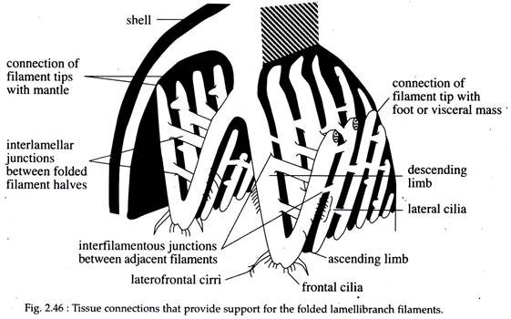
Two ctenidia are situated, each on either side of the body. Each ctenidia is composed of many gill filaments arranged one after another, forming gill lamellae (Fig. 2.46). The ctenidia are bipectinate, i.e., filaments are arranged on a central axis like the feather of birds (Fig. 2.46).
Each gill filament becomes folded or U-shaped. The arm of the U, that is attached to the axis of the gill is called the descending limb and the arm next to the mantle or visceral mass, is called the ascending limb.
The lengthened, folded filaments and their attachment to one another give the ctenidia a sheet-like form. The two ctenidia thereby provide four large, broad, filtering surfaces. At the angle of flexure, the frontal surface of each filament has well developed indentation, or notch, which, when lined up with the notches of adjacent filaments, forms a food groove that extends to the entire length of the underside of each ctenidia.
ADVERTISEMENTS:
The internal space between the ascending and descending limbs of the filament is subdivided by inter-lamellar junctions. Between the gill filaments, bounded by inter-lamellar junctions, there are minute apertures called the ostia.
Through the ostia water comes to the inner chamber of the ctenidia. The two adjacent filaments are connected by inter- filamentous junctions. The tip of the filaments are bounded with mantle or foot by a special type of muscular tissue (Fig: 2.46).
The inner side of the filament has few ciliated cell linings called abfrontal cilia. On the outer side of the filament, there are ciliary tracts — the lateral cilia, frontal cilia and laterofrontal cirri (Fig. 2.46). Each cirrus is a bundle of many adhering cilia.
(b) Labial Palps:
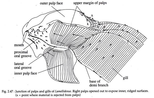
There are two pairs of leaf like outgrowths from the upper and lower border of the mouth called the labial palps. The labial palps, are ciliated on their internal faces, and are also crossed by a series of diagonal folds. These folds overlap each other in the direction of mouth. All, except the uppermost part of one fold, are covered by the next adjacent one.
3. Feeding Mechanism in Lamellidense:
(a) Collection of Food by Gills (Fig. 2.47):
The water current coming inside the mantle cavity through inhalant siphon contains suspended and deposited particles. The current is maintained by the lateral ciliary beat of the gills.
From the mantle cavity the water moves into the inter-lamellar and supra-branchial cavities. As the water passes between the filaments, the food particles are filtered out by laterofrontal cirri. The cirri are all beating together at right angles to the long axis of the gill filament.
ADVERTISEMENTS:
The distance between adjacent cirri is about 2 to 3.5 µm, but the effective space is smaller than this, since the cilia bend at regular intervals along the cirrus. Thus, they form a meshwork between the cirri and also between adjacent filaments. This arrangement permits the retention of a high proportion of incoming particles with the size ranging from 1-3 µm, and virtually a 100% retention occurs of those of 4 µm and above.
The particles thus trapped by the laterofrontal cirri are thrown onto the frontal cilia. Here the particles are entangled in mucus. Then they pass to the food groove and ultimately reach the labial palps.
(b) Sorting of Food Particles by Labial Palps (Fig. 2.47):
The sorting mechanism in the labial palp depends solely on the weight and not on the size of the particles, as the heavier ones settle down into the grooves. In this position the particles come under the influence of a powerful ciliary current that sweeps them onto the upper margin of the palp. Lighter particles avoid this current because they do not sink in the same way.
As a result, they are swept from the slope to the next fold and pass towards the mouth. The heavier particles, which move towards the upper margin of the palp, form pseudo- faeces and are rejected. From both the palps and the gills, pseudo-faeces travel to the mantle cavity, and are thrown out through the exhalant siphon. The food that reaches the mouth are subjected to digestion in the digestive system.
4. Digestive System in Lamellidense:
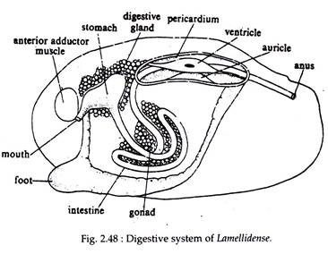
The digestive system is composed of alimentary canal and digestive glands (Fig. 2.48).
Alimentary Canal:
This canal starts from mouth and ends in anus. The different parts of the alimentary canal are mouth, gullet, stomach, intestine, rectum and anus. Mouth is a transverse slit, situated below the anterior adductor muscles and bounded by the lips of the labial palps. The mouth leads to a short tube called gullet. The gullet empties into a spacious stomach.
The stomach is sac-like and receives the secretion of the digestive glands, through several pores on its wall. From the posterior end of the stomach, starts the intestine. The intestine is a long tube. After emerging from the stomach it enters into the visceral mass and forms a coiled loop. It then goes up, takes the level of the stomach, enters into the pericardium and moves posteriorly through the ventricle as rectum.
The inner lumen of the rectal portion of the intestine is provided with a longitudinal ridge known as typhlosole. The typhlosole forms two longitudinal grooves, one on each side. They increase the absorptive area of the intestine. From one such groove, a gelatinous rod-like structure known as crystalline style projects into the stomach. Finally the rectum opens into the exhalant siphon through the anus.
Digestive Gland:
A pair of irregular digestive glands surrounds the stomach. The glands are composed of several digestive lobules. Each lobule is composed of single layer of secretory cells. The secretory cells show rhythmic activity. The secretion of the cell accumulates within the lumen of the lobule, from where they are transported into the stomach via digestive gland ducts. The alimentary system is highly adapted for microphagy.
5. Digestion in Lamellidense:
The mucus bound food, enters the mouth from the oral grooves of the palp and moves to the stomach via a short gullet. Here it encounters the crystalline style, rotating anticlockwise, which winds the mucus string in the stomach.
The principal features and functions of the stomach are:
a. It sorts fine food particles from the mucus string by means of the ciliary sorting areas and rejects the debris to the intestine.
b. Here primary digestion by amylase occurs. The amylase is supplied by the crystalline style from the intestine.
c. It conveys the partly digested material to the intestine.
There is no proteolytic enzyme in the stomach. The partly digested food from the stomach enters the intestine along with the secretion of digestive glands. In the intestine, protease and lipases are present, which complete the digestion.
The long intestine provides longer retention of food particles for extracellular digestion. After digestion the food is absorbed through the typhlosolar region of the intestine. The undigested materials come out through the anus.


