The entire nervous system of Rattus norvegicus may be divided into three groups:
(A) Central nervous system,
(B) Peripheral nervous system and
(C) Autonomic nervous system.
ADVERTISEMENTS:
(A) Central Nervous System:
It includes the:
(1) Brain, and
(2) Spinal cord
ADVERTISEMENTS:
(1) Brain:
In spite of general similarity in the pattern of brain with other vertebrates, there is considerable amount of specialisation of this organ in mammals. This specialisation is responsible for their advancement over other vertebrates. The brain consists of five divisions, telencephalon, diencephalon, mesencephalon, metencephalon and myelencephalon.
Following are the special features of the brain of rat:
(i) Meninges or coverings of the brain are three-layered. In-between outer Dura mater and inner Pia mater, there is a distinct middle third layer called arachnoid layer.
ADVERTISEMENTS:
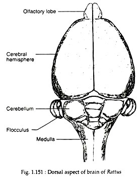
(ii) Size of the brain is large and the cerebral hemispheres and cerebellum are much convoluted so as to increase the area.
(iii) Olfactory lobes are small and club- shaped.
(iv) Cerebral hemispheres are much enlarged and cover the diencephalon and mesencephalon. Each hemisphere is subdivided into four lobes—frontal, parietal, temporal and occipital, by grooves called sylvian fissure (Fig. 1.151). The growth of cerebral hemisphere is due to growth of its roof called neopallium.
(v) Transverse bands of nerve fibres called corpus callosum connect the two cerebral hemispheres.
(vi) The ventral side of diencephalon is well-developed and known as hypothalamus. It bears optic chiasma, pituitary body (Fig. 1.151) and a pair of small round mass called mammillary bodies. The dorsal side of diencephalon carries pineal body or epiphysis and a vascularized fold called anterior choroid plexus.
(vii) Mesencephalon or midbrain is thick and contains four optic lobes called corpora quadrigemina.
(viii) Cerebellum is enlarged, folded and divided into a median vermis and two lateral lobes. Each lateral lobe is with a short flocculus. A broad band called pons varolii is present on the ventral side of the cerebellum.
(ix) Medulla oblongata is prominent and carries a vascularized posterior choroid plexus on its non-nervous roof.
(2) Spinal cord:
ADVERTISEMENTS:
The spinal cord runs up to the posteriormost end through the neural canal of vertebral column. Posteriorly, the spinal cord forms a narrow, triangular cone, called conus terminalis, from which a bunch of nerves arises. These are called filum terminale. In the brachial and lumbar regions, the spinal cord is slightly swollen.
(B) Peripheral Nervous System:
It includes nerves which are given out from brain and spinal cord. The nerves from the brain are called cranial nerves and those from the spinal cord are called spinal nerves. Twelve pairs of cranial nerves are present in Rattus Norvegicus besides the terminal nerve. The origin and distribution of the cranial nerves in rat are basically similar to the cranial nerves of Bufo and Calotes already described.
The tenth or vagus nerve, after forming the vagus ganglion, sends a branch called cardiac depressor to the heart, and a branch anterior laryngeal to the larynx. The main trunk runs posteriorly through the neck region.
Near thorax, the main trunk sends a branch called recurrent laryngeal, which in the left side curves around aorta and around subclavian in the right side, and, finally, turns anteriorly to supply the larynx. After entering the thoracic cavity, the vagus sends usual branches to lungs, heart and other visceral organs.
The eleventh cranial nerve or spinal accessory originates from the lateral side of medulla oblongata and innervates the muscles of neck region. A branch of it called ramus internus supplies nerves to the muscles of pharynx and larynx. This is a motor nerve. The twelfth cranial nerve or hypoglossal begins from the mid-ventral region of the medulla oblongata and innervates the tongue muscles through a number of branches. This is a motor nerve.
Thirty-two pairs of spinal nerves are present. They are built up in the same plan as that of toad. On each side, the fourth and fifth spinal nerves of the cervical region unite as phrenic nerve to supply the muscles of the diaphragm.
The brachial plexus is formed by the participation of first four nerves in the cervical region and first thoracic nerve. It innervates the forelimbs. Hind limb is innervated by sciatic plexus, which is formed by last two lumbar nerves and sacral nerves.
(C) Autonomic Nervous System:
It consists of a pair of sympathetic nerve cords, one on each side of the aorta. Each cord bears one anterior and one posterior cervical ganglia.
Sense Organs:
To receive different types of stimuli, there are different categories or receptors. The stimuli in the form of touch, pain and temperature are received by numerous free nerve endings or encapsulated corpuscles which remain scattered within the superficial layer of the skin. Taste is determined by group of specialised cells which remain within the papillae on the surface of the tongue.
These sensory papillae are called taste buds. Smell is perceived by specially sensory olfactory cells which are distributed in the mucous membrane of nasal cavity. Eyes and ears are much specialised receptor organs for receiving stimuli in the form of light and sound respectively. Ears, in addition to hearing, are also responsible for maintaining balance.
(i) Eyes:
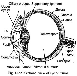
Eyes are built up in typical vertebrate plan (Fig. 1.152). The upper and lower eyelids are provided with small fine hairs. The edges of the eyelids are devoid of eye lashes.
Special features in the eyes of Rattus Norvegicus are:
(1) Presence of lacrymal gland on the upper side of each eyeball. It secretes a fluid called tear which cleans and lubricates the surface of the eye.
(2) Lens is biconvex and focusing is done by changing the curvature of lens through the ciliary muscles.
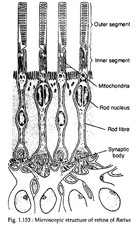
(3) Retina possesses both rod and cone cells. The former determines the intensity of light while the latter deals with colours. Fig. 1.153 gives an idea of the microscopic structures of retina.
(ii) Ear:
Each ear is divisible into:
(a) External ear,
(b) Middle ear, and
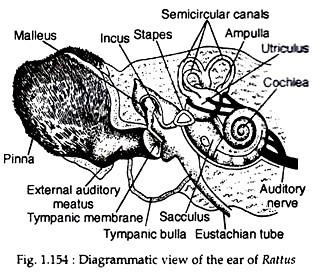
(c) Internal ear (Fig. 1.154).
External ear:
It consists of a movable, flap-like pinna and a canal called external auditory meatus. Pinna is responsible for collecting sound waves and sending it inside the ear through external auditory meatus.
Middle ear:
The external ear is separated from the middle ear by a tightly stretched membrane called tympanum. The middle ear is in communication with buccal cavity by a canal called eustachian tube. Within the middle ear, tympanum is connected with the internal ear by three ear ossicles—elongated malleus, slightly bent incus and triangular, ring-like stapes.
Malleus is attached with the tympanum and stapes is attached with the opening in the wall of internal ear, called fenestra ovalis. The incus is present in between the two. The ear ossicles are responsible for carrying the sound waves to the internal ear and the Eustachian tube is for regulating the equilibrium of atmospheric pressure.
Internal ear:
It consists of a bony labyrinth which is filled up with a fluid called perilymph. Within the perilymph a membranous labyrinth is suspended which contains another fluid called endolymph. The membranous labyrinth consists of following parts — lower sacculus, at the top of which lies the utriculus with which three semicircular canals are connected.
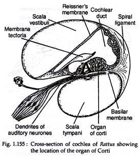
The sacculus is drawn into a spirally coiled cochlea (Fig. 1.155) which contains special receptor cells called organ of Corti. The bony cochlear canal encloses three cavities. The scalavestibuli is bounded by the vestibular membrane and located above the cochlear duct.
The scala tympani is situated below the cochlear duct. The scala media is the cavity of the cochlear duct itself. The scala vestibule and scala tympani are the perilymphatic spaces, while the scala media is filled with endolymph. The Reissners membrane and basilar membrane are the demarcating partitions between the three cavities as shown in figure 1.155.
The organ of Corti, the receptor apparatus for hearing, is supported on the basilar membrane inside the cochlear duct. The organ of Corti is composed in differentiated cells arranged in orderly rows. The semi-circular canals, utriculus and sacculus are responsible for balancing.
