Coelom:
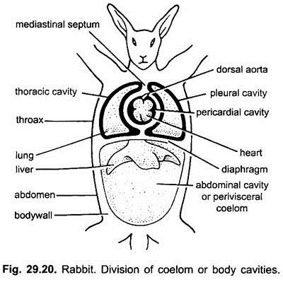
The body cavity of rabbit is divided into anterior thoracic and posterior abdominal cavities which are separated from each other by a transverse muscular partition, the diaphragm. Both these cavities are derived from the original perivisceral body cavity or coelom of the rabbit.
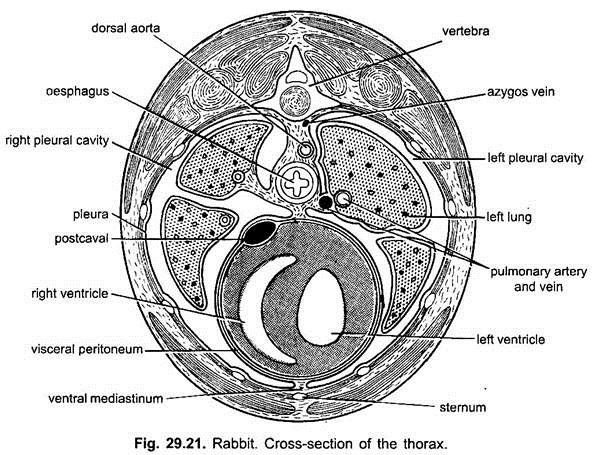
Unlike frog, the thoracic cavity is further divided into three cavities, two of them are situated laterally and lodge soft spongy lungs known as pleural cavities, while median one situated between the two pleural cavities is known as pericardial cavity lodging the heart and the roots of the great vessels.
Thoracic cavity also contains the trachea, bronchi and posterior part of oesophagus. The oesophagus perforates the diaphragm and enters the abdomen. The abdominal or the peritonial cavity is large and contains all the remaining visceral organs, e.g., stomach, intestine, liver and pancreas, spleen, urinogenital organs, etc.
The coelom is lined by outer parietal epithelium and inner visceral epithelium, which covers and in fact encloses all the organs. The pleural cavity is lined by a moist membrane, the pleura, which can be divided into parietal and visceral parts. The parietal pleura lines the inner side of the pleural cavity, while the visceral pleura is closely applied to the lungs.
ADVERTISEMENTS:
The pleural cavities are separated from each other by a mediastinal space which contains oesophagus and trachea. The viseral pleura of two sides are closely applied in the ventral part of the mediastinal cavity to form a median mediastinal septum.
In the pericardial cavity the outer parietal membrane (parietal pericardium) is transparent and the inner visceral membrane (visceral pericardium or epicardium) is closely applied to the heart. The parietal pericardium is separated from the heart by the pericardial cavity.
ADVERTISEMENTS:
The membranes of the abdominal cavity are parietal and visceral peritoneum. The peritoneum forms double layered mesenteries or ligaments which support the alimentary canal and organs of the viscera.
Viscera:
The heart, lungs, oesophagus and trachea are contained in the thoracic cavity. The abdominal cavity has a dark brown five-lobed liver fitted into the concavity of the diaphragm, and a saccular dark blue gall bladder is situated in one of the lobes of the liver. The oesophagus opens into the stomach which lies dorsally to the left lobe of the liver.
The spleen is attached to the left border of the stomach.The stomach is continued into a long coiled intestine, occupying largely the whole abdominal cavity. A pinkish irregular structure, the pancreas, is found in the ‘U’-shaped duodenum. Two bean-shaped kidneys are found attached to the dorsal wall of the abdomen outside the coelom. The right kidney is situated a bit anterior to the left one.
Ureters are seen coming out from the kidneys to join a thin-walled sac, the urinary bladder. A small rounded yellow coloured adrenal gland is found in front of each kidney. The adrenal glands are endocrine in nature. In a male rabbit the two testes are situated in two pouches of the bodywall known as the scrotal sacs lying outside the abdominal cavity. The scrotal sacs are situated one on either side of the penis.
ADVERTISEMENTS:
The abdominal cavity is continued into the cavity of the scrotal sacs by inguinal canals. The vas deferens of the testes unite with the neck of the urinary bladder to form a common urinogenital canal or urethra which passes in the penis. The penis is the copulatory organ of the male rabbit. In a female rabbit, two small oval ovaries are situated behind the kidneys.
From behind each ovary a convoluted duct, the oviduct, arises which dilates to form a uterus. The two uteri thus formed unite together to form a vagina. The neck of the urinary bladder joins the vagina to form a urinogenital canal or vestibule which opens out by a slit-like vulva. The vulva is the external aperture of the female reproductive system.

