In this article we will discuss about:- 1. Distinctive Features of Branchiostoma 2. Respiratory and Feeding Structures of Branchiostoma 3. Excretory Organs.
Distinctive Features of Branchiostoma:
1. Branchiostoma is a transparent, fish-like animal occurring near the shore, burrowing in rocks.
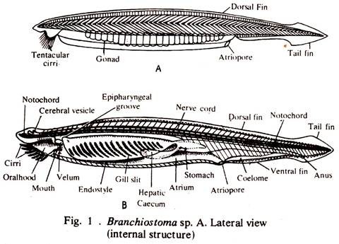
2. The body of Branchiostoma is narrow, 2.5-5.8 cm long, laterally compressed and pointed at both the ends. The anterior two-thirds of the body is triangular in cross section.
3. Along the mid-dorsal line a dorsal fin extends the whole body length. It is joined posteriorly to a somewhat broader caudal fin around the tail (Fig.1A).
ADVERTISEMENTS:
4. A ventral fin is situated mid-ventrally running from the caudal fin to a median opening, the atriopore.
5. The muscles of Branchiostoma are arranged in sixty two V-shaped segments or myotomes.
6. Three unpaired apertures are present:
(a) The mouth overarched by the median, ventral oral hood, and fringed with tentacle-like cirri. (Fig. 1B).
(b) The atriopore in myotome thirty six to expel water from the pharynx which enters through the mouth.
(c) The anus, ventral and slightly to the left behind the atriopore but at some distance from the posterior end of the body.
7. The notochord of Branchiostoma is a flexible, un-segmented rod, pointed at both the ends and runs from one to the other end of the body.
8. The pharynx is supported by gill-rods, which border the numerous gill slits.
ADVERTISEMENTS:
9. The intestine of Branchiostoma is straight and without any loop.
10. The liver diverticulum is simple in Branchiostoma.
11. The circulatory system of Branchiostoma is ill developed. A definite heart is absent but the ventral or the branchial artery is rhythmically contractile.
12. The blood of Branchiostoma is colourless. A few amoeboid cells are present in it.
13. About ninety pairs of segmentally arranged nephridia are present on the dorsolateral walls of the pharynx.
14. The dorsal nerve cord is shorter than the notochord, above which it lies.
15. A definite brain is absent although anteriorly the central canal of the nerve cord widens to form the so-called cerebral vesicle.
16. Two sets of nerve roots are present, the ventral roots far outnumber the dorsal roots. The two sets of roots do not unite as in vertebrates. Spinal and sympathetic ganglia absent.
Respiratory and Feeding Structures of Branchiostoma:
Branchiostoma dwells in the shallow waters of tropical and semitropical seas and has apparently retained a great many relatively simple features, despite the fact that it has probably evolved a long way from the actual ancestors of chordates.
ADVERTISEMENTS:
This small animal shows the chordate plan reduced to almost its lowest terms and its development and structure point the way to the rise of complicated conditions found in higher vertebrates.
Structure of Pharynx:
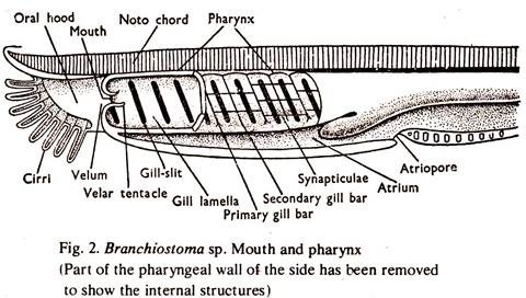
1. The mouth is situated at the base of a vestibule overarched by a median ventral oral hood containing tentacle-like structures, called buccal cirri. The oral hood contains a complex set of ciliated tract, the wheel organ. The mouth is surrounded by a membrane called the velum, and is fringed by twelve velar tentacles and leads posteriorly into the pharynx.
2. The pharynx is a high, compressed chamber and occupies anterior half of the body (Fig. 2).
3. The walls of the pharynx are perforated by nearly 200 oblique vertical slits, the gill slits or branchial apertures.
4. The portions of the pharyngeal wall separating the clefts are known as branchial lamellae.
5. Each lamella is supported towards its outer edge by a rod made of elastic fibres, known as branchial rod.
6. The rods are united with one another dorsally by loops but free ventrally.
7. The bars at their free ends are alternately forked and simple and known as primary and secondary branchial rods respectively.
8. The branchial lamellae bearing the primary rods are called primary lamellae and those lodging the secondary rods are called secondary lamellae.
9. Transverse branchial lamellae supported by branchial rods connect the primary lamellae at regular intervals.
10. The inner and lateral faces of each lamella are provided with long cilia.
11. The pharynx ends posteriorly into a narrow intestine.
12. On the ventral wall of the pharynx is a longitudinal groove, the endostyle. The groove is composed of four tracts of mucous glands, each separated from the other by tracts of ciliated cells.
13. A longitudinal groove, the epipharyngeal or hyperpharyngeal groove, lined with cilia is present on the dorsal surface of the pharynx.
14. A pair of lateral grooves, the peripharyngeal bands encircle the mouth at the anterior end of the endostyle. They meet dorsally and join with the epipharyngeal groove.
Respiratory and Feeding Mechanism:
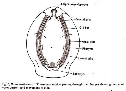
Cilia of the wheel organ and pharyngeal wall maintain a continuous flow of water, which enters the pharynx through the mouth and passes to the exterior through the gill slits, atrium and atriopore. The branchial lamellae are highly vascular and a gaseous exchange takes place there.
Minute organic particles, chiefly microscopic organisms, are brought into the pharynx by the water current. During feeding, a continuous stream of water enters the pharynx through the mouth and passes to the atrium through the pharyngeal slits, from where it is expelled to the exterior through the atriopore.
The cilia oriented along the anterior and posterior faces of each gill lamella, known as lateral cilia, work in unison with the cilia distributed over the inner (pharyngeal) surface of the gill lamella, known as frontal cilia, and thus form an efficient ciliary feeding machine (Fig. 3), and as such, the Branchiostoma is known as a ciliary feeder (Grove and Newell).
The endostyle as well as the pharyngeal epithelium secrete a cord of mucus in which food particles are entangled. By the beating movement of the cilia, food particles entangled in the twisted rope of mucus in the endostyle are driven forwards, then upwards through the peripharyngeal bands and backwards along the epipharyngeal groove and finally into the opening of the oesophagus.
Summary of the Feeding Mechanism:
The cilia in the gill lamellae set up an water current, which enters the pharynx → gill slit → atrium. Food particles are entangled in the mucous layer of the pharyngeal surface, the frontal cilia help in driving the food into the oesophagus.
Excretory Organs of Branchiostoma:
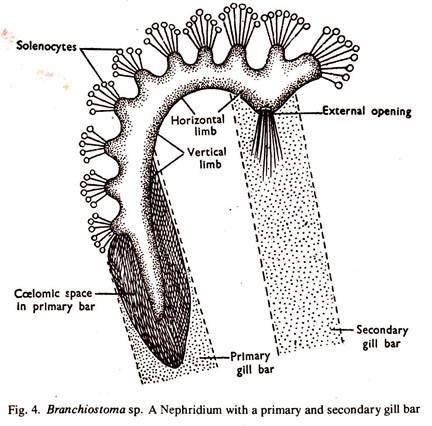
The excretory organs of Branchiostoma consist of about 90 pairs of true nephridia and a pair of brown funnels. Some parts of the atrial wall also serve as excretory organ. Of the different excretory structures, the nephridia are supposed to play the most important role. The nephridium does not obtain wastes from the coelom, instead, it collects the same from the blood vessels that surround it (Fig.4).
Structure of Nephridia:
1. The nephridia are situated between the atrial epithelium and the epithelium of the dorsopharyngeal coelom, on the dorsolateral wall of the pharynx.
2. Each nephridium is a bent tube lined with cilia, and consists of an anterior vertical and a posterior horizontal limb in relation with the primary gill lamella.
3. The nephridium opens into the atrium opposite the dorsal end of a branchial rod, in one of the dorsal pouches:
4. Close to the opening, it is divided into two canals, the horizontal limb passes forward and then turns ventral and the vertical limb passes backwards.
5. The vertical limb terminates in a large group of fibres ending in small knobs.
Each fibre projects into the coelom and contains a long vibratile flagellum and its is called solenocyte. The movement of the flagellum flushes out the wasted from the cell stalk. Several smaller groups of solenocytes are also present on the horizontal limb. These solenocytes have a close similarity with those found in polychaete annelids.
6. A tuft of specially long cilia project into the atrium through the renal opening.
7. The nephridia receive special blood supply from the pharyngeal vessels and the extracted wastes are discharged into the atrium. Branchiostoma is the only chordate with true nephridia as excretory organs.
Brown Funnels:
A pair of funnel-shaped structures known as brown tubes, are present on the hinder part of the coelom and open into the atrium at their wide ends. The brown funnels are presumably of excretory function.
Atrial Wall:
Some parts of the atrial wall are provided with groups of columnar excretory cells and are supposed to be excretory in function.
The development of the nephridia in Branchiostoma indicates that they are ectodermal in origin and as such they are in no way comparable to the pronephros of vertebrates.


