Learn about the comparison of foetal membranes in Gallus and Oryctolagus.
Foetal Membranes: Comparison # Gallus:
A. Formation of Amnion:
The amnion begins to form on the second day of incubation and is completed on the fourth day.
1. A fold of blastoderm occurs just in front of the head of the embryo. It consists of ectoderm and endoderm. With the appearance of mesoderm, it takes part in the same and finally the head-fold of the amnion is constituted by an extra embryonic ectoderm and a somatic mesoderm. The fold gradually grows back over the embryo.
ADVERTISEMENTS:
2. At the beginning of the third day, another fold, the amniotic tail-fold appears at the posterior end of the body and it gradually grows towards the head-fold. It consists of ectoderm and somatic mesoderm from the beginning.
3. At the beginning of the fourth day the amniotic folds meet and fuse at the centre and the amebryo is surrounded by a cavity, the amniotic cavity, which is bounded by two membranes, the outer and the inner.
4. The cavity is called amniotic cavity and is filled up with a fluid, amniotic fluid. The fluid acts as a shock absorber and protects the embryo. The outer layer is known as the chorion and the inner layers as the amnion. The space between the amnion and chorion is known as the extraembryonic coelomic cavity. Both the layers consist of an outer ectoderm and an inner mesoderm facing the amniotic cavity.
5. Muscle fibers in the amniotic wall contract rhythmically which rocks the embryo in the amniotic fluid and the adhesion of the embryo to the amniotic wall is prevented.
ADVERTISEMENTS:
B. Formation of Allantois:
The allantois begins to form on the third day and becomes well developed by the end of the fifth day.
1. The allantois arises as an outpushing from the ventral wall of the hind gut and consists of an inner endodermal and an outer mesodermal layer.
2. The allantois gradually pushes out through the coelomic space between the somatic umbilicus and the yolk-stalk, and spreads out in the extra embryonic coelom. The neck of the allantois is known as allantoic stalk and two arteries and one vein pass through it.
ADVERTISEMENTS:
3. It grows rapidly and covers the amnion within two days and coming very close to the shell acts as a respiratory organ due to the presence of ramified blood vessels. By this time it fuses with the chorion.
4. The allantois finally encloses the whole embryo and yolk-sac with the remains of the albumen. It absorbs the white of the egg and acts as an organ of nutrition. It is also excretory and acts as the urinary bladder of the embryo, receiving wastes from the kidney.
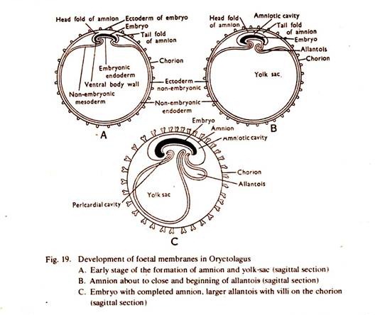
Foetal Membranes: Comparison # Oryctolagus:
A. Formation of Amnion:
The amnion begins to form after the appearance of the coelom as a result splitting of the mesoderm into two layers, the outer somatic and the inner splanchnic layer.
1. The amnion arises by two folds, one in front of the embryo, the head-fold and the other at the posterior end of the embryo, the tail-fold. Both the folds grown upward and inward. Each fold consist of an outer trophoblastic ectoderm and an inner somatic mesoderm. These constitute a part of the extra embryonic layers.
3. The two amniotic folds meet at the middle over the embryo and unite to from two layers. The embryo is surrounded by a cavity, the amniotic cavity, which is bounded by two membranes, the outer and the inner.
4. The cavity is called amniotic cavity and is filled up with a fluid, the amniotic fluid. The fluid acts as a shock absorber and protects the embryo. The outer layer is known as chorion and the inner layer is amnion. Bothe the layers consists of an outer trophoblastic ectoderm and an inner somatic mesoderm facing the amniotic cavity.
The chorion becomes closely associated with the mucous membrane of the uterine wall and send out processes or primary villi by which attachement and nourishment are effected.
5. The rhythmic contractions of the muscle fibers in the amniotic wall rock the embryo in the amniotic fluid and prevent the adhesion of the embryo to the amniotic wall.
ADVERTISEMENTS:
B. Formation of Allantois:
The allantois begins to appear at the time of establishment of the amnion and grows out rapidly.
1. The allantois arises as an outpushing from the ventral wall of the hind gut, and consists of an inner endodermal and an outer splanchnic layer of mesoderm.
2. The allantois gradually pushes into the extra embryonic coelom between the chorion and aminion. The neck of the allantois is known as allantoic stalk and two arteries and one vein pass through it.
3. It grows rapidly covers the amnion and fuses with the chorion and trophoblast is small area. The wall of the allantois is highly vascular.
4. The allantois finally encloses the whole embryo and the yolk-sac. The region where the chorion, trophoblast and allantois unite, sends vascular outgrowths or villi which burrow into the corresponding crypts or depressions on the uterine wall and an allantoic placenta is formed, through which nutrition, excretion and respiration are performed.
Prior to the formation of allantoic placenta, nourishment is temporarily drawn by yolk-sac placenta. Part of the yolk-sac wall unites with the chorion and trophoblast and forms the foetal part of the yolk-sac placenta.