In this article we will discuss about the application of genetic engineering in diagnosis and treatment of diseases.
Diagnosis of Diseases:
1. Parasitic Diseases:
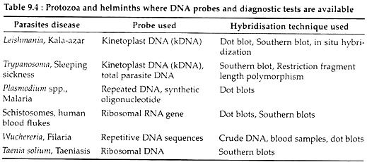
In contrast to conventional diagnostic methods, discovery of DNA probes have proved to be very effective and most sensitive tools for the diagnosis of a variety of diseases. Probes are now available for a number of human parasites like protozoa and helminths (Table 9.4).
DNA probes can also be used for the diagnosis of many other diseases. In such cases, the DNA is labeled radioactively, which is hazardous. Therefore, non-radioactive labelling of probes, methods for quick preparation of target DNA and suitable protocols for hybridisation are being developed which will allow quick and accurate diagnosis of different parasites.
ADVERTISEMENTS:
2. Genetic Disease:
In human, genetic diseases are caused by enzyme or other protein defects that are the result of mutations at the DNA level. DNA molecular tests are performed for the presence of mutations associated with several genetic diseases like Huntington disease, haemophilia, cystic fibrosis, Tay-Sachs disease and sickle-cell anaemia. DNA molecular testing is a type of genetic testing that focuses on the molecular nature of mutations associated with disease.
It can only determine whether an individual has symptoms of, or is at a high risk of developing a genetic disease. Thus, it differs from conventional diagnostic tests which reveal whether a disease is present or to what extent the disease has developed.
ADVERTISEMENTS:
Genetic testing is done for three main purposes:
(1) Prenatal diagnosis,
(2) Newborn screening,
(3) Carrier (heterozygote) detection.
ADVERTISEMENTS:
1. Prenatal diagnosis:
It is done to assess whether a foetus is at high risk for a genetic disease. Amniocentesis or chorionic villus samples can be taken and analysed for a specific gene mutation or for biochemical or chromosomal abnormalities. Recently, for in vitro fertilization, embryos are being tested for genetic disorders. Embryos containing mutated genes that could lead to serious genetic disease can then be discarded before implantation.
2. Newborn screening:
It is done for diagnosis of specific mutations in the newborn that lead to severe diseases. For example, in United State, all newborns are tested for phenylketonuria and African-American newborns are tested for sickle-cell anaemia. These tests are done on blood samples.
3. Carrier detection:
This test, identifies recessive genetic disease carriers (heterozygotes), who can pass on a deleterious gene to their offspring.
Examples of DNA Molecular Testing:
For DNA molecular testing, DNA samples taken from the patients are subjected to analysis by restriction enzyme digestion and Southern hybridisation, or by PCR.
1. Restriction Fragment Length Polymorphism Analysis:
ADVERTISEMENTS:
It has already been discussed that a restriction map is independent of gene function, so an RFLP is detected whether the DNA sequence change responsible does or does not affect a detectable phenotype. A number of RFLPs are associated with a gene known to cause a disease.
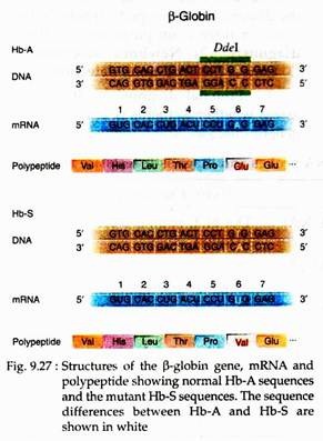
In sickle-cell anaemia, an AT base pair is changed to TA base pair, so the sixth codon for β-globin is changed from GAG to GUG. As a result of this SNP allele (single nucleotide polymorphic), valine instead of glutamic acid is inserted into the polypeptide (Fig. 9.27).
This mutation change also generates an RFLP for the restriction enzyme Ddel which is:
5′ – CTNAG – 3′
3′ – GANTC – 5′
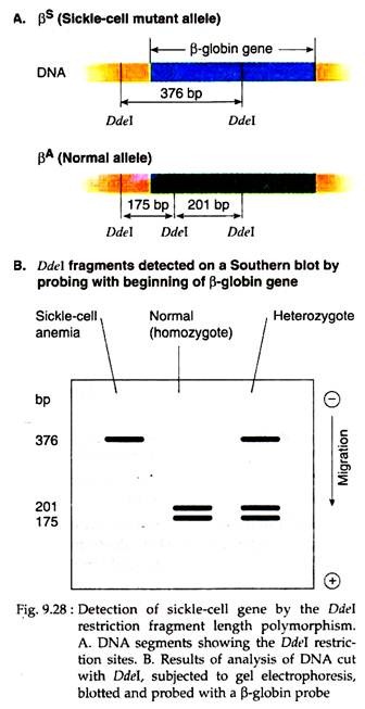
Here the central base pair may be any of the four possible base pairs. The AT to TA mutation changes the fourth base pair in the restriction site. As a result, in the normal β-globin gene, PA, there are three Ddel sites (Fig. 9.28A), but in the sickle-cell mutant β-globin gene, βs, the mutation removes the middle Ddel site (Fig. 9.27), leaving only two Ddel sites. For testing normal individual’s DNA is cut with Ddel and resulting fragments are separated by gel electrophoresis.
It is then transferred to a membrane filter by Southern blot technique and then probed with the 5′ end of a cloned β-globin gene. Two fragments of 175 bp and 201 bp are seen (Fig. 9.28B). When DNA from sickle-cell anaemia patients are analysed in the same way, only one fragment of 376 bp can be seen because of the loss of the Ddel site. However, heterozygotes (carriers) can be detected by the presence of three bands, of 376 bp, 201 bp and 175 bp.
In this way, RFLP helps to detect genetic diseases. Some RFLPs result from changes to the DNA flanking the gene (e.g., Phenyl ketonuria) and sometimes recombination between the RFLP and the gene of interest can occur causing difficulties in the interpretation of the results.
2. Testing by PCR:
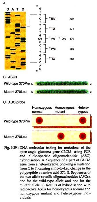
The most common genetic test using PCR is allele specific oligonucleotide (ASO) hybridisation. In open-angle glaucoma, the GLCIA (one of several genes responsible for glaucoma) is mutated. The GLCIA gene has been sequenced and primers were designed for PCR amplification of the region of gene containing the mutation. One of the mutations involves a change from CG to TA in the DNA, resulting in a codon change from CCG (Pro) to CUG (Leu) (Fig. 9.29A).
DNA samples from glucoma patients, normal individual and carrier were amplified by PCR, then separated by agarose gel electrophoresis, transferred from the gel and dotted onto two membrane filters under conditions that denatured double stranded DNA to single stranded. On the other hand, ASOs were made both from the normal and mutant alleles (Fig. 9.29B) keeping the mutation approximately in the middle.
Following radioactive labelling, each ASO was hybridised with the DNA immobilised on one of the filters. The resulting autoradiograms indicated which DNA was from glucoma patient, which from normal or heterozygous individual (Fig. 9.29C). Thus, PCR is used to analyse affected members of glucoma families for the presence of particular mutations.
Another approach called reverse ASO hybridisation PCR is useful to detect the disease that is caused by numerous mutations in the gene. This technique uses radiolabeled PCR product as a probe for hybridisation with many different ASOs bound to a membrane filter. In cystic fibrosis, there are hundreds of mutations in the gene (F).
For detecting this disease, multiplex PCR is used to amplify several regions of the gene in DNA samples from patients. The resulting PCR products are labeled radioactively and hybridised with wild type or mutant oligonucleotides bound to membrane filter.
The resulting autoradiograms indicate the dot to which PCR product binds was of patient’s DNA. It also indicates whether the individual is homozygous or heterozygous for the mutant allele.
Treatment of Diseases:
A. Drugs:
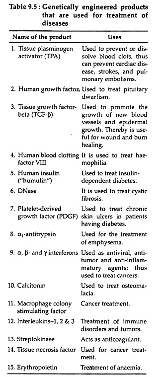
Cloning and other DNA manipulation techniques are abled to yield a variety of medically useful substances (Table 9.5). Some of such substances previously only could be obtained from human tissues. For example, human growth hormone for the treatment of pituitary dwarfism was extracted from pituitary obtained during autopsites and thus was available only in limited quantities.
But now, with the help of genetic engineering, the gene encoding growth hormone has been cloned and expressed and thus could be used for synthesis of growth hormone in a large scale. In the same way insulin could be produced.
Production of insulin by recombinant DNA technology:
Insulin is a protein hormone, synthesised by the β cells of islets of human gene product manufactured using recombinant DNA technology and licensed for therapeutic use was human insulin. The functional insulin protein contains two polypeptide chains, A and B.
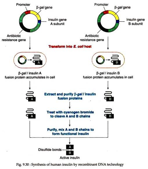
The A has 21 amino acids and the B subunit consists of 30 amino acids. Synthetic genes for the A and B subunits were constructed by oligonucleotide synthesis (63 nucleotides for the A chain and 90 nucleotides for the B chain). Each such synthetic oligonucleotide was inserted into a vector at a position adjacent to a gene encoding the bacterial form of the enzyme, β-galactosidase gene.
Therefore, when transferred to bacterial host, the β-galactosidase gene and the synthetic oligonucleotide were transcribed and translated as a unit. The product was a fusion polypeptide, consisting of the amino acid sequence for β-galactosidase attached to the amino acid sequence for one of the insulin subunits (Fig. 9.30).
The fusion protein then purified from bacterial extracts and was treated with cyanogen bromide which cleaved the fusion protein from the β-galactosidase. The two subunits of insulin thus produced, when mixed together, unite spontaneously forming an intact, active insulin molecule. It then can be used for the treatment of diabetic patients.
Similarly, a number of genetically engineered proteins with therapeutic importance have been produced.
Production of human protein of phermaceutical importance by second generation eukaryotic host:
Sometimes bacterial cells are unable to process and modify eukaryotic proteins which may require addition of sugars and phosphate groups for their full biological activity. To overcome this difficulty and to increase the yields, second- generation methods now use eukaryotic hosts.
In the heritable, fatal respiratory disorder emphysema, a deficiency in the synthesis of the enzyme α-1-antitrypsin is found. To synthesise this enzyme by genetic engineering, the human gene encoding this enzyme was cloned into a vector at a site adjacent to a sheep promoter sequence that regulates the expression of milk-associated proteins.
As a result this gene will be expressed only in mammary tissues. Now the fusion gene was microinjected into sheep zygotes fertilized in vitro, which, in turn, implanted into foster mothers. Following mating, this transgenic sheep produced milk that contained high concentration of functional human α-1-anti- trypsin. It was found that its concentration was up to 35 g/litre of milk.
Similarly pig embryos injected with human haemoglobin gene develop into transgenic pig that synthesise human haemoglobin. Current plans are to purify the haemoglobin and use it as a blood substitute. A pig could produce 20 units of blood substute per year.
Similar techniques have produced transgenic goats whose milk contains up to 3 gm. of human tissue plasminogen activator that dissolves blood clots and is used to treat cardiac patients. In this way herds of different transgenic animals can act as bio-factories and might become part of pharmaceutical industry.
B. Vaccines:
Another most beneficial application of recombinant DNA technology is the production of vaccines that could prevent a number of diseases. Vaccines stimulate the immune system to produce antibodies against a disease-causing organism and thereby confer immunity against that disease.
Vaccines may be of different types and these can be prepared variously. However, the advent of recombinant DNA and monoclonal technologies coupled with the recent advances in immunology has opened a new era into the production of vaccines.
Some of such vaccines are:
(i) Subunit vaccine:
These vaccines consist of one or more surface proteins of the virus or bacterium. These proteins act as antigen U. stimulate the immune system to make antibodies against the virus or bacterium. For example, the first licensed subunit vaccine was for a surface protein of hepatitis B virus which causes liver damage and cancer.
The gene for the hepatitis B surface protein was cloned into a yeast expression vector and produced using yeast as a host. The protein following extraction is purified from the host cells and used as vaccine.
(ii) Edible vaccines:
Recombinant DNA technology is currently being used to produce hepatitis B surface protein in edible plants. Usually plant leaves or fruits serve as the source of an oral vaccine which would be less expensive and would not require injection. In a model system, the antigenic subunit of hepatitis B vaccine has been transferred to tobacco plants and expressed in the leaves.
Leaves from such transgenic plants were treated with antibodies against hepatitis B antigen and was shown to produce the antigens. Similar tests are underway using genetically engineered potatoes, bananas etc. Thus, genetically engineered edible plants are used to vaccinate people against many diseases.
(iii) Genetically engineered vaccinia virus:
With the help of genetic engineering methods, vaccinia viruses are now being used as a popular vaccine expression system. The vaccinia genome is very large and it is possible to delete large quantities of its genome and still maintain a viable virus. The non-essential vaccinia genes can be replaced with a number of genes coding for other proteins from other viruses.
Recently, this system has been used in case of wildlife rabies, where vaccinia virus containing the rabies virus glycoprotein is incorporated in bait and seeded in rural areas by dropping the bait from planes. Foxes and raccoons, which can be carriers of this virus, would eat the bait and be immunized against this virus. Using this approach, vaccines against HIV are being produced using the gene for HIV-1 envelope glycoprotein.
Examples of other genetically engineered vaccines include:
(i) Foot-and-mouth disease vaccine which is produced through cloning the vaccine genes into E. coli.
(ii) E. coli vaccines for pigs with extra plasmids encoding the antigen for k88 strain of E. coli;
(iii) Vaccines for malaria etc.
3. Gene therapy:
Methods for the introduction of specific genes into mammalian cells, originally developed as a research tool, are now being used to treat genetic disorders. This process is called gene therapy. From the above discussion it appears that recombinant DNA techniques are playing an important role in research on the molecular basis of different diseases.
The transfer from research to practical application often is slow because it is very difficult to treat a disease effectively unless its molecular mechanism is known. Thus, genetic engineering may aid in the fight against a disease by providing new information about its nature, as well as by aiding in diagnosis and therapy.
