In this article we will discuss about the reproductive system of balanoglossus with the help of suitable diagrams.
Sexual Reproduction:
In Balanoglossus, the sexes are separate and are indistinguishable externally except in case of the colour of the ripe gonads shown through the body wall in the living animal. The gonads occur in one or more longitudinal rows to the sides of the alimentary canal lying within the genital pleurae in the anterior part of the trunk. Gonads develop from the coelomic wall, though they have no connection with coelom in the adult.
The gonads are generally sacciform bodies but may be elongated or lobulated and secondary gonads may arise by subdivision of the primary ones through lobulation. Each gonad is a sac, it continues into a short ductule which opens to the exterior by a gonopore. The gonopores are generally located to the lateral (external) side of the gill-pores in the same branchiogenital groove.
The saccular gonads are lined with germinal epithelium which is continuous with the ectoderm. By the proliferation of cells from the germinal epithelium sperms or ova are produced. The mature sperms and ova are discharged outside through the genital pores.
ADVERTISEMENTS:
The sperm has a rounded head and a flagella-like tail but the ova are of two types. The small ovum measures about 0.06 mm in diameter and undergoes indirect development with a pelagic larva known as tornaria larva, while the larger ovum measures about 0.4 mm in diameter and undergoes direct development without larval stage. Fertilisation is external.
Development:
In Balanoglossus, during sexual reproduction zygote develops either directly or indirectly depending on the size. The small fertilised eggs develop indirectly but large fertilised eggs develop directly. The indirect development is through a larval form called tornaria larva. In both the cases early phases of development are same.
Fertilisation:
ADVERTISEMENTS:
During breeding season (May to June) mature ova and sperms are discharged in the surrounding water where fertilisation takes place. First the ova, egg-mass, are discharged by the female from its burrow and then the sperms are discharged by the male from its burrow. The number of eggs discharged at a time varies from few dozens to more than a thousand. Normally one to three hundred eggs are shed at a time.
According to available evidences maturation starts some four hours before ovulation and that the egg is generally in the metaphase of the first meiotic division when shed. It is at this condition the egg is fertilisable. Fertilisation of eggs within 6 to 7 hours after shedding yields a high percentage of normal development. The spermatozoon is able to enter the eggs at any point over the entire surface. Thus, fertilisation is external. After fertilisation, the cleavage starts.
Indirect Development:
Indirect type of development is studied in Balanoglossus clavigerus by workers like Heider (1909), Stiansy (1914) and Payne (1937).
ADVERTISEMENTS:
Embryonic or Prelarval Development:
Cleavage:
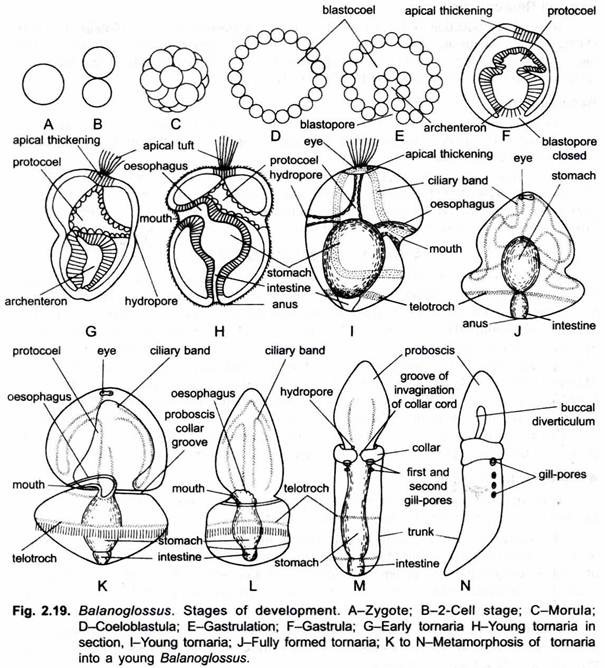
The zygote, produced as a result of fertilization, undergoes cleavage. The cleavage is holoblastic, almost equal and mostly of the radial type. The first cleavage starts about two hours after fertilisation and produces two generally, but not invariably, equal cells.
The second cleagave is like the first and produces usually (but not invariably) four approximately equal cells. As a result of third and subsequent cleagaves a sphere of equal blastomeres is produced, it is called morula.
The morula undergoes the reorganisation of its blastomeres and takes the form of a single-layered hollow and spherical blastula or coeloblastula. Its central fluid-filled cavity is called the blastocoel. As the cells multiply the volume of blastula increases. Blastula results in about 6-15 hours after fertilization. Within 12-24 hours, an invagination starts in the blastula which deepens to form the archenteron. The archenteron opens to the outside through a blastopore. The blastopore marks the posterior end of the embryo.
The blastopore soon closes and the embryo now called gastrula. The gastrula elongates along the antero-posterior axis. Now the anterior tip of the archenteron is differentiated as a coelomic vesicle called the protocoel. Thus, origin of coelom is enterocoelic. The remaining posterior part of the archenteron marks the future gut or alimentary canal. The protocoel becomes triangular in shape.
One end of the protocoel gets attached to the underside of the apical thickening and another end opens to outside through an aperture, the hydropore, towards the dorsal side of the embryo. The protocoel and hydropore represent the future proboscis coelom and proboscis pore respectively. The collar and trunk coelom develop as solid invaginations of the hindgut, independent of the formation of protocoel.
Larval Development:
After the formation of the protocoel, the inner end of the early gut moves towards the ventral surface and opens to the outside through a mouth. The gut is now differentiated into the oesophagus, stomach and intestine. The intestine opens to outside through an anus, formed at the place of closed blastopore. By this time (after a day or so) the embryo becomes uniformly ciliated and escapes from fertilisation membrane to lead a free swimming larval life. It is called tornaria larva.
ADVERTISEMENTS:
Tornaria Larva:
Tornaria larva was first of all discovered by J. Muller in 1850 and was considered by him as the larva of echinoderms. Later on in 1869 it was Metschnikoff who established that it is a larva of Balanoglossus clavigerus. The name tornaria is given to it because of its habit of rotating in circles. Tornaria larva is usually oval in shape and is excessively transparent. The size of tornaria larva varies from 1 mm to 3 mm.
It has a ventral mouth near the equatorial plane of the body, a posterior terminal anus and gut differentiated into an oesophagus, stomach and intestine. The cilia form two bands on the body surface. The anterior ciliary band or circumoral band takes up a winding course over the preoral surface and forms a postoral loop. Its cilia are short and serve to collect the food. The posterior ciliated band or circumanal ring or telotroch lies as a ring in front of the anus.
The cilia in this band are long, powerful and act as chief locomotor organ of tornaria. A ciliary wave passing along the telotroch causes the larva to rotate constantly in swimming. At the anterior end is an apical plate of thickened epidermal cells. The apical plate bears a pair of eye spots or ocelli and a tuft of sensory cilia called apical tuft or ciliary organ.
The protocoel (proboscis coelom) is present in the form of a thin-walled sac and opens to the exterior through a hydropore (proboscis pore). To the right of the hydropore lies a pulsating heart vesicle which develops in the later stages of tornaria larva. The collar and trunk coeloms appear in the older larva.
Metamorphosis:
The tornaria larva swims freely, leads a planktonic life feeding on minute organisms. After swimming for some time the tornaria larva sinks to the bottom and metamorphoses into an adult. During metamorphosis, the size of larva is reduced probably due to loss of water. Transparency, ciliary bands, sensory cilia and eye spots are lost.
The body elongates and is distinguished into proboscis, collar and trunk by the appearance of two constrictions, and the trunk region is elongated. The hydropore persists as proboscis pore. Simultaneously the buccal diverticulum and gill-slits appear as outgrowth of the alimentary canal. The reproductive organs make their appearance, probably from the mesoderm. Thus, the larva gradually changes into the adult. The adult leads a benthonic life.
Direct Development:
Direct development is found in the genus Saccoglossus. It is found in the large eggs provided with good amount of yolk. The embryo elongates in the fixture antero-posterior axis, develops cilia, and after rotating for a time inside the egg membrane, escapes at about 24 to 30 hours and leads a planktonic existence for a time. It is provided with an apical tuft of cilia and the telotroch. Saccoglossus kowalevskii, however, does not hatch until the seventh day and at once takes up a benthonic life.
After hatching the larva swims for a short time (24-26 hours) and soon elongates and develops in equatorial constriction marking the proboscis-collar boundary. The second constriction marks the collar-trunk boundary. The larva soon elongates and leads a benthonic life. It looses its apical tuft of cilia and the telotroch (posterior band of cilia). The larva now becomes worm-like juvenile Balanoglossus.
Asexual Reproduction:
Asexual reproduction is known to occur in Balanoglossus capensis. During summer the juvenile phase of this, at first considered a distinct species for it lacks hepatic sacculations, reproduces by cutting off small pieces from the tail end forward. These regenerate completely into the adult sexual type found in winter.
Regeneration:
Balanoglossus has great power of regeneration, small pieces are constricted from the posterior end, each of which regenerates into a complete individual. Other broken pieces of the animal also regenerate into new individuals.
A detailed study of regeneration was made by Dawydoff (1902, 1907, 1909) for Gloss, minutus and by Rao (1955) for Ptychodera flava. The isolated proboscis, with or without the collar, lines and moves about for some time but appear incapable of regenerating posterior structures. Pieces of trunk regenerate completely in both species.
