In this article we will discuss about:- 1. Description of Sponges 2. Structure of Sponges 3. Canal System 4. Histological Elements 5. Reproduction and Development 6. Commercial Sponge 7. Classification.
Contents:
- Description of Sponges
- Structure of Sponges
- Canal System of the Sponges
- Histological Elements Constituting the Wall of Sponges
- Reproduction and Development in the Sponges
- Commercial Sponge
- Classification: Phylum Porifera or Sponges
1. Description of Sponges:
The Porifera (L. parous = pore + ferre = bear) or sponges are lowly organised group of plant-like sessile animals remaining attached to the substratum. Of the described sponges, about 5,000 species are marine and very few are fresh water. They grow mainly in shallow waters but their habitat may extend up to a depth of 5,600 metres.
Sponges are of varying shapes, sizes and colours. They are distinct from the Protozoa in having a cellular construction but the cellular grade is at the lowest. Absence of true tissues and extensive system of pores and canals powered by the action of peculiar flagellated cells, the choanocytes, make them distinct from other metazoan.
ADVERTISEMENTS:
Very often they are considered as a subkingdom, Parazoa. Sponges are distributed in all seas—from the equator, to the poles.
Sycon, recently named Scypha, is the simplest form of sponge, marine in habitat. It remains attached to sea shore rocks just below the tide line. The colour of a live specimen is a combination of shades of grey and light brown. Scypha belongs to subclass Galcoronea, class Calcarea, phylum Porifera.
2. Structure of Sponges:
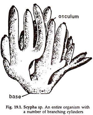
The Scypha is either a simple solitary cylinder or a number of cylindrical branches (Fig. 19.1) connected together to form a colony. Solitary or colonial, it is anchored to rock by a structure; called stolon at the base. The cylinder is vase-shaped, swollen at the middle and narrows down at both the ends, being a little more narrow at the free or distal end.
ADVERTISEMENTS:
Due to protrusion of numerous monaxon spicules from the surface, it appears bristly.
Osculum:
The osculum or the exhalant pore is a wide opening, present at the free end of the cylinder. It establishes direct communication between the Para gastric cavity or the spongocoel and the exterior. The osculum is surrounded by numerous straight, monaxon, calcareous spicules arranged in a circlet, imparting the appearance of a delicate fringe to it.
Dermal pores:
The body surface bears numerous regularly arranged, polygonal elevations separated by depressed lines or furrows. Numerous microscopic apertures are present in the furrows. These are Ostia or inhalant pores or dermal pores leading to the incurrent canals.
3. Canal System of the Sponges:
ADVERTISEMENTS:
It is evident from the term ‘porifera’ that the surface of the body bears a large number of pores, minute in size and inhalant in function. These pores open into a system of channels, which, after penetrating almost all the portions of the body, open to the exterior by an opening known as osculum, at the tip of the branch.
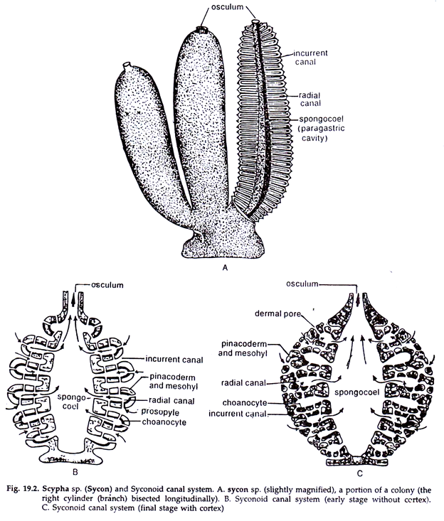
All the canals are collectively called canal system. The canal system in Scypjia (Sycon) is known as syconoid type. It establishes a continuous passage for the inflow and outflow of water within the body of a sponge (Fig. 19.2).
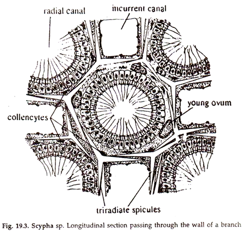
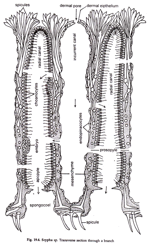
The following structures are found in association with the canal system of Scypha. (Fig. 19.3, 19.4):
ADVERTISEMENTS:
1. Spongocoel:
At the free end of a branch, a fairly large aperture known as osculum is present. Osculum leads internally into a canal, the spongocoel, which runs along the middle of the branch and receives a large number of ex-current canals of the branch.
The Para gastric cavities or spongocoels of different branches in a colonial form, are in communication with one another through a broad chamber at the base. The cavity is lined by flattened cells of ectodermal origin, the pinacocytes.
2. Incurrent Canal:
The surface of each branch of Scypha is provided with alternate elevations and depressions. At each depression a group of minute pores known as inhalant pores or Ostia are present. Ostia lead internally into the incurrent canal. The canal is dilated towards the outer end and narrower and blind at the inner end. It is lined by flattened cells of ectodermal origin, the pinacocytes.
3. Radial Canal:
Lying alternate and parallel to the incurrent canals are the radial canals. The radial canals are situated opposite to the points of elevations on the surface. Each canal is narrower and blind towards the outer end but broad and open at the inner end. It is lined by flagellated choanocyte cells of endodermal origin. The radial canals are connected with the incurrent canals by narrow passages known as prosopyles.
4. Ex-Current Canal:
These are short but wide canals. Each canal is situated along the same long axis of the radial canal. It is lined by flattened cells of ectodermal origin. The ex-current canal is connected at the outer end with the radial canal by an aperture, the apopyle, and at the inner end with the Para gastric cavity by a large aperture, the gastric or internal ositum.
Course of Water Current:
Due to the rapid backward movement of the flagella of the cells lining the radial canals, a constant water current is maintained in the canal system. The rate of flow of water can, of course, be regulated by increasing or decreasing the diameter, of the different apertures in the canal system.
Water from outside enters the incurrent canals through Ostia and from there to the radial canals through the prosopyles. It passes from radial canals to ex-current canals through the apopyles and from there to the spongocoel through gastric Ostia and, thence, to the exterior through the osculum.
Role of the Canal System:
The canal system serves various functions:
1. It serves the purpose of nutrition. The food such as diatoms, protozoans, etc., are carried by the water current and reach the radial canals where they are picked up and digested by the flagellate cells.
2. Oxygen for respiration is carried by the streaming current of water.
3. It functions for excretion. Current of water, passing out of the osculum, also remove the carbonic acid and other nitrogenous waste matters.
4. The outward current takes away the reproductive units from the body of the sponge.
5. The complicated canal system also increases the surface area of the animal which is directly exposed to water.
4. Histological Elements Constituting the Wall of Sponges:
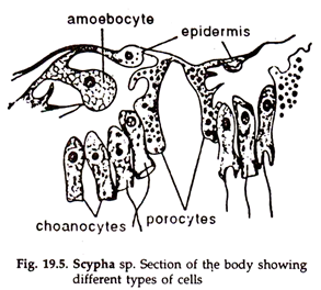
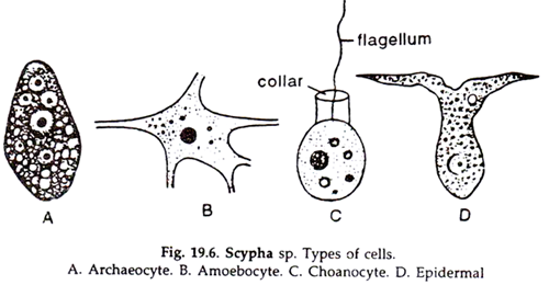
The different microscopic elements constituting the body wall of Scypha are known as histological elements. The elements may be divided into three groups—cell elements, skeletal elements and mesenchymal substance (Figs. 19.3-19.6).
1. Cell Elements:
The cells of the sponges are fairly differentiated, but, excepting choanocytes, others seem to be only modified forms of undifferentiated amoeboid cells corresponding to the primitive connective tissue cell of higher animals.
Following different types of cells are found in the body wall of Scypha (Fig. 19.5-19.6):
a. Ecotodermal cells:
These are flattened scale-like cells with inconspicuous nuclei and their edges are closely cemented together to form an epithelium. The cells of the dermal layer covering the outer surface of the sponge are known as pinacocytes.
Each pinacocyte has a thickened central bulging containing a nucleus. Some pinacocytes (endopinacocytes) also line the incurrent canals and spongocoel. They are highly contractile and those lining the incurrent canals are called skeletogenous cells.
b. Endodermal cells:
These are columnar cells, each with a large nucleus, one or more vacuoles and bears flagellum at the inner end, the base of which is surrounded by a delicate, transparent, collar-like up growth. Such cells are known as choanocytes and are restricted to the radial canals.
c. Myocytes:
These are fusiform contractile cells around the ostia, prosopyles and apopyles effecting the closure of the apertures, and are usually arranged in a circular fashion to form a sphincter.
2. Mesenchyme Substance:
The mesenchyme consists of a gelatinous, transparent matrix generally known as mesogloea, supposed to be protein in nature. It affords rigidity to the animal. The mesogloea contains—to some extent—free wandering cells or amoebocytes.
Following types of amoebocytes are found in Scypha:
a. Amoeboid wandering cells:
Amoeba-like and can move from place to place. They are concerned with the nutrition of the animal.
b. Collencytes:
The cells bear slender branching pseudo- pods and connect the different elements of the body. If bipolar, collencytes are termed desmacytes or fibre cells.
c. Chromocytes:
These are with lobose pseudopods and contain pigments. The colouration of the sponge is dependent on these pigments.
d. Thesocytes:
Cells with lobose pseudo- pods and store reserve food materials.
e. Scleroblasts:
The cells secrete the skeletal elements or spicules.
They are of two types:
i. Calcoblasts:
Amoebocytes secreting calcareous spicules.
ii. Silicoblasts:
Amoebocytes secreting siliceous spicules.
f. Reproductive cells:
These are modified amoebocytes and possibly also modified choanocytes.
g. Porocytes:
Also known as pore cells, they are tubular and with a central canal acting as an incurrent passage. The pores can be closed by a thin cytoplasmic sheet, the pore diaphragm. Porocytes are possibly transformed pinacocytes. The mesenchyme is responsible, after all, for one of the most important characters in sponges, that is, secreting the skeletal elements.
3. Skeletal Elements:
These are hard structures connected together in such a way as to support and protect the soft parts of the body. These structures are pointed and are made of calcium carbonate and known as spicules. Spicules develop from the scleroblasts and one scleroblast is required to form each arm of a spicule.
The spicules may be needle-like or club-shaped and range from one to many axes,—or monaxon to polyaxon,—of which triaxon is the most abundant type (Fig. 19.4) There are spicules where the body is round and the growth is concentric.
These are known as spheres while the crepis—on being deposited with layers of silica in an irregular fashion is termed Desma. The club-shaped spicules projecting on the outer surface beyond the ectoderm are known as oxeote spicules.
Spicules are responsible for the framework of Scypha. Although the primary function of the calcareous skeleton is to support, it also serves to buffer the mesenchyme against any drop in pH that could cause hardening of the ground substance.
5. Reproduction and Development in the Sponges:
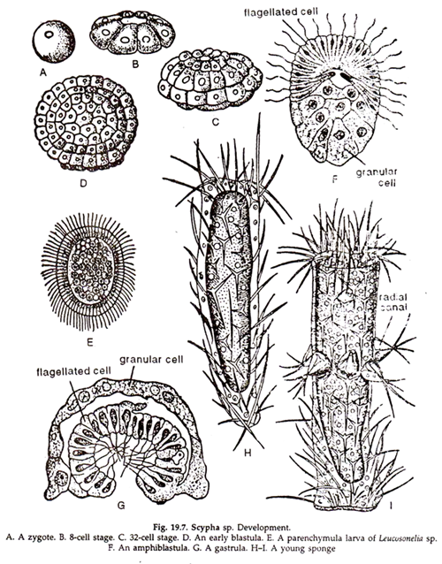
Reproduction in sponges takes place both by asexual and sexual means (Fig. 19.7):
A. Asexual Reproduction:
It is effected by internal buds or gem- mules. Some of the amoebocytes come to lie in the mesogleal layer around the lining cells of the radial canals, and after repeated division a mass of cells or the gemmule is formed. This process is known as gemmation.
A gemmule cannot escape to the outside until the branch in which it grows is separated from the main body of the sponge and lost. For this reason, gemmules are formed in older branches. By the time of its liberation the; gemmule is transformed into an amphiblastula, the subsequent development of which is like that of the same in sexual reproduction.
B. Sexual Reproduction:
Sponges are monoecious, i.e. ova and sperms develop in the same individual. Sexual reproduction is effected by the formation of the archaeocytes (specialized amoebocytes). Sex organs are absent. The mesenchyme around the gastral layer of the flagellated chamber is the seat of the sex cells.
Spermatozoon is with a round head and a long tail. Spermatozoa from another sponge are carried to the radial canal with water current. The ovum is large and round and remains attached to the maternal tissue. Fertilization takes place within the body.
The fertilized egg of Scypha divides vertically and eight concial cells are formed. These cells then undergo horizontal cleavage, with the result the eight long cells and eight small cells are formed enclosing a blastocoel.
The long cells are destined to form the future epidermis while the small cells form the future choanocytes. The eight small cells increase in size, elongate, and each acquires a flagellum on its inner side. The larger cells do not divide, instead, they assume a round form and become granular.
The mouth opening appears at their middle which absorbs the neighbouring cells. This stage of the blastula is known as stomoblastula. The blastula-undergoes inversion and the flagellated cells are brought outside.
The embryo at this stage is known as amhiblastula (Fig 19.7F). This is a typical calcareous larva. It escapes from the parent and has two types of cells—the small narrow flagellated cells and the large round granular cells.
After a brief period of free existence which may last for a few to several hours, the larva undergoes gastrulation, in which the flagellated half is invaginated into or overgrown by the large granular cells (Fig. 19.7G.).
It attaches itself to some solid object by the blastoporal end and is gradually transformed into a narrow cylinder and then to a small sponge. The flagellated cells become the collared cells, while the granular cells become numerous and form the dermal epithelium.
The mesenchyme cells are formed from both the layers. Gradually, the cylinder (larva) increases in thickness. With the growth of the intermediate layer the radial and other canals gradually appear.
6. Commercial Sponge:
Sponges are mostly beneficial to man. Skeleton of some sponges are used to manufacture commercial sponge, which is of great economic importance. The sponging skeleton is treated with hydrochloric acid and the spicules dissolve.
The residue left after the acid treatment is further subjected to chemical treatment, and the skeleton becomes marketable as the ‘bath sponge’. Sponges are extensively used in bath rooms, laboratories and in surgical operations.
7. Classification: Phylum Porifera or Sponges:
1. Sponges are usually small, plant-like animals that are radially symmetrical or irregular in shape, occurring singly or in colonies.
2. Multicellular body with two loosely arranged and differentiated germ layers and an intermediate gelatinous layer cantaining amoeboid cells and skeletal pieces known as mesohyl or mesenchyme (not truly diploblastic), hardly formed in tissues.
3. There are no organs, head, mouth or gut. The body structure is organised around a system of canals and chambers through which water flows.
4. Flagellated coller cells, or choanocytes, which line the chambers, not only create the water current but also filter out fine food particles.
5. The supporting framework or the skeleton consists of fine, flexible fibres made of proteinous spongin or spongin fibres and siliceous spicules; or siliceous spicules alone; or spicules of calcium carbonate.
6. The body surface is provided with pores and, at the top, a large opening, the osculum. The interior cavity, the atrium or spongocoel opens to the outside through osculum.
7. The nerves and sensory cells are altogether absent; the cells act independently. Most cells can transmit stimuli slowly.
8. Both hermaphrodite and unisexual forms exist. Reproduction occurs asexually by the development of buds and gemmules, and sexually by development of typical ova and sperms; specialised gonads absent; the ova and sperms develop from the cell type archaeocytes.
9. Development direct; the embryo leaves the sponge body as an active, free-swimming ciliated larva, which finally attaches itself to a substratum and undergoes metamorphosis to reach the plant-like adult stage. The larvae are of two types: the Amphiblastula and Parenchymula.
10. The sponges have power of regeneration.
Because of their highly variable characteristics, number of intermediate forms and immense collateral affinities, it is difficult to classify sponges. The peculiarities of the skeleton is the mam basis of classification and the sponges are grouped into four classes accordingly.
Class 1. Calcarea or Calcispongiae:
a. Calcareous sponges; skeleton solely of calcareous spicules which may be one, three or four-rayed and not distinguishable into mega-and microscleres.
b. Body form is asconoid, syconoid or leuconoid.
c. Mostly coloured, size not exceeding 10 cm in height.
Subclass i. Calcaronea:
a. Triradiate spicules usually having one long ray.
b. Choanocyte nucleus is apical in position and the flagellum arises directly from the nucleus.
c. Larvae are ampliblastula. Examples: Leucosolenia, Scypha (Sycon), Grantia, etc.
Subclass ii. Calcinea:
a. Triradiate spicules with equal rays.
b. Choanocyte nucleus is basal in position and flagellum arises independently.
c. Larvae are parenchymula. Examples: Clathrina, Leucetta, Minchinella, Petrobiona, etc.
Class 2. Hexactinellida or Hyalospongiae:
a. Glass sponges; skeleton purely siliceous, composed of six-rayed spicules (hexactines). The skeleton is beautiful, glass-like, hence glass sponge.
b. The internal structure consists of a network, the trabecular net, made of thin strands forming open meshes.
c. Without jelly and osculum, may be covered by a sieve plate of silica.
d. Canal system simple and choanocytes small.
e. Mostly radially symmetrical; strongly individualized; may be cylindrical or shaped like a vase, urn, funnel, attached directly at the base or more usually by way of root tufts of spicules (basal stalk).
f. Exclusively marine, deep-sea sponges occurring at depths of 20-5,000 metres. Examples: Euplectella aspergillum (Venus flower basket of Philippines), Staurocaptus (cup-shaped glass sponge, without roof spicules), Hyalonema (glass-rope sponge), etc.
Class 3. Demospongiae:
a. Either devoid of skeleton or with skeleton made up of spongin fibres alone (horny sponges) or in having combination of both siliceous spicules and spongin fibres.
b. Canal system mostly complicated with usually small rounded flagellated chambers having small choanocytes.
Subclass i. Tetractinomorpha:
a. The body is often radially constructed and cortical or axial development is advanced.
b. Usually lack spongin fibres; having tetraxonid and monaxonid megascleres. Examples: Oscarella (without skeleton), Plakina, Geodia, Tethya, Axinella, Aciculiteos, etc.
Subclass ii. Ceractinomorpha:
a. Extremely variable quantity of spongin present; in most cases spongin fibres incorporate some spicules or detritus.
b. Monaxonid megascleres and sigmoid or chelate microscleres present.
c. Body form mostly flattened with growth and looks like crumb of bread. Examples: Haliochondria, Clathria, Spongilla (fresh water type), Spongia, Aplysilla, Dysidea, etc.
Class 4. Sclerospongiae (Boring Sponges):
A small group of tropical sponges with siliceous spicules and spongin fibres but encased within a solid external skeleton of calcium carbonate. Found within marine caves and tunnels associated with coral reefs. Boring sponges are found throughout the world and play an important role in breaking down coral and shell. Example: Cliona.
