The blood vascular system of rabbit (mammal) consists of a circulatory media, called the blood, channels through which the blood flows, called blood vessels, and a central pumping organ, the heart, which pumps the blood in the blood vessels.
However, like that of the other vertebrates, the blood vascular system of rabbit is of closed type. This system is generally concerned with the distribution of materials (digested food, water, oxygen, hormones, excretory products, etc.) from one part of the body to the other.
Heart:
The heart lies in between the two lungs within the median space, the mediastinum slightly to the left side of the thoracic cavity.
1. External Features:
ADVERTISEMENTS:
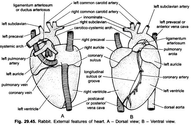
The heart is a pear-shaped, muscular, four-chambered pumping organ. The pointed apex of the heart is directed posteriorly and broad base towards the anterior side.
(i) Pericardium:
Heart is found completely enclosed within a thin-walled, double layered membranous sac, the pericardium. It is connected to the ventral thoracic wall and posterior diaphragm. In between the two layers of pericardium, outer parietal and inner visceral layer is a narrow space, known as pericardial cavity filled with a water pericardial fluid. The pericardial fluid protects the heart from external injuries and allows its free movement.
External Divisions:
ADVERTISEMENTS:
A transverse groove distinctly separates the heart into an anterior smaller auricular part and posterior larger ventricular part. This groove is called coronary sulcus or auriculo-ventricular groove.
(ii) Auricles:
The auricular part consists of right and left auricles. The left auricle is smaller than the right. The sinus venous is absent and merged in the right auricle. The posterior part of each auricle is swollen flap-like, called auricular appendix, slightly covering the corresponding ventricles.
(iii) Ventricles:
ADVERTISEMENTS:
The ventricular part consists of right and left ventricles. The ventricle is divided into right and left parts by an oblique interventricular groove, extending from the top of the heart backwards towards the right but not reaching the apex. Therefore, left ventricle is bigger than the right ventricle and includes the apex of the heart. The conus or truncus arteriosus is absent and merged with the ventricles.
2. Internal Structure:
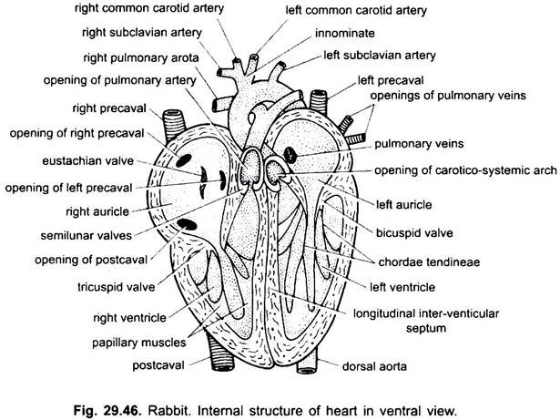
The internal structure of the heart can be easily seen by dissecting it in a longitudinal fashion. Internally the heart is divided into four chambers-anterior two auricles and posterior two ventricles.
(i) Auricles:
The auricles are thin-walled, separated from each other by a thin, muscular and vertical inter-auricular septum. A small oval area, called fossa ovalis, is found on the septum, which has an opening, the foramen ovale in the embryo through which the two auricles are communicated with each other.
The blood goes from right auricle to left auricle without entering in the lungs, which are not yet functional. The foramen ovale is closed in the adults when lungs become fully functional. The inner lining of the auricular wall forms a network of low muscular ridges, which are called musculi pectinati.
The right auricle receives venous blood from all parts of the body except lungs by two superior vena cavae (precavals) and an inferior vena cava (postcaval). All open separately into the right auricle. The opening of postcaval is guarded by a rudimentary eustachian valve. The left auricle receives oxygenated (aerated) blood through the pulmonary veins through a common pulmonary opening. Both the auricles open behind into the ventricle of their side through the auriculo-ventricular aperture.
The right auriculo-ventricular aperture is guarded by right auriculo-ventricular valve, called tricuspid valve made of three triangular flaps or cusps. The left auriculo-ventricular aperture is guarded by a bicuspid or mitral valve made of two flaps.
ADVERTISEMENTS:
(ii) Ventricles:
Like the auricles, the ventricle is also divided completely by an oblique vertical inter-ventricular septum into two chambers, the right and left ventricles. The left ventricle is thick-walled and larger than the right and is circular in shape in transverse section. It also forms the apex of the heart. The smaller right ventricle is crescentic in transverse section.
The inner wall of the ventricles is raised into irregular small ridges, called columnae carneae. Besides these, there are large conical ridges, known as papillary muscles. The free edges of tricuspid and bicuspid valves hanging freely in the ventricle are attached with the papillary muscles or columnae carneae of the ventricle through long, tough connective tissue strands, the chordae tendineae.
The tricuspid and bicuspid valves between right and left auriculo- ventricular apertures respectively allow the passage of blood only in one direction, i.e., from the auricles into the ventricles and not from the ventricles to the auricles.
A pulmonary aorta arises from the right ventricle which carries venous blood to the lungs for oxygenation. The opening of pulmonary aorta into the right ventricle is guarded by three semilunar valves which allow the passage of blood from the ventricle into the aorta and prevent the reflux of blood into the ventricle.
Similarly, a carotico-systemic aorta arises from the left ventricle which carries oxygenated blood to the different parts of the body. Its opening into the left ventricle is also guarded by three semilunar valves allowing the passage of blood only in one direction, i.e., from left ventricle to aorta and prevents the backward flow of blood in the ventricle.
At the place where carotico-systemic aorta crosses the pulmonary aorta, a muscular strand ligamentum arteriosum, is present in adults but during embryonic condition both the aortae are connected with each other by an artery called ductus arteriosus or Botalli.
Working of the Heart:
The heart performs the function of a force pump beating continuously throughout life. It pumps blood into the arteries from the ventricles and also acts as a suction pump by drawing blood into the auricles. During heartbeats, once the auricles contract and the ventricles relax or dilate and then auricles relax and the ventricles contract.
Contraction and relaxation goes on simultaneously. Contraction is called systole and the relaxation is called diastole. The heart muscles are of cardiac type which has the inherent tendency for continuous beating.
The right auricle receives venous or impure blood from different parts of the body through precaval and postcaval veins. The left auricle receives oxygenated or pure blood from the lungs through pulmonary veins. The venous and oxygenated blood are forced into their respective chambers of ventricles by the contraction of the auricles.
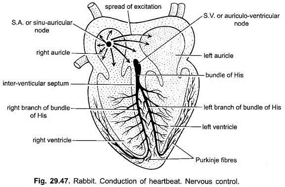
The wave of contraction in the heart originates from sinu-auricular node (SA-node) or pacemaker, which is a group of specialised cardiac muscle cells with nerve fibres. It is situated in the wall of the right auricle between the openings of right precaval and postcaval. It represents the remnants of sinus venosus of lower vertebrates.
The wave of contraction spreads like concentric rings into the two auricles and, thus, both the auricles contract at the same time. At the same time the tricuspid and bicuspid valves of the right and left auriculo- ventricular apertures respectively are forced to open towards the ventricles and, thus, forcing the venous blood from right auricle into the right ventricle and oxygenated blood from left auricle into the left ventricle.
The auricles then relax and another wave of contraction begins from the proximal end of the ventricles. This wave of auricular contraction travels over a bridge called the auriculo- ventricular node (AV-node). It is situated on the lower end of inter-auricular septum and is made of specialised cardiac muscle fibres. From AV-node arises the bundle of His which bifurcates in the inter-ventricular septum into auriculo-ventricular bundles. Their fibres called Purkinje fibres ramify into the ventricular wall.
Wave of contraction from SA-node stimulates the AV-node, which do not immediately sends impulses of contraction to the Purkinje fibres. Thus, ventricles contract, a little time later than the auricles. When the ventricles begin to contract the right and left auriculo- ventricular apertures are closed by their tricuspid and bicuspid valves. Therefore, blood cannot go back into the auricles.
The semilunar valves of the aortic arches are still closed and the blood is blocked in all directions, finally resulting into an increase in the blood pressure inside the ventricles. When this pressure is more than those of the arteries, the semilunar valves of the aortic arches open and the blood is forced in them.
Thus, the oxygenated blood from the left ventricle goes into the carotico-systemic aorta for the distribution in the different parts of the body and venous blood from right ventricle goes into the pulmonary aorta carrying it into the lungs for oxygenation.
Then the ventricles relax and the auricles are again filled with blood, similar wave of contraction starts again after a very short rest or pause. This action goes on continuously as long as the animal is alive. A complete contraction (systole) and relaxation (diastole) of the heart constitutes a heartbeat or heart cycle.
Double Circulation:
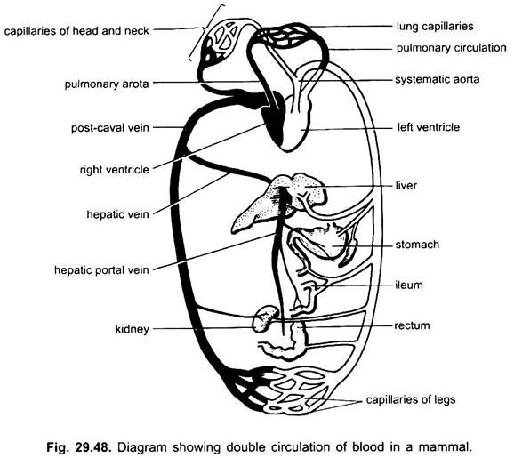
Now it is clear from the above descriptions that there remains a complete separation of venous and oxygenated blood in a mammalian heart. The right part of the heart is concerned with venous blood and the left part with the oxygenated blood. In one complete circuit in the body, the blood passes twice through the heart, once through its right side and then through its left side. This type of circulation of blood is known as double circulation. The complete separation of ventricles into two chambers enables the venous and oxygenated blood not to mix.
Course of Blood Circulation:
The circulation of blood in the body parts from the heart can be represented as given below:
Oxygenated blood from lungs → left auricle → left ventricle → carotico- systemic aorta → arteries → arterioles and → arterial capillaries → various organs of the body → venous capillaries and venules (venous blood) → veins → precavals and postcaval → right auricle → right ventricle pulmonary aorta → pulmonary arteries → lungs (for purification) → pulmonary veins → left auricle (oxygenated blood). Thus, the blood passes through the heart twice – once through pulmonary circuit and second time through systemic circulation.
Blood Vessels:
The blood vessels in rabbit are a system of closed channels through which blood flows. The blood vessels are the arteries and veins. The arteries are those vessels which carry blood away from the heart and veins are those which carry blood towards the heart. The arteries, thus, supply blood to the various parts of the body and constitute the arterial system.
The veins similarly collect impure or deoxygenated blood from the different parts of the body and together constitute the venous system. In the closed blood vascular system the artery supplying a particular organ ramify to form arterioles finally forming arterial capillaries, which transform into venous capillaries. Both types of capillaries are united with each other. The venous capillaries unite together to form venules so as to form veins.
1. Arterial System:
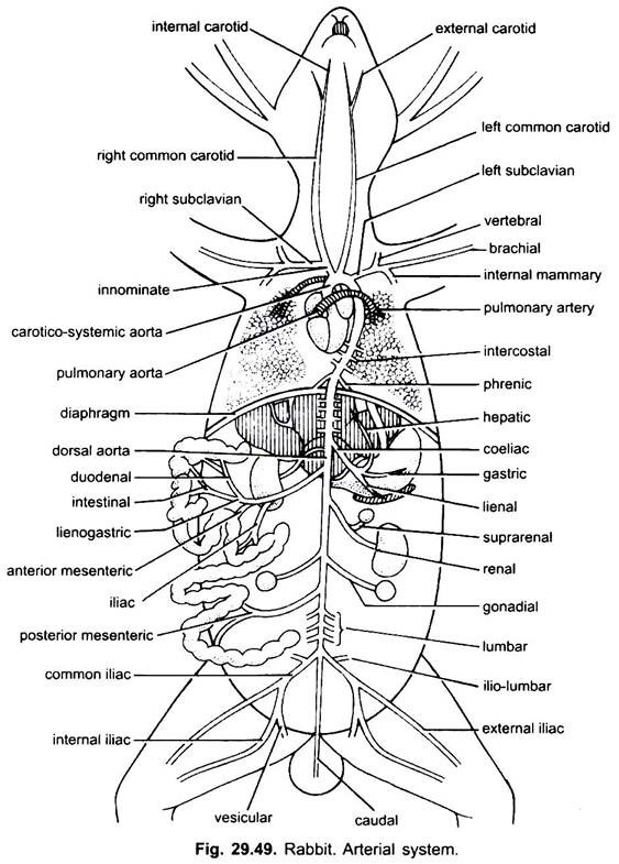
In rabbit, the arterial system consists of two large vessels, called the aortic arches directly originating from the ventricles, known as – (i) the pulmonary aorta and the (ii) the left carotico systemic aorta.
(i) Pulmonary aorta:
The pulmonary aorta originates from the right ventricle and runs dorso-posteriorly and then divides into two- right and left pulmonary arteries, each of them going to the lungs of their sides. The pulmonary aorta close to its bifurcation is connected which carotico-systemic aorta by a vessel called ductus arteriosus in the embryo, but in the adult these are connected by a cord of connective tissue, the ligamentum arteriosum. It is remnant of ductus arteriosus.
(ii) Carotico-Systemic Aorta:
The carotico-systemic aorta originates from the left ventricle and arches towards the left side within pericardium. It gives off two small right and left coronary arteries supplying blood to the wall of the heart. The carotico-systemic aorta now arches to the left and passes ventral to trachea and gives off two main arteries- innominate and left suclavian.
(a) Innominate Artery:
Innominate artery soon divides into right and left carotids which travel anteriorly through the neck along each side of trachea and near the angle of jaws each divides into external and internal carotids. The internal carotid supplies blood to the different parts of the brain and the external carotid supplies to the back of the head, tongue, lips, salivary glands, pinna and muscles of the jaw.
(b) Right Subclavian:
The right subclavian artery originates from the base of the right carotid and soon divides into three main branches- (i) vertebral artery supplying to the vertebral column and brain, (ii) internal mammary artery supplying to the diaphragm, pericardium, mammary glands, etc., and (iii) brachial artery supplying blood in the forelimbs.
(c) Left Subclavian:
The left subclavian artery originates from a little left of the innominate from the aorta and divides into three branches like that of the right subclavian.
iii. Dorsal Aorta:
The carotico-systemic aorta now turns to the left and runs posteriorly above the heart and then runs backward mid-dorsally below the vertebral column as a dorsal aorta. The dorsal aorta pierces the diaphragm and continues in the abdomen up to the tip of the tail. The part of the dorsal aorta in the thorax is known as thoracic aorta, while its part in the abdomen is known as abdominal aorta.
The following arteries arise from the dorsal aorta in the order given below:
(a) A series of paired (5-7) intercostal arteries which supply blood to the ribs.
(b) A pair of small phrenic arteries supplying blood to the muscles of the diaphragm. These two arise from the thoracic aorta. The later ones arise from the abdominal aorta.
(c) A median coelic artery originates just behind the diaphragm which goes to the left side and divides into two- a hepatic artery supplying to the liver and a lineogastric artery supplying to the stomach and spleen.
(d) Anterior mesenteric artery. It arises from the dorsal aorta just behind the coelic artery and divides to form a number of branches supplying to the duodenum, pancreas, ileum, caecum and colon.
(e) Renal arteries. A pair of renal arteries arise from the dorsal aorta supplying to the kidneys. The right renal artery may be situated a bit below to the anterior mesentric. Each renal artery gives off a suprarenal branch to the suprarenal gland.
(f) Gonadial arteries. The paired gonadial arteries arise from the dorsal aorta which supply the gonad of its side. The gonadial artery in male is known as spermatic and in female the ovarian.
(g) Posterior mesenteric artery. The median posterior mesenteric artery originates from the dorsal aorta behind the gonadial and supplies blood to the lower part of the colon and the rectum.
(h) Lumbar arteries. A series of lumbar arteries originating from the dorsal aorta behind the posterior mesenteric supply blood to the dorsal body wall.
(i) Common iliac arteries. The dorsal aorta then divides into two common iliacs each supplying blood to the hindlimb of its side. Each common iliac gives off an iliolumbar for the dorsal abdominal wall. Then, each common iliac divides into internal iliac and vesicular supplying to the organs of the pelvis, e.g., urinary bladder, uterus, rectum, anus, etc., and an external iliac which continues as femoral artery supplying to the hindlimbs.
(j) Caudal artery. The caudal artery supplies blood to the tail region. It may arise mid-dorsally from the dorsal aorta, a little in front of its bifurcation.
2. Venous System:
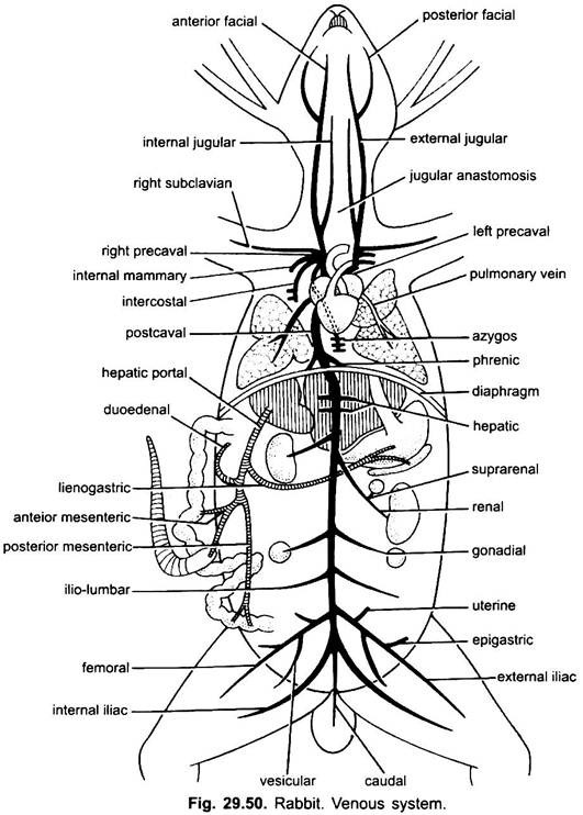
The blood supplied from the heart in the different parts of the body is collected by the veins as deoxygenated blood and finally poured into the heart. The venous system consists of a pair of pulmonary veins, right and left precavals and a postcaval vein, coronary veins and hepatic portal system.
a. Pulmonary Veins:
The pulmonary veins bring oxygenated blood from both the lungs. Both of them unite to form a common pulmonary vein which opens in the left auricle by a single opening.
b. Precaval Veins or Right and Left Anterior Vena Cavae:
The anterior right and left precavals collect blood from the anterior regions of the body, e.g., head, neck, shoulders, forelimbs and thoracic wall.
Each precaval is formed by the union of following branches:
(i) External Jugular Veins:
The external jugular veins collect blood from the head, tongue and shoulder region through anterior and posterior facials, deep external jugular and cephalic veins. Both the external jugulars are long veins running through the neck and are connected together by a transverse jugular anastomosis.
(ii) Internal Jugular Veins:
The internal jugular veins collect blood from the brain and occipital region of skull. The internal jugulars are connected with the external jugulars posteriorly.
(iii) Subclavian Veins:
The subclavian veins collect blood from the shoulder and forelimbs and are connected with the external jugulars.
(iv) Internal Mammary Vein:
It drains blood from the mammary glands and joins the precavals.
(v) Anterior Intercostal Vein:
The right precaval also receives an anterior Intercostal vein collecting blood from the anterior intercostal spaces.
(vi) Azygos Vein:
An azygos vein brings blood from the posterior intercostal spaces (i.e., posterior 8 or 9 ribs) and lumbar region. The azygos and intercostal veins are not found in the left precaval but a few small veins on the left side form a hemiazygos vein which meets with the azygos vein of the right side through transverse anastomosis.
(vii) Coronary Veins:
A pair of small coronary veins collecting blood from the wall of the heart open in the left precaval only.
c. Postcaval vein:
The postcaval vein is a large, median vein formed by the union of a number of veins from the abdominal region, caudal region and hindlimbs. It receives a number of veins running along the corresponding arteries.
(i) Caudal Vein:
It drains blood and marks the beginning of postcaval.
(ii) External Iliac Veins:
A pair of large veins and each is formed by the union of femoral vein collecting blood from the outer side of hindleg, a vesicular vein from urinary bladder, seminal vesicle in male and uterus in female, and a posterior epigastric vein from pubic region.
(iii) Internal Iliac Veins:
These collect the blood from the inner region of thighs.
(iv) Ilio-Lumbar Veins:
They are paired veins bringing blood from the muscles of the back.
(v) Genital Veins:
A pair of gonadial veins bringing blood from the gonads (spermatic and ovarian in male and female respectively).
(vi) Renal Veins:
A pair of renal veins bringing blood from the kidneys (renal portal system being absent in rabbit). Each renal vein receives a suprarenal vein from the corresponding suprarenal gland.
(vii) Hepatic Veins:
Two pairs of hepatic veins arise from the liver lobes.
(viii) Phrenic Veins:
A pair of phrenic veins bringing blood from the diaphragm. These pierce the diaphragm and join the postcaval vein. Thus, the postcaval receiving these veins during its course, travels towards the anterior sides and finally opens in the right auricle.
3. Hepatic Portal System:
The blood from the different parts of the alimentary canal are not directly carried to the heart but collected by the hepatic portal vein which brings blood into the liver.
The hepatic portal vein is formed by:
(i) Lineo-Gastric Vein:
Collecting blood from stomach and spleen.
(ii) Duodenal Vein:
Collecting blood from duodenum.
(iii) Anterior Mesentric Vein:
Collecting blood from the ileum, caecum and colon.
(iv) Posterior Mesentric Vein:
Collecting blood from the rectum and anus.
The hepatic portal vein, thus, enters in the left lobe of the liver where it branches into capillaries. The blood from the liver is collected by hepatic veins which meet with the postcaval.
Blood:
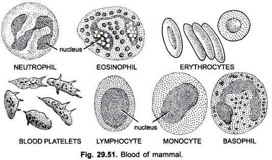
The blood is a fluid connective tissue which flows in the blood vessels. It consists of a fluid matrix the plasma, and formed elements, the blood corpuscles which are found floating in the fluid matrix. These are red blood corpuscles (erythrocytes), white blood corpuscles (leucocytes), and platelets.
The red colour of the blood is due to red blood corpuscles containing haemoglobin, a red coloured iron pigment. It carries oxygen.
The blood of rabbit (mammal) resembles with that of frog in all the structural and functional details but, however, it exhibits certain differences:
(a) The erythrocytes of rabbit are small, nearly 7.5 µ in diameter, circular, biconcave, non- nucleated and more in number, but in frog they are large, nearly 23 -26 µ in diameter, oval, nucleated, biconvex and less in number.
(b) The number of erythrocytes is more in rabbit, e.g., 5 x 10⁶/cu mm but in frog they are less, nearly 4 x 10⁶/cu mm.
(c) The thrombocytes or blood platelets of rabbit are irregular, non-nucleated and more in number, while they are spindle-shaped, nucleated and less in number in frog.
Leucocytes are of two types on the basis of presence of granules in their cytoplasm- granulocytes and non-granulocytes.
Granulocytes are of three types on the basis of the reaction of granules to dyes:
(i) Acidophil (eosinophil),
(ii) Basophil and
(iii) Neutrophil.
Their nuclei are also many lobed. Agranulocytes are lymphocytes with large nucleus and monocytes with saucer-shaped nucleus and abundant cytoplasm. These are phagocytic, destroy the bacteria, etc.
