
In this article we will discuss about the blood vascular system of garden lizard with the help of suitable diagrams.
Heart:
1. External Features:
Heart of the garden lizard (Calotes) is placed in the anterior part of the pleuroperitonial cavity. The heart is enclosed in a thin transparent membrane, the pericardium, which is lined with endocardium, thus, it is two-layered. The space between the heart and pericardium is filled with pericardial fluid. The heart is triangular in shape and three-chambered-two auricles and a ventricle.
The right auricle is larger than the left auricle. Both the auricles are clearly marked from the posterior ventricle by a transverse auriculo-ventricular groove. Truncus arteriosus is absent. The thin-walled and bilobed sinus venosus is reduced and placed transversely and dorsal to the right auricle. It is formed by the fusion of the three venae cavae.
ADVERTISEMENTS:
These three venae cavae empty independently into the sinus venosus. There are no valves. The smaller left lobe of sinus venosus is formed by the left precaval, while the larger right lobe is formed by the union of right precaval and the postcaval. The apex of the ventricle is attached with the liver by a thin, white cord of tissue, the gubernaculum cordis.
2. Internal Structure:
Both the auricles are internally separated by a thin, complete inter- auricular septum. The left auricle is smaller and receives the single pulmonary vein on its dorsal wall. Its aperture has no valve. The right auricle is larger and its inner wall is raised internally into smaller ridges, the musculi pectinati.
Sinus venosus opens into the dorsal wall of right auricle by a semicircular sinu-auricular aperture guarded by sinu-auricular valves. These valves develop from the upper and lower margins of the aperture and their frilled free ends project into the lumen of right auricle.

The ventricle is thick-walled, muscular, spongy and triangular in shape. Its cavity is incompletely divided by an inter-ventricular septum into right and left halves or cavum pulmonale and cavum dorsale respectively. Its inner layer is projected into interlacing ridges called columnae carneae.
Both the auricles open into ventricle through right and left auriculo- ventricular apertures guarded by auriculo-ventricular valves. The flaps of these valves are attached to columnae carneae by thread-like muscles, the chordae tendineae. From the ventricle arise three arches- right and left systemic and pulmonary. The openings of these arches are guarded by paired semilunar valves to check the return of the blood.
Working of Heart:
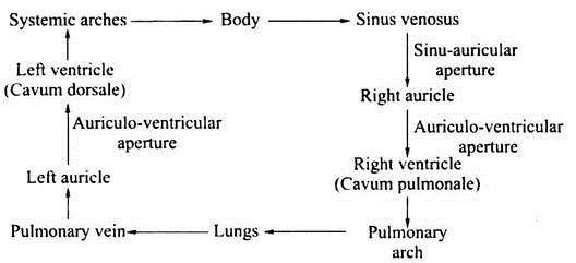
In the garden lizard (Calotes), the circulation of blood is double. The deoxygenated blood of the sinus venosus enters the right auricle through sinu-auricular aperture. The oxygenated blood from the lungs enters the left auricle by the pulmonary vein.
ADVERTISEMENTS:
The left auricle pours its blood into the left part of cavum dorsale of the ventricle through the auriculo-ventricular aperture. The right auricle also pours its blood into the right portion of cavum pulmonale or ventrale of the ventricle through the auriculo-ventricular aperture.
The oxygenated blood from the cavum dorsale goes principally through both carotid arches and the right systemic arch. Some of the right systemic blood may be added via the ductus caroticus to that of the internal carotid.
While the deoxygenated blood from the cavum pulmonale passes into the pulmonary arteries for reoxygenation in the lungs. The blood in the right part of the cavum dorsale goes into the left systemic arch and probably passes through the ductus caroticus on that side into the carotid.
Arterial System:
From the ventricle of the garden lizard (Calotes) arise three aortic arches:
1. A pulmonary arch,
2. A pair of systemic arches.
These three arches are twisted around each other at the base and covered by a fibrous sheath.
1. Pulmonary Arch:
ADVERTISEMENTS:
It arises from the right portion of the ventricle (i.e., cavum pulmonale) and soon splits into two pulmonary arteries, each entering into a lung. It carries deoxygenated blood.
2. A pair of Systemic Arches:
Right systemic arch arises from the left side of the ventricle (i.e., left part of cavum dorsale) and carries pure blood. Left systemic arch originates from the right side of the ventricle and carries mixed blood. Both the systemic arches cross each other, curve backwards round the oesophagus and unite behind the heart to form the dorsal aorta.

Right Systemic Arch:
(i) Innominate:
From the apex curvature of right systemic arch arises a single common carotid artery or innominate. It runs anteriorly and soon divides into right and left carotid arteries, that run parallel to the systemic arch of that side. Each carotid artery again divides into internal and external carotid arteries supplying blood to the head region and before division each carotid gives off a thyroid artery. Each external carotid artery also gives off one laryngo-tracheal artery and three buccal arteries.
Each internal carotid artery at its tip bifurcates into palatine artery and stapedial artery. The internal carotids supply blood to the brain. The internal carotids are also connected with the systemic arches of their side by a small ductus caroticus which are the remnants of embryonic lateral aorta.
(ii) Subclavian:
The common subclavian arises from the right systemic arch and it divides into right and left subclavian arteries which supply blood to the forelimbs.
(iii) Vertebral:
It also arises from the right systemic arch supplying blood to the vertebral column.
(iv) Oesophageal:
These arise from both right and left systemic arches. Right systemic arch gives off 3 and left systemic arch gives off 4 oesophageals.
3. Dorsal Aorta:
It runs backwards mid-dorsally and gives off the following branches:
(i) Anterior Oesophageal Artery:
It arises from the central surface of the dorsal aorta.
(ii) Parietal Arteries:
Several pairs to dorsal muscles and vertebral column.
(iii) Gastric Arteries:
These vary from 4 to 8 pairs to stomach.
(iv) Anterior Mesenteric Artery:
It runs obliquely backward to supply the intestine.
(v) Coeliac Artery:
It runs anteriorly to supply the pyloric stomach.
(vi) Posterior Mesenteric or Hepato-Intestinal Artery:
It arises from the right side of dorsal aorta and runs obliquely backward to supply the gall-bladder and intestine after bifurcation.
(vii) Genital Artery:
A pair, right and left, to the gonads (testes or ovaries).
(viii) Renal Arteries:
More than one pair of renal arteries to the kidneys.
(ix) Iliac Arteries:
One pair, right and left, runs backward to the hindlimb of its own side. From each a pelvic artery arises to supply the pelvic girdle. Later, each branch bifurcates into external and internal iliacs to the leg. Before bifurcation it gives off a slender vesicular branch to the bladder.
(x) Caudal Artery:
Dorsal aorta finally enters the tail as caudal artery.
Venous System:
The deoxygenated blood from the various parts of the body is brought by three great veins into the sinus venosus. These three great veins are two anterior, left and right precavals and a posterior postcaval.
Precavals:
Each precaval is formed by the union of 2 veins:
(i) External jugular brings the blood from the floor of the mouth and tongue,
(ii) Internal jugular drains the blood from the brain and
(iii) Subclavian draining the blood from the forelimb.
The right precaval also receives an azygos vein from the ventral thoracic wall.
Postcaval:
Postcaval is formed by the union of two right and left efferent renal veins draining the blood from the kidneys. Each efferent renal vein receives genital veins from gonads before their union with each other. The postcaval before entering the sinus venosus receives a pair of stout hepatic veins from the liver.
Renal Portal System:
A median caudal vein brings blood from the tail and after entering the body bifurcates into a renal portal vein which capillaries into the kidneys. From each renal portal vein arises a pelvic vein, which receives femoral and sciatic veins from the hindlimb. Both the pelvic veins unite in front of kidneys to form a median epigastric or anterior abdominal vein that enters into, liver and breaks up into capillaries.

Hepatic Portal System:
The hepatic portal vein is formed by the branches from stomach, intestine, pancreas, etc., and it enters into the left lobe of liver to break Pulmonary vein. From each lung the blood are carried pulmonary veins unite to form a common pulmonary vein to veins carry oxygenated blood to the heart.
Blood:
The blood of garden lizard (Calotes) is red in colour and contains plasma and blood corpuscles. The red blood corpuscles are elliptical, biconvex and nucleated. The white blood corpuscles are like other vertebrates. The red cells of reptiles survive for much longer than those of mammals. The total haemoglobin concentrations and oxygen carrying power of the blood are only half those of mammals.
The Garden Lizard (Calotes), like other reptiles, is cold blooded or poikilothermous.
Lymphatic System:
The lymphatic system is exceptionally well developed and with large vessels. There are no lymph nodes but large cisterns are found at the sites where nodes are found in birds and mammals. Lymph is pumped by paired lymphatic hearts to a large cisterna chyli in the abdomen and from there passes through thoracic ducts, forming sheaths around the aortae, to enter the base of the subclavian and jugular veins.