In this article we will discuss about the external morphology of rabbit with the help of suitable diagrams.
Shape, Size and Colour:
The rabbit is about sixteen inches (40 cm) in length from mouth to anus and weighs two to four pounds. Its body is pointed anteriorly and broad posteriorly, which is covered with soft uniform fur or hairs. It keeps the body temperature constant, i.e. 38.8°C., acting as heat insulator.
The colour of the European wild rabbit, Oryctolagus cuniculus, is dusty brown above but the ventral side and lower part of tail remains white. Its colouration is protective camouflaging with the surroundings. The colouration of the domestic varieties of rabbits varies greatly, e.g., it may be pure white, pure black or white with brown or black patches, etc.
Divisions of Body:
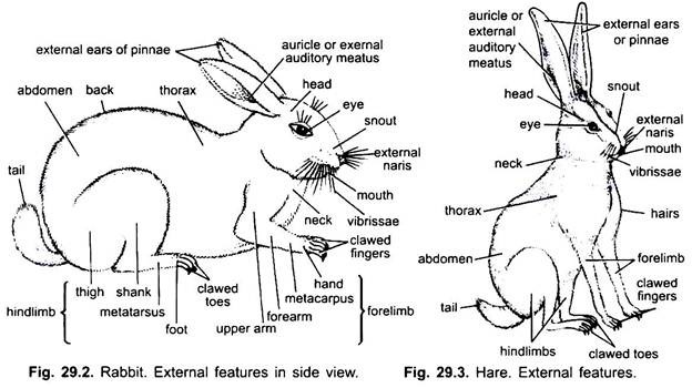
The rabbit shows a typical mammalian form of the body which consists of head, neck, trunk and tail. The trunk is further divisible into thorax and abdomen.
ADVERTISEMENTS:
Head:
The head is large and spherical posteriorly but produced anteriorly into a large pointed blunt snout or muzzle.
The head bears the following structures:
1. Mouth:
ADVERTISEMENTS:
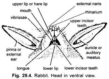
The snout has a terminal transverse slit-like mouth, which is surrounded by two soft, fleshy movable lips. The upper lip is divided in the middle into right and left halves due to a vertical cleft extending up to the nostrils. Such a divided lip is known as the hare lip, due to which the upper front incisors are exposed.
2. External Nares:
Just above the mouth are two oblique slit-like opening, the nostrils. The nostrils are surrounded by a bare moist skin, the rhinarium and lead into nasal or olfactory chambers.
ADVERTISEMENTS:
3. Vibrissae:
From the sides of the upper lip thick tactile hairs vibrissae or whiskers project outwards. The hairs are stiff, long and sensory in function, because they have nerve endings around their bases.
4. Eyes:
A pair of eyes are situated at the sides of the head, each having movable upper and lower eyelids with very fine, short eyelashes. A small white coloured third eyelid, the nictitating membrane, is also present in the inner anterior comer of the eye. The nictitating membrane is also movable and stretched across the cornea and used for cleaning cornea.
5. External Ears or Pinna:
A pair of large, movable trumpet-shaped external ears or pinnae is found situated at the posterior lateral side of the head. The long pinnae are movable in all directions to receive the sound waves. Each pinna has an external auditory meatus at its base, which is closed below by the tympanic membrane. Both the pinnae remain upright when the rabbit is on the alert and laid back on frightening and running.
Neck:
The neck is an extension of the body which connects the head with the trunk at a slight angle. It enables the head to move in all directions. The neck of the rabbit is short and flexible. Its short neck is advantageous in its burrowing and fast running habits.
ADVERTISEMENTS:
Trunk:
The neck is followed by large, cylindrical trunk, which is divided into anterior thorax and posterior broad, soft-bellied abdomen. The thorax or the chest forms a bony cage having ribs at the sides and sternum ventrally. The abdomen is without ribs and sternum. The cavity of the thorax is known as thoracic cavity in which tender body parts like heart and lungs are protected.
1. Teats:
On the ventral side between the thorax and abdomen are situated 4-5 pairs of well-developed and functional teats or nipples in females and rudimentary in males. The 4 or 5 pairs of mammary glands open on the outside at the teats.
2. Anus:
At the posterior end of the abdomen, at the base of tail is found the anus, which is the external opening of the digestive tract. A pair of hairless depressions is found in both the sexes, one on each side of the anus, called the perineal pouch into which the ducts of the perineal glands open. The secretion of the perineal glands has a strong odour, characteristic of rabbit.
3. Urinogenital Aperture:
Urinogenital opening is situated at the tip of the penis in male in front of anus. The penis is cylindrical, muscular and covered by the skin. In the male a pair of scrotal sacs are situated, one on either side of the penis, in which testes are lodged. The scrotal sacs are thin-walled bags of skin.
In the female the slit-like urinogenital aperture or vulva is present beneath the anus and at the anterior margin of vulva is present a rod-like clitoris which is similar to the penis of the male.
Limbs or Appendages:
The trunk bears two pairs of pentadactyle limbs. Both pairs of limbs take part in locomotion and support the weight of the body. Forelimbs are shorter than the hindlimbs.
1. Forelimbs:
The forelimbs are short and are held rigid to take the shock at the end of a leap. Each forelimb has a proximal upper arm or brachium, a middle forearm or antebrachium and the distal hand or manus with a wrist or carpus, palm or metacarpals and five fingers or digits with sharp, curved claws. The forelimbs are used for digging the burrow. The palm is hairy.
2. Hindlimbs:
The hindlimbs are longer and more powerful than the forelimbs. Each hindlimb has a proximal thigh, a middle shank or crus and a distal foot or pes with an ankle or tarsus, metatarsals and four clawed digits. Hallux (first toe) is absent. The hindlimbs are main locomotory organs. The sole is hairy.
Tail:
A short, bushy tail is found at the hind end of the trunk. The lower surface of the tail has a white hairy patch in wild rabbit, which is used for warning signal to other rabbits when danger approaches.
Locomotion:
The mode of locomotion in rabbits may be of three types- viz., walking, running and leaping. For walking, firstly, the forelimbs are moved forward as a whole nearly through 90 degrees therefore, the hands are directed in the same direction as the head and the pre-axial end of the limb remains situated near and parallel to the side of the body.
Then the upper arm is bent backwards, the forearms forward, while the hand rests with palms downward on the ground. Exactly in the same way, the hindlimbs are moved forward, hence, the preaxial side is near the body but the thigh remains directed forwards, while the shank is bent backwards and the sole of the toot comes in contact with the ground. So in such movements in rabbit, the limbs he under the body forming vertical levers.
However, during running the body is not stable. When the speed of walking increases, each foot is raised off the ground before the previous one is kept. At the time of leaping only two feet touch the ground, actually the hind feet hit the ground at a point in front of which the forelimbs have just left. Thus, its locomotion is well adapted for its terrestrial mode of life.
Integument:
The bodies of all vertebrates are invested by an outer protective covering, the integument or skin, which is partly responsible for retaining the body shape. The skin is comparatively thick and covered with hairs. It consists of two main layers; the epidermis which is ectodermal in origin and the dermis which is mesodermal in origin. Several other structures which are derived from the skin are found associated with it, e.g., hairs, claws, nails, various glands, etc.
1. Epidermis:
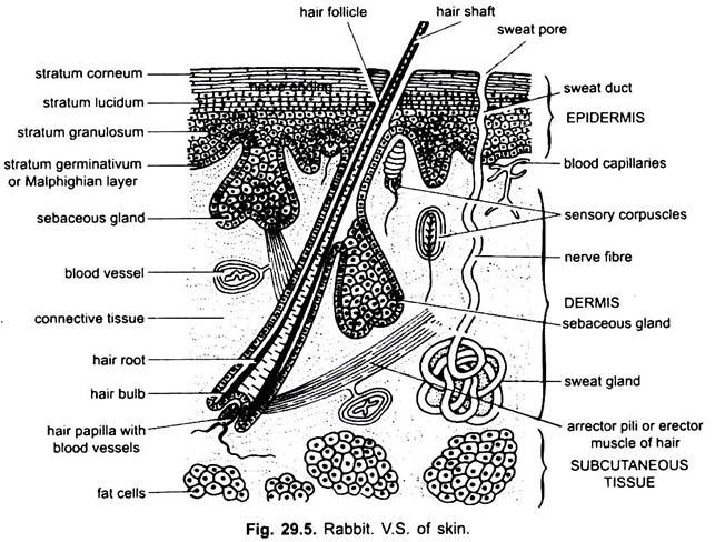
The epidermis is the outermost, thick and more complex layer formed of stratified epithelium. The epidermis is mainly formed of two layers, the superficial outer cornified layer and deeper inner Malpighian layer.
Cornified Layer:
The outermost layer of cells is hard, scale-like, dead, fully keratinized and flattened. They form the straturm corneum. It is thick on palms and soles. This layer contains hard keratin which prevents the passage of water and solutes. In places where there is considerable friction this layer may become very thick.
This layer is cast off in shreds periodically which are called dandruff. New cells are constantly added from below when granular cells become horny and dead. Epidermis contains openings of sweat glands and hair follicles.
(i) Stratum Germinativum:
It is the innermost layer based on basement membrane secreted by the dermis. It is formed of a single layer of columnar cells. These cells continuously divide mitotically and, thus, new cells are added into the epidermis. All the glands are derived from this layer.
(ii) Stratum Spinosum:
It lies above the stratum germinativum layer. Its cells are polyhedral in shape and arranged in several layers. These cells are gradually pushed upwards and become flattened and keratinized due to deposition of keratin (scleroprotein).
(iii) Stratum Granulosum:
It is present above the stratum spinosum. Its cells contain granules of keratohyalin. This layer is especially developed in palms and soles, etc., where the epidermis is thick.
(iv) Stratum Lucidum:
This layer is formed of thin transparent and hard shiny and refractile cells. A chemical substance eleiden is found in this layer. The stratum lucidum is transformed into stratum corneum when it is removed. It is the innermost layer of the epidermis which lie over the dermis. The cells of this layer are very active and divide to form new cells.
As the new cells are formed, the older ones are pushed upwards and at the same time they become flattened, their protoplasm becomes granular and horny due to the formation of keratin which is a protein. Finally they come to the surface and become perfectly flat and horny.
2. Dermis:
The epidermis is followed by the dermis, which is thick, tough, flexible and elastic. The dermis is formed of dense connective tissue fibres, unstriped muscles, blood capillaries, lymph vessels, nerves, pigment cells, and fat cells. The nerve endings, receptors and blood capillaries are distributed throughout the dermis.
Nerves and sensory organs of touch, pressure, temperature and pain are abundantly found in dermis. A few nerve endings penetrate the epidermis. Pigment cells are present in the outer layer of dermis. These cells contain melanin. Some of the epidermal glands, e.g., sweat gland, sebaceous gland, etc., are found in the dermis. The deeper part of the dermis is formed of fatty or adipose tissue consisting of groups of fat cells.
Fat is found stored in the lobules of the fat cells in the deeper parts of dermis and in the subcutaneous tissue. The fatty layer acts as a reserve food supply and as a heat insulator. The functions of the dermis are to provide firmness and flexibility to the skin, protecting and supporting the body and carrying blood to the general body surface.
Skin Derivatives:
1. Hair:
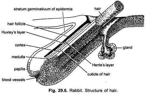
The hairs are epidermal in origin and found in the mammals only. A hair develops as a thickening of the stratum germinativum of epidermis. Later on, it is differentiated into two parts: the basal part deeply situated in the dermis is called the root, which is bulb-like and the upper part that traverses the dermis and projects out of the epidermis is called the shaft.
The hair is enclosed in a tube-like invagination of the epidermis formed by stratum Malpighii, the hair follicle. At the base of the follicle in the dermis a small swelling of dermal tissues is found which is evaginated to form the dermal papilla or hair papilla. The hair papilla is richly supplied by nerves and fine blood vessels carrying nourishment which diffuses into the root of the hair.
The epidermal cells situated above the hair papilla multiply actively to form the hair bulb. From the hair bulb develops a cylindrical shaft of cornified cells, the base or root of the hair. Constant addition of cells causes the hair base to extend through the follicle and pierce the skin as a column of cells.
The hair base is only living, while the cells of hair become horny and die beyond the skin. The hair is made only by the secretion of sebaceous gland opening into the hair follicle. With each hair follicle is attached a small smooth muscle, the erecter pili, for erecting the hair due to fear, excessive cold, etc.
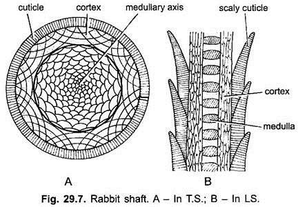
In a cross section the hair has three regions. The outer is a cuticular layer formed of overlapping microscopic scales, which is followed by a pigmented fibrous cortex consisting of elongated horny epidermal cells. In the centre of the hair is the pith or medulla formed of rounded cells. In larger hairs it contains air spaces.
Functions of Hairs:
The principal function of hair is the part it plays in temperature regulation, but it can be tactile, protective and possesses sexual functions as well. In man and certain other mammals (cetaceans, sirenians) that have become relatively hairless, heat loss is retarded by a compensatory layer of hypodermal fat (blubber) that arises in the superficial fascia.
Vibrissae on the facial region and sometimes near carpus and tarsus are richly innervated and tactile in nature.
Spines in spiny anteaters, hedgehogs and porcupines are the modified hairs and used as defensive organs.
Bristles (hairs) in the nostrils and auditory meatus of many mammals, including man, serve to block the entrance of foreign substances and insects.
Hairs in specialised areas assist in the retention of animal odours significant in courtship.
2. Cutaneous or Epidermal Glands:
These are abundant and peculiar in the skin of mammals. The epidermal glands are sebaceous, sweat, mammary, lacrimal, Meibomian, scent and wax glands. These glands are multicellular and present in the dermis.
(a) Sebaceous Glands:
The sebaceous glands are large glandular, branched alveoli-like saccular glands found attached with the upper part of the hair follicle by a small duct. The sebaceous glands are actually outpushings of the wall of the upper part of hair follicles. It secretes an oily secretion called sebum, which lubricates the hair and makes the skin waterproof and greasy. Sebum is also bacteriocidal in nature.
(b) Sweat Glands:
The sweat or sudorific glands are thin, long coiled tubes formed as tubular invagination of the stratum germinativum. The sweat glands found deep in the dermis and their long spiral ducts open at the surface of the skin through minute pores. The glandular part of sweat glands formed of large columnar cells and their ducts are simple epithelial cells.
This gland separates a salty, watery solution from the blood called sweat which is a secretion as well as an excretion. It is conducted along the ducts and passed outside on the general surface of the skin. The sweat glands help in removing the metabolic wastes of the body in form of sweat which contains water and dissolved excretory wastes and inorganic salts.
This gland plays an important role in the regulation of body temperature. When the body temperature rises too much, the sweat glands are stimulated to take up water from blood capillaries surrounding the sweat glands and pour out their secretion on the general surface of the skin.
Evaporation of sweat from the body surface uses up latent heat of vaporisation from the skin, thus, the extra heat of the body is used up and the body cools down reducing the temperature. Sweat glands also act as excretory organs because they excrete urea dissolved in water. Sodium chloride, a useful salt, is also excreted dissolved in water. People living in Tropical countries, thus, need more salt in their diet since they perspire profusely in summers. In rats sweat glands are not found. In arboreal animals they occur on plantar and palmar surfaces.
(c) Mammary Glands:
These are modified sweat glands and one of the most important characteristics of placental mammals. The mammary glands consist of branching system of tubules and alveoli secreting milk, and their ducts open on small projection of the skin surface, called nipples or teats. The mammary glands arise along the mammary lines which run from axilla to inguinal region on each side of the trunk.
They differ considerably in number and distribution from one pair (e.g.. most primates, giraffes) to eleven pairs occur in eutherians. Rabbit generally possesses four pairs of mammary glands. Mammary glands may be restricted to the thoracic region (e.g., Chiroptera, Sirenia, and all primates except lemurs) or to the inguinal area (e.g., cetaceans, ungulates).
In females they secrete milk for the nourishment of the young after birth. In males they remain undeveloped and functionless. Milk is cellularly synthesised from precursors in the blood stream and passes through the membranes as alveolar cells.
Other Glands:
A number of other glands are also found associated with the skin of mammals.
They are as follows:
(i) Perinaeal Glands:
These glands are situated under the skin near the anus and open into hairless depressions, called perinaeal pouches. These glands are in fact modified sebaceous glands which secrete a secretion with characteristic odour of rabbit.
(ii) Meibomian Glands:
These glands are found on the margins of eyelids just beneath the conjunctiva and open on the free edges of the eyelids. These are also modified sebaceous glands having long ducts into which several alveoli open. An oily secretion is secreted by these glands tor the lubrication of the eyelashes which make them soft and flexible.
(iii) Scent Glands:
These glands with strongly smelling secretions open on various parts of the integument are regarded as modified sebaceous glands or more rarely sweat glands. Examples of such glands are the occipital glands of camel, cerumen glands of antelopes and deer temporal glands of elephant, pedal glands of ruminants, preputial glands of musk deer and beaver, etc. These glands are often found near anus or in inguinal region and open into special cutaneous pits,
(iv) Lacrymal Gland:
It has several openings on the conjunctival surface beneath the upper eyelid towards the outer side of the eye.
(v) Harderian Gland:
It lies at the inner side of eyeball and mainly on its lower surface. It opens in connection with the nictitating membrane at the inner angle. Harderian gland is absent in primates.
(vi) Wax Glands:
These are present in the auditory canal and secrete fatty earwax or cerumen. It lubricates and protects the tympanum.
Other Derivatives of the Skin:
Claws, nails, horns and hooves are the other derivatives of the skin found in most of the mammals.
Functions of the Integument:
The integument or the skin performs the following functions:
1. Shape:
It helps in maintaining a characteristic shape of the body.
2. Protection:
It protects the body organs from mechanical injuries due to function and blows. It is a germ-proof covering of the body which prevents the entry of bacteria and the other germs. It prevents an undue evaporation of water.
3. Defence:
Claws, horns, nails and hooves are the derivatives of the skin which serve as useful tools, such as digging, weapons of offence and defence.
4. Body Temperatures:
The sweat glands of the skin help in regulating the body temperature by evaporating the sweat. The dermis gives rise to the dermal bones of the skull. In some reptiles it forms dermal bony plates.
5. Excretion:
The skin also helps in removing the metabolic wastes of the body along with the sweat. The shedding of the keratinized dead cells of the epidermis is a kind of excretion.
6. Secretion:
The skin serves as an organ of secretion. The milk is produced in the mammary glands and the sebaceous gland secretes an oily secretion, the sebum, which is responsible for water-proofing of the skin and hairs.
7. Sexual Function:
It helps in sexual selection. The colour of the integument and various integument structures serve to attract the opposite sex.
8. Sensation:
The fine nerve endings and special tactile corpuscles in the integument provide the sensory function to the skin. It receives stimuli of touch, pain, pressure, temperature, etc.
9. Synthesis of Vitamin D:
The skin is also capable of synthesising vitamin D in the presence of sunlight.
10. Other Uses:
(i) In some mammals like bats, the skin forms patagium to help the animal in flight. The patagium looks like a wing which is merely fold of skin. The flying-squirrels are also provided with such patagium.
(ii) It also serves to absorb oils and ointments to some extent, thus, serving as an organ of absorption and checks excessive absorption of injurious substances.
(iii) The dermis gives rise to dermal bones of the skull. In some reptiles it forms dermal bony plates.
(iv) It acts as an organ of storage of reserve food. Fats are usually stored in the liver and muscles, but a considerable amount of fats are stored in the form of subdermal fat in the skin. In whales such a fatty layer, called blubber, is several centimetres thick.
