In this article we will discuss about the external morphology of uromastix with the help of suitable diagrams.
Shape, Size and Colour:
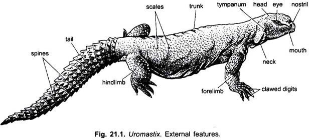
The body of Uromastix is long, massive and depressed. The length of the body may vary from 20 to 30 cm. The general colour of the body on the dorsal surface is yellow-brown with dark spots, while the ventral surface is light pale.
The whole body surface is covered with granular epidermal scales. The crest of spines present on the neck and back found in some members of family Agamidae, e.g., Calotes to which Uromastix also belongs, is in this species are entirely absent.
Division of Body:
The body of Uromastix is distinctly divisible into four regions, viz.,
ADVERTISEMENTS:
a. Head,
b. Neck,
c. Trunk and
d. Tail.
ADVERTISEMENTS:
a. Head:
The head is small, depressed and roughly triangular and produced in front into a short and blunt snout. The mouth is anterior and like a wide slit bounded by immovable lips covered with scales. Lower lip is rounded but the margin of upper lip is sharp covering the lower lip.
A pair of oval openings, the external nares, is located at the anterior end of snout above the mouth. During digging, the nostrils become closed by a prominent swelling of their posterior mucous linings which serve as valves. A pair of small bead-like eyes is situated dorso-laterally almost in the middle of the head. The pupil is round and the iris is light orange in colour.
Each eye has two thick movable eye-lids and a nictitating membrane. Eye-lids are well developed. The lower eye-lid is much more mobile than the upper one. The two eye-lids, completely close the eye. In addition to the two eye-lids, the nictitating membrane is a transparent thin membrane and is placed in the anterior comer of the eye. It also acts like a third eye-lid.
ADVERTISEMENTS:
The nictitating membrane cleans the cornea at the time of need. Behind the eyes are two shallow pits called the external ear openings or auditory meatus. At the base of each auditory meatus lies an oval tympanic membrane. The meatus has a denticulated anterior border but the posterior border is not denticulated.
b. Neck:
Head is followed by the neck. The neck is short and devoid of gular pouches. There are prominent folds of skin on the ventral and lateral sides of the neck when the animal is at rest but these folds disappear when the animal stretches out its neck.
c. Trunk:
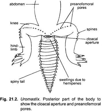
The neck is followed by the long, broad and depressed trunk to which are attached two pairs of limbs. The trunk is smooth and flattened ventrally but rough and convex dorsally. Its lateral sides skin is loosely folded. On the ventral surface a thick-lipped transverse cloacal aperture is placed at the junction of the trunk and the tail.
d. Tail:
The tail is long, thick and strongly depressed, widest at the base and tapers posteriorly. There are about twenty transverse rows of hard and strongly keeled scales covering the dorsal surface of the tail. These scales form a sort of armour for self-defence and, thus, check the loss of water. Due to the presence of spiny scales on the tail, the Uromastix is also called the “spiny-tailed lizard”.
Caudal autonomy is lacking. Just behind the cloacal aperture, on the ventral side at the broad base of the tail is present a pair of nodule-like swellings, which are due to the presence of hemipenes in male.
Keith (1955) has given an interesting account of scales found on the dorsal, ventral and lateral surfaces of the body. According to her, the whole body is covered with scales. The scales present on the dorsal surface of the head are unequal, smooth or keeled. These are larger on the snout but become smaller towards the eye. The scales on the cheeks are oval and 12-14 in number. These are called upper labials.
The scales present on the dorsal surface are small, numerous with or without larger scales lying between them. The ventral scales are subquadrangular in shape and they are bigger even than the bigger dorsal scales. The gular scales are rounded and smaller than ventrals. On the tail are present spinose scales. Due to the presence of spinose scales Uromastix is called spiny-tailed lizard.
Sexual Dimorphism:
In the male, the cloaca possesses a pair of protrusible copulatory organs, called hemipenes. These open separately into the proctodaeum. These are absent in females. In the male, in breeding season, a pair of nodule-like swellings appears ventrally at the base of the tail just behind cloacal aperture. These are also absent in females. Besides these, preanofemoral pores on the ventral surface of thighs are conspicuous in male than in the female.
Appendages:
There are two pairs of limbs emerging laterally from the trunk- one pair of forelimbs and one pair of hindlimbs. They are pentadactyle type and adapted for burrowing.
1. Forelimbs:
The forelimbs arise laterally from the trunk just behind the neck. These are shorter than the hindlimbs. Each forelimb consists of the usual three parts, viz., upper arm or brachium, the forearm or antebrachium and the hand possessing the five digits or fingers. The first finger or pollex is the smallest and the fourth is the largest. All the digits end in pointed curved claws. The lower surface of hand bears transverse rows of leaf-like adhesive pads.
2. Hindlimbs:
The hindlimbs arise from the posterior end of the trunk. Each hindlimb consists of a proximal thigh, middle shank or crus and distal foot or pes provided with five clawed toes. The digits of the hindlimbs gradually increase in length- the first or the hallux is the smallest and the fourth is much longer, while the fifth is again small. All the digits are provided with strongly curved claws.
3. Preanofemoral pores:
There are about 9 to 18 prominent pores arranged in a curved row on the ventral surface of both thighs from cloaca to the knee-joint. These are called preanofemoral pores and more prominent in male. Each pore is surrounded by a ring of scales, amongst which one is larger than the others. Each pore leads into a tubular gland producing a horny secretion which foams temporary spines which help in copulation.
Skin or Integument:
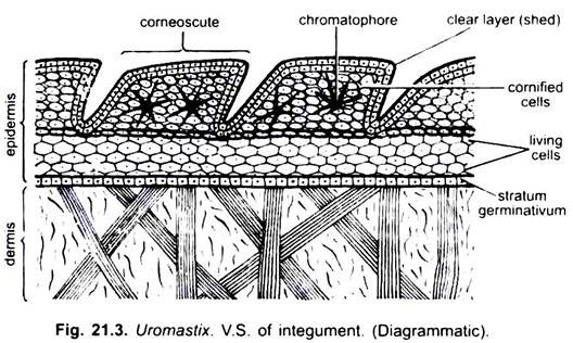
The skin of Uromastix is rough, dry and scaly and has almost no glands. It prevents the loss of water. This is an adaptation to prevent evaporation of water. The histological structure of skin shows two layers- an outer epidermis and an inner dermis.
1. Epidermis:
The epidermis is stratified and derived from ectoderm. The epidermis comprises a well developed stratum corneum or horny layer which serves admirably to protect the lizard from desiccation.
The stratum corneum is composed of thin, flattened and dead cells due to the deposition of horny scleroprotein or keratin. Due to accumulation of keratin in the outermost stratum corneum cells, they become keratinized or cornified and then they die.
The epidermis produces horny scales covering the body. This layer is sloughed off periodically in flakes (ecdysis). Below this is a layer of chromatophores, which are not found in the skin of those areas which are covered with scales.
At the base of epidermis lies a single-layered germinal epithelium with prominent nuclei. The germinal epithelium proliferates cells which move outward and form the stratum corneum. The horny layers covering the terminal ends of digits form the claws.
2. Dermis:
The dermis lies beneath the epidermis. It is a well developed thick layer of dense fibrous connective tissue. It contains muscle-fibres, blood vessels, nerves and chromatophores. The scales, covering the entire surface of the animal, are formed from special folds of dermis.
There are no glands in the skin except a few femoral glands which are found on the ventral surface of the thigh. These glands secrete a substance in males that hardens into temporary spines which help in clasping the female at the time of copulation.
3. Muscles:
Below the body wall or skin is present a layer of muscles which help in locomotion, etc. Beneath the muscle layer is the peritoneum which is formed of closely fitting mesodermal cells. It encloses the body cavity.


