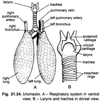
The respiratory system of Uromastix consists of two main portions, viz., respiratory tract and respiratory organs.
Respiratory Tract:
The respiratory tract starts from the external nares. These are a pair of small oval apertures on the snout slightly above the mouth. On its inner side each naris possesses a prominent swelling of mucous membrane which serves as a valve to close the aperture during burrowing. The external nares lead into small sacs, the nasal chambers or olfactory sacs, lying in the skull.
The nasal chambers open posteriorly by a pair of small apertures, the internal nares, into the anterior part of a spacious buccopharyngeal cavity. On the floor of buccopharyngeal cavity between the basal bifurcations of the tongue lies a small median longitudinal slit called the glottis which leads into a short median chamber called larynx.
The wall of larynx is supported by a pair of arytenoid and a median cricoid cartilages. The arytenoid cartilages are situated in the antero-lateral wall of the larynx and give support to the lips of glottis. Posteriorly these two cartilages articulate with the antero-lateral sides of the cricoid cartilage. In Uromastix, the vocal cords are lacking.
ADVERTISEMENTS:
The larynx is provided with two pairs of muscles- the inner musculus compressor laryngis which serves as a sphincter of the glottis and the outer musculus dilator laryngis which serves as a dilator of glottis. The larynx leads posteriorly into the trachea; which is an elongated cylindrical tube running backwards through the neck ventral to oesophagus.
The wall of the trachea is supported by a series of complete cartilaginous rings throughout its length which prevent the trachea to collapse. Some of the tracheal rings are bifurcated. After entering the thorax the trachea almost reaches the base of the heart, where it bifurcates into two very short, narrow tubes, the bronchi, which are also supported by complete cartilaginous rings. Each bronchus enters the lung of its side and does not branch into intrapulmonary extensions or bronchioles.
Respiratory Organs:
The respiratory organs of Uromastix are a pair of lungs. They are situated one on either side of the heart in the thoracic cavity. Both the lungs are symmetrical in shape and size. They are elongated, fusiform, thin-walled elastic hollow sacs of orange colour. The posterior ends of lungs appear pointed when they are empty but rounded when they are inflated.
ADVERTISEMENTS:
The inner lining of the lungs is raised into a network of low ridges which enclose shallow depressions or alveoli. The cavity of the lung is continuous. These ridges are more numerous and closer in the anterior part of the lung than in the posterior part. The ridges increase the respiratory surface of lungs. The walls of the lungs and alveoli have a rich supply of blood-capillaries.
The blood supply is more abundant in the anterior part of the lungs than in the posterior part. Therefore, the anterior parts of the lungs are more respiratory than the posterior parts. The bronchi do not branch into bronchioles in the lungs. The blood entering the lungs through pulmonary artery is deoxygenated and the oxygenated blood is carried to the heart by pulmonary vein.
Mechanism of Respiration:
In Uromastix, the mechanism of respiration is different from that of the frog. The process of respiration is effected only by ribs and their inter-costal muscles. The inter-costal muscles, which extend between the ribs, pull the ribs backward and forward and, thus, the pleuro-peritoneal cavity is decreased or increased in size.
When the cavity is increased, the lungs expand and fresh air rushes into the lungs from outside through the nostrils, buccal cavity, glottis, trachea and bonchii. When the cavity is decreased, the lungs are pressed and the air is forced out through the same above passage. Exchange of gases takes place in the alveoli through their thin moist walls. In Uromastix, the respiration is only pulmonary.
