Learn about the comparison of circulatory system in various arthropods.
Comparison: Arthropod # Macrobrachium:
1. Heart of Pericardium:
The heart (Fig.41) is a hollow organ, somewhat triangular in outline, and with thick muscular walls. It is situated dorsally at the posterior end of the cephalothorax. It is lodged in a special haemocoel, the pericardial cavity, the walls of which form the pericardium. A horizontal pericardial septum forms the floor of the pericardial sinus. A median cardiopyloric strand and 2 lateral strands support the heart in the pericardium.
5 pairs of valved ostia are present on the walls of the heart; one pair a little behind the middle on the ventral surface, one on each side; second pair opposite to the first pair on the dorsal surface; third pair on the posterior border; fourth pair behind the apex and the fifth or the last pair, one on each side of the lateral angle of the heart. The heart is traversed by a large number of interlacing muscle fibres, the interstices of which is the cavity of the heart.
ADVERTISEMENTS:
2. Blood:
Blood of the prawn is a clear fluid having a number of colourless leucocytes. The respiratory pigment is proteid-haemocyanin. The oxygenated blood is shining blue, but colourless when deoxygenated.
3. Arteries:
These are thick-walled vessels, through which the heart pumps out its contained blood.
ADVERTISEMENTS:
From the apex of the heart proceeds anteriorly a slender, median ophthalmic artery up to the root of the oesophagus.
Two antennary arteries arise from the inner lateral sides of the heart and run anteriorly, slightly obliquely. Behind the eyes, the arteries of the two sides anastomose and form a loop, the circulas cephalicus, with which the median ophthalmic artery joins. From this loop comes off a rostral artery on each side.
Each antennary artery on its way gives off a pericardial branch to the pericardium, a gastric branch to the cardiac stomach, a mandibular branch to the mandibular muscles, and finally an optic artery supplying the eye of the side/Before giving off the optic artery, the antennary artery sends a common artery, which divides into renal, antennal and antennular branches and supply the respective organs.
A pair of small hepatopancreatic arteries arises from the heart, ventrolateral to the roots of the antennary arteries. They end in branches in the hepatopancreas.
ADVERTISEMENTS:
A short and stout dorsomedian artery arises from the posterior and ventral region of the heart. It divides immediately into a supraintestinal and a sternal artery. The supraintestinal artery runs up to the posterior tip of the abdomen lying dorsal to the alimentary canal. In its course, it gives off a number of small branches to the intestine.
The sternal artery is a large vessel. It runs obliquely to the ventral region of the body either through the right or left side of the mid gut. It then pierces through the thoracic ganglionic mass of the ventral nerve cord and divides into two branches. One proceeds anteriorly lying below the nerve cord and is known as ventral thoracic, while the other runs posteriorly below the nerve cord and is known as ventral abdominal.
They branch profusely, and the former supplies blood to the thorax, first three pairs of walking legs, the maxillipeds, maxillae and the maxillulae, while the latter supplies blood to the ventral region of the abdomen, fourth and fifth pairs of walking legs, the abdominal appendages and the mid gut.
4. Blood Sinuses:
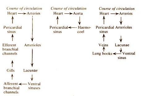
Blood from the arteries is finally received into minute intercommunicating body spaces known as lacunae, which ultimately open into two large spaces known as ventral sinuses, situated lengthwise in the ventral region, beneath the hepatopancreas. The two sinuses are connected with each other at several places.
For aeration, blood from the ventral sinuses is sent through six pairs of afferent branchial channels to the gills. After aeration, blood from the gills is returned to the pericardial sinus through six pairs of efferent branchial channels.
Comparison: Arthropod # Periplaneta:
1. The heart and pericardium
The heart (Fig.42) is an elongated tube, which runs through the whole length of the thorax and abdomen as a dorsomedian contractile vessel. The cavity of the vessel is divided into 13 segmentally arranged chambers, of which 3 are thoracic and 10 are abdominal. Externally, each chamber is marked by constrictions and internally communicates with the adjacent chambers through narrow passages guarded by valves.
ADVERTISEMENTS:
The wall of each chamber is perforated by two openings known as ostia. The whole structure is enclosed in a space known as the pericardial sinus bounded by a thin membraneous pericardium. The pericardium also bears a number of perforations and as a result, the pericardial cavity is in communication with the perivisceral cavity or the haemocoel.
2. Blood:
Blood is a watery fluid having no colour. White blood corpuscles, the amoebocytes float in it. The blood bears no respiratory pigment and as such it is not concerned with respiration. It carries the absorbed nutriment from gut for distribution and nitrogenous wastes from the site of origin to the excretory organs for elimination.
3. Arteries:
The heart is anteriorly continued into the head as a slender tube, the aorta (Fig.42). The aorta runs forward along the dorsal surface of the oesophagus and ends in front of the peripharyngeal nerve ring in the form of a funnel. By the successive contraction of the chambers of the heart blood is driven forward and is poured in the haemocoel through the aorta. Here, the blood bathes all the visceral organs.
4. Blood Sinuses:
The haemocoel remains filled up with blood coming from the heart. From there it enters the pericardial sinus through apertures in the pericardium.
Comparison: Arthropod # Palamnaeus:
1. The Heart and Pericardium
The heart (Fig.43) is a dorsal elongated tube lying in a groove in the liver mass, entirely in the preabdominal region, extending from the seventh to the thirteenth segment. It is clearly demarcated in its anterior and posterior limits by valves. The heart is single chambered.
Seven pairs of ostia, guarded by valves are present on the walls of the heart. The heart is suspended by eight groups of ligaments arranged metamerically. The pericardium is thin, membraneous and horizontal in position. The pericardial cavity is divided into four sinuses, two laterals, one ventral and one dorsal. The cardiac nerve extends from one to the other end of the heart.
2. Blood:
The freshly shed blood is indigo in colour. White blood corpuscles, the amoebocytes float in it. The blood contains a blue coloured respiratory pigment, proteidhaemocyanin. The oxygenated blood is bright blue, but colourless when deoxygenated.
3. Arteries:
The heart continues anteriorly as the anterior aorta for a considerable distance. Afterwards, it divides into two branches, which embrace the oesophagus on both the sides and unite below in the form of a short arch connecting the aorta with the right and left thoracic sinuses. In the ventral region, from the arch comes out a common blood vessel that runs over the nerve cord. This blood vessel is known as the supraneural artery.
On reaching the anterior end of the su bo esophageal ganglionic mass it turns downward and then runs backward as a subneural artery ending behind the ganglionic mass.
The supra- and sub- neural arteries are connected by nine vertical arteries.
A pair of large cephalic arteries arises from the aortic arch. Each gives off a number of branches, of which the ophthalmic artery and the cheliceral artery are more important. The ophthalmic supplies blood to the eye and the cheliceral to the chelicera.
Each thoracic sinus sends a small vessel to the supra- neural artery and four large vessels to the appendages supplying blood to the pedipalp and legs of the side. The heart continues posteriorly as posterior aorta. It runs straight backward up to the posterior tip and supplies blood to the intestine as well as various organs by a number of delicate branches.
4. Blood Sinuses:
The ventral sinus is a large space where blood from various parts of the body is collected as well as sent to the respiratory organs or lung books for aeration. Between the roof of the ventral sinus and the floor of the pericardial sinus a special arrangement of muscles is found.
The contraction of these special muscles causes the ventral sinus to enlarge and thereby venous blood is drawn into it. The relaxation of these special muscles results in the pumping of blood into the book-lungs. A series of segmental vessels or veins help in the conduction of blood from the lung- books to the pericardial sinus.