The circulatory system of scoliodon consists of the circulatory fluid called blood, the heart, the arteries and the veins.
(i) Blood:
The blood consists of a colourless plasma and corpuscles suspended in the plasma. Two kinds of corpuscles are encountered the RBC (or erythrocytes) and the WBC (or leucocytes). The erythrocytes are oval bodies containing a nucleus. Haemoglobin is present in the erythrocytes. The leucocytes are amoeboid in structure.
ADVERTISEMENTS:
(ii) Heart:
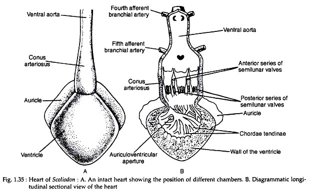
Heart is a bent muscular tube and consists of the receiving parts, comprising of a sinus venosus and a dorsally placed auricle and the forwarding parts, consisting of a ventricle and a conus arteriosus (Fig. 1.35A). The heart is situated on the ventral side of the body between two series of gill pouches.
The sinus venosus is a thin-walled tubular chamber. It is highly contractile and the beating of the heart originates from this part. Two great veins, the ductus Cuveiri, open into the sinus venosus, one on each lateral side. Two hepatic sinuses enter the sinus venosus posteriorly. The sinus venosus opens into the auricle by sinuauricular aperture which is guarded by a pair of valves.
ADVERTISEMENTS:
The auricle is a large, triangular and thin walled chamber situated dorsal to the ventricle but in front of the sinus venosus. The auricle communicates with the ventricle through a slit-like auriculo-ventricular aperture guarded by two lipped valves. The receiving chambers (sinus venosus and auricle) receive the venous blood from all parts of the body.
The ventricle has a very thick muscular wall. Its inner surface gives many muscular strands, which gives it a spongy texture (Fig. 1.35B). It is an oval chamber and constitutes the most prominent part of the heart. The conus arteriosus is a stout median muscular tube arising from the ventricle.
The lumen of the conus arteriosus is provided with two transverse rows of semilunar valves. To keep the valves in position the free ends of the valves are attached to the ventricular wall by fine tendinous threads called chordae tendinae. The conus arteriosus is continued forward as the ventral aorta.
The function of the heart is to receive the deoxygenated blood from all parts of the body and to pump it for aeration to the gills. Such a type of heart is designated as the venous or branchial heart, because only the deoxygenated blood circulates through it.
ADVERTISEMENTS:
(iii) Arterial System:
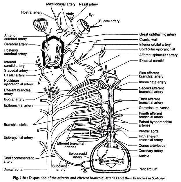
The arterial system of Scoliodon is divided into two distinct categories of arteries. The afferent branchial arteries arising from the ventral aorta which bring the deoxygenated blood to gills for oxygenation and the efferent branchial arteries which originate from gills and convey the oxygenated blood to the different parts of the body (Fig. 1.36).
(a) Afferent branchial arteries:
The ventral aorta is situated on the ventral surface of the pharynx and extends up to the posterior border of the hyoid arch. The ventral aorta divides into two branches called innominate arteries, which again bifurcates into the first and second afferent branchial arteries.
The third, fourth and fifth afferent arteries arise from the ventral aorta. Each afferent branchial artery arises from the ventral aorta by independent opening except the anterior- most pairs which arise by a common opening (Fig. 1.36).
(b) Efferent branchial arteries:
The afferent branchial arteries break up into capillaries in the gills. From the gills the blood is collected by efferent branchial arteries (Fig. 1.36). There are nine pairs of efferent branchial arteries and these are equally distributed on each side.
The first eight arteries form a series of four complete loops around the first four gill slits. The ninth efferent branchial artery collects blood from the demi branch of the fifth gill pouch.
ADVERTISEMENTS:
In addition to short longitudinal connectives connecting the four loops, these are further connected with each other by a network of longitudinal commissural vessels called the lateral hypobranchial chain. From each efferent branchial loop arises an epibranchial artery. The four pairs of epibranchials join in the mid-dorsal line to form the dorsal aorta.
Anterior arteries:
The head region gets the blood supply from the first efferent branchial artery and partly from the proximal end of the dorsal aorta.
Arteries from the first efferent branchial (hyoidean efferent) are:
(a) The external carotid,
(b) The afferent spiracular, and
(c) The hyodean epibranchial which in turn receives a branch from dorsal aorta.
The external carotid artery originates from the first collector loop and divides into a ventral mandibular artery giving branches to the muscles of the lower jaw and a superficial hyoid artery which supplies the second ventral constrictor muscle, the skin and the subcutaneous tissue beneath the hyoid arch.
Dorsal aorta and its branches:
The dorsal aorta is formed by the union of epibranchial arteries. It runs posteriorly and is situated ventral to the vertebral column. It is continued up to the tip of the tail as the caudal artery.
Along the anteroposterior direction the following arteries have their origin from the dorsal aorta:
1. Several buccal and vertebral arteries are given off anteriorly.
2. A pair of small subclavian arteries arise from near the origin of the fourth epibranchial arteries.
The subclavian artery gets the epicoracoid artery on its way and divides into:
(i) A branchial artery to the pectoral girdle and pectoral fin,
(ii) An anterolateral artery to the body musculature and
(iii) A dorsolateral artery to the dorsal musculature.
3. A large coeliacomesenteric artery arises just behind the origin of fourth epibranchial artery. It divides into a smaller coeliac artery and a larger anterior mesenteric artery.
4. A lienogastric artery originates posterior to the coeliacomesenteric artery and gives off:
(i) An ovarian (in females) or spermatic artery (in males),
(ii) A posterior intestinal artery,
(iii) A posterior gastric to the posterior part of the cardiac stomach and
(iv) A splenic artery to the spleen.
5. Series of paired parietal arteries emerge out behind the subclavian artery. Each parietal gives a dorsal parietal artery and a ventral parietal artery. The dorsal parietal artery supplies to the dorsolateral musculature, the vertebral column, the spinal cord and the dorsal fin. The ventral parietal artery supplies to the ventral muscles and the peritoneum. The ventral parietal gives renal branches to the kidneys.
6. A pair of iliac arteries extend to the pelvic fin as femoral arteries.
Hypobranchial chain:
A lateral hypobranchial chain is formed by a network of slender arteries arising from the ventral ends of the loop of the efferent branchial arteries. Four commissural vessels arise from the lateral hypobranchial chain which on the ventral wall of the ventral aorta unites to form a pair of median hypo-branchials which communicate with one another by transverse vessels.
Posteriorly the median hypo-branchials unite to form a median coracoid artery which gives rise to the coronary artery and a pericardial artery. The pericardial artery gives off the common epicoracoid artery which in turn divides into left and right epicoracoid arteries each joining one subclavian artery.
(iv) Venous System:
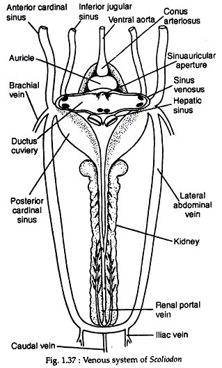
The deoxygenated blood from the different parts of the body is returned to the heart by veins which form irregular blood sinuses throughout their courses (Fig. 1.37). The existence of extensive blood sinuses is a characteristic feature of the venous system of Scoliodon.
The venous system is extremely complicated and is described under the following heads:
(A) Cardinal system:
The blood from the anterior region of the body is returned to the heart by paired jugular and anterior cardinal sinuses. The blood from the posterior region is collected by a pair of posterior cardinal sinuses. The anterior and posterior cardinals unite on each side to form a transverse sinus called ductus Cuvieri.
Anterior cardinal system:
This system of veins returns blood from the head region and consist of a pair of internal jugular veins. Each internal jugular vein is composed of the olfactory sinus, the orbital sinus, the post-orbital sinus and the anterior cardinal sinus.
The blood from the rostral region is drained by the anterior facial vein to the olfactory sinus and from there to the orbital sinus. The orbital sinus opens into the anterior cardinal sinus through the postorbital sinus. The anterior cardinal sinus enters the ductus Cuvieri.
Posterior cardinal system:
The caudal vein collects blood from the tail region and proceeds forwards through the haemal canal. In the abdominal cavity, the caudal vein divides into left and right renal portal veins which break up into sinusoid capillaries in the kidneys.
Throughout its length, the renal portal vein receives small parietal veins. The renal veins collect blood from the kidneys and unite to form the posterior cardinal sinuses. Two posterior cardinal sinuses open into the ductus Cuvieri.
(B) Hepatic portal system:
A large number of small veins carrying blood from the alimentary canal and its associated glands unite to form the hepatic portal vein. The hepatic portal vein receives the lienogastric vein, and anterior and posterior gastric veins.
Actually the hepatic portal vein is formed by the confluence of the anterior and posterior intestinal veins. The hepatic portal vein breaks up into capillaries in the liver. From the liver, blood is collected by another set of capillaries which unite to form two large hepatic sinuses opening into the sinus venosus.
(C) Cutaneous system:
This system consists of a dorsal, a ventral and two paired lateral cutaneous veins. The inferior lateral cutaneous vein joins with the lateral cutaneous vein near the anterior end of the pectoral fin. Each lateral cutaneous vein ultimately opens into the brachial vein.
(D) Ventral system:
This system comprises of two sets of veins—anterior ventral veins and posterior ventral veins. The anterior ventral veins pour blood into the ductus Cuvieri through inferior jugular sinuses. The posterior veins discharge through the subclavian vein.
Each inferior jugular sinus is formed by the union of the sub-mental sinus from the lower jaw, the hyoidean sinus and the ventral veins from the gills. Each inferior jugular vein opens to the ductus Cuvieri. The subclavian vein also opens to each side of the ductus Cuvieri.
Two large lateral abdominal veins are formed by a small caudal vein and two iliac veins. The lateral abdominal veins are connected posteriorly by a commissural vein. Anteriorly the lateral abdominal vein joins the brachial vein to form the subclavian vein which in turn opens into the ductus Cuvieri.


