In this article we will discuss about the respiratory system of fishes.
Respiratory System of Shark (Scoliodon):
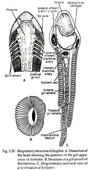
The respiratory organs are the gills which are borne by the gill-pouches. The structure of gill-pouches differs in different dogfishes. Figure 2.35B shows the organisation of gill-pouch of Brachaelurus, a related genus of Scoliodon. There are five pairs of gill-pouches, each of which communicate with the pharyngeal cavity by a large internal branchial aperture and opens to the outside by exterior gill-slits (Fig. 2.35A).
The mucous membrane lining the gill- pouches gives a series of horizontal branchial lamellae. The branchial lamellae are highly vascularized structures. Each gill-pouch has an anterior and a posterior set of branchial lamellae. The gill-pouches are separated by interbranchial septum which projects beyond the branchial lamellae (Fig. 2.35C).
ADVERTISEMENTS:
The pharyngeal end of each inter-branchial septum is supported by a visceral arch. Each arch supports the anterior set of lamellae of one gill-pouch behind and the posterior set of lamellae of the next gill-pouch. The first gill- pouch lies between the hyoid and the first branchial arches and the last one is present between the fourth and fifth branchial arches.
There are two types of gills:
(i) Holobranch or complete gill when a branchial arch bears two sets of gill lamellae.
(ii) Demi branch or hemi branch or half gill when single set of gill lamellae is present. The hyoid arch supports only a demi branch and the first four branchial arches support holobranchs. The last branchial arch is gillless.
ADVERTISEMENTS:
Mechanism of Respiration:
During respiration the floor of the buccal cavity is lowered and the mouth is opened. Then the water rushes in to fill the greatly expanded buccal cavity. The mouth is now closed and the pharynx contracts.
The water then enters the gill-pouches and goes out after gaseous exchange through gill-slits. The spiracles are occasionally used as accessory pathways for the entry of water for respiration, instead of the mouth when it is otherwise occupied.
Respiratory System in Bony Fishes [Lates sp. (Bhetki)]:
Respiration is performed by four pairs of gills, situated in the gilichambers. These two chambers are covered externally by the operculum and the branchiostegal membrane which is attached to the posterior margin of the operculum. The wall of pharynx is perforated by five gill-slits on each side and are separated by four gill-arches or interbranchial septa.
ADVERTISEMENTS:
Each gill-arch is provided with teeth- like gill-rakers on the inner concave border and two rows of comb-like gill-filaments on the outer convex border. The gill-rakers present in the inner borders of the gills prevent the escape of food materials from the pharyngeal cavity to the gill-chamber. The gills are holobranchs, i.e., each gill-arch bear’s two rows of gill-filaments.
Physiology of respiration:
Bhetki utilizes the oxygen dissolved in water.
The physical mechanism of respiration can be described under the following sequences:
(a) Inspiration:
During inspiration, the outer opening of the gill-chamber remains tightly closed to the body wall by the branchiostegal membrane and the two opercula bulge out to increase the accommodating capacity of the pharyngeal and buccal cavities. As a consequence, water from the exterior rushes inside through the opened mouth and fill in the buccopharyngeal cavity.
(b) Expiration:
Immediately with the entry of water, the pharyngeal and the buccal cavities contract and exert pressure to the contained water. As the mouth, by this time, becomes closed by oral valves, the contained water finds the way out through the gill-slits.
The operculum as well as the branchiostegal membrane are lifted by this time and the water from the gill-chambers goes out through its openings. The dilatation and contraction of the pharyngeal cavity are caused by the alternate retraction and protraction of the hyoid arch supporting the buccopharyngeal cavity.
ADVERTISEMENTS:
(c) Physiology of gaseous exchange:
The gills are highly vascular structures and are supplied by afferent and efferent branchial arteries. The afferent branchial artery carrying the deoxygenated blood is situated very superficially on the outer edge of the gill. The afferent branchial artery breaks-up into capillaries in the gills.
During the transit of water through the gill-slits, the deoxygenated blood in the capillaries of the gill-filaments takes up the oxygen dissolved in water and gives out carbon dioxide by diffusion. The blood thus aerated is collected by efferent branchial arteries and is conveyed to the different parts of the body.
