In this article we will discuss about:- 1. External Features of Cuttle Fish 2. Locomotion in Cuttle Fish 3. Digestive System 4. Respiratory System 5. Circulatory System 6. Excretory System 7. Nervous System 8. Reproductive System 9. Fertilization and Development.
Contents:
- External Features of Cuttle Fish
- Locomotion in Cuttle Fish
- Digestive System of Cuttle Fish
- Respiratory System of Cuttle Fish
- Circulatory System of Cuttle Fish
- Excretory System of Cuttle Fish
- Nervous System of Cuttle Fish
- Reproductive System of Cuttle Fish
- Fertilization and Development in Cuttle Fish
1. External Features of Cuttle Fish:
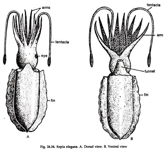
1. Body divisible into a head and a trunk (Fig. 26.34) joined by a short, narrow neck.
2. Ten arms are present in the head region surrounding the mouth, of which eight are comparatively shorter and stout, provided with suckers, arranged in longitudinal rows.
3. The remaining pair of arms (the fourth pair) are known as tentacles. They are also provided with suckers. The tentacles may be retracted in large basal depressions.
4. The fifth arm of the left side, the hectocotylysed arm, has suppressed suckers and acts as intromittent organ in males.
5. A pair of large eyes are present on the lateral sides of the head.
ADVERTISEMENTS:
6. The free extremity of the head bears the mouth.
7. The shield-shaped trunk is flat below and slightly convex above with fringed fin on either side. The fin extends from the oral to the aboral end where a notch separates them.
8. The trunk is covered by the thick mantle, forming a ridge at the oral end and ventrally it forms the oral lip of the opening of the mantle cavity.
9. The transverse slit-like pallial aperture is located at the anterior end of the trunk, on the ventral surface communicating the pallial or mantle cavity with the exterior.
ADVERTISEMENTS:
10. A ventral funnel is present that opens externally behind the neck and internally in the mantle cavity by a wide aperture, the pallial aperture and serves as the main outlet of the mantle cavity.
11. The skin is provided with chromatophores containing three kinds of pigments and iridocytes.
12. A shell is present that serves as an internal skeleton and float, and is placed within a sac. Calcareous particles are arranged in the form of lamellae in the shell enclosing spaces for gas.
Mantle:
1. The mantle forms a thick integument covering the trunk. Towards the oral end it terminates in a ridge around the neck.
2. Dorsally, the ridge projects forward as a prominent, round lobe, partially covering the head. Ventrally, it forms the oral lip, or collar, which surrounds the neck, between the head and visceral mass.
3. A pair of fins, extending from the oral to aboral end, are formed by lateral extensions of the mantle.
4. The mantle encloses a mantle or pallial cavity.
Pallial complex:
ADVERTISEMENTS:
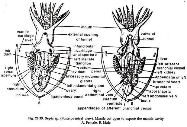
The mantle cavity or pallial cavity is a spacious chamber (Fig. 26.35) communicating with the exterior anteriorly through the funnel and the pallial aperture between the neck and mantle collar.
It encloses:
1. A pair of plume-shaped ctenidia or gill on either side.
2. The rectum in the middle line of the ventral surface, the anus opening close to the internal opening of the funnel.
3. A narrow tube, the renal sac with a terminal opening is present on each side of the rectum.
4. The genital duct, spermiduct or oviduct opens on the left side.
Endoskeleton:
The internal shell, the cranial cartilage and other cartilages constitute internal skeleton.
Shell:
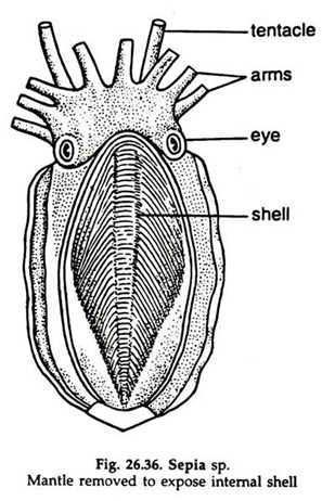
A calcareous shell enclosed in a sac is present in the dorsal wall of the mantle.
1. The shell is leaf-like, bilaterally symmetrical, broad at the oral and narrower at the distal end, drawn into a sharp, dorsally projected spine (Fig. 26.36).
2. The ventral surface of the shell is convex.
3. The dorsal surface is convex anteriorly and bounded laterally by thin wing-like ridges, converging to meet at the aboral end, which is deeply concave.
4. The body of the shell consists of numerous closely arranged, thin calcareous laminae, enclosing spaces for gas.
5. A chitinoid layer, thin on the surfaces and slightly thicker along the margins encloses the shell.
Cranial and Other Cartilages:
The cranial cartilage encloses closely aggregated nerve ganglia and statocyst. In addition, the eyes are protected by cartilaginous cups; a thin, shield-shaped nuchal cartilage lies in the ventral wall of the neck; a thin plate of cartilage in each of the paired elevations of the ventral wall of the funnel and the corresponding depressions on the dorsal surface of the body; thin cartilages supporting the fins and bases of the arms.
2. Locomotion in Cuttle Fish:
The locomotors organs are arms and the funnel, which are modifications of foot, and the lateral fins. The mantle also helps in locomotion.
1. Undulating movement of arms and fins help in slow movement. The fins also help in changing direction.
2. Darting rapidly through water is effected by rhythmic contraction of the muscular mantle, causing jets of water forced out through the opening of the funnel.
The funnel is used to expel water only from the mantle cavity:
a. With the relaxation of the mantle, water enters the mantle cavity through the cleft, the pallial aperture, between the neck and mantle collar.
b. The cleft is tightly closed, the mantle contracts and water from the mantle cavity is forced out in the form of a jet through the opening of the funnel.
c. The thrust of water shoots the animal backwards when the tip of the funnel is directed forward. By bending the tip backwards, Sepia can dart forward. Forward movement, which helps in catching prey, is slower than that of the backward movement helping to escape from predators.
3. Digestive System of Cuttle Fish:
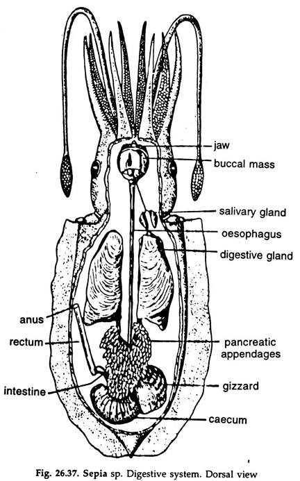
The digestive system consists of an alimentary canal, a pair of salivary glands, a digestive gland and a pancreas (Fig. 26.37):
Alimentary canal:
The alimentary canal is divisible into a foregut consisting of a buccal mass with an odontophore and an oesophagus; the midgut constituted by the stomach and the hindgut by intestine and rectum.
Mouth:
A round aperture at the anterior end Surrounded by oral arms. A thin, lobed, peristomial membrane surrounds the mouth. The lip is circular and beset with many papillae. A pair of jaws are lodged in the lip. The mouth leads to the buccal cavity. The jaws somewhat resemble the beak of a parrot, the ventral one larger and strongly bent than the dorsal one, which it encloses partly.
Buccal mass:
It is a fairly large, pyriform muscular structure containing a spacious chamber, the buccal cavity. A large odontophore is present in the buccal cavity.
Odontophore:
A fairly large, pyriform, muscular body containing buccal cartilages and a radula. The radula bears numerous teeth.
Oesophagus:
A straight, narrow tube, runs posteriorly from the buccal mass, along the middle line, between the lobes of the digestive gland and joins the stomach posterior to the middle of the trunk.
Stomach:
The stomach has two interconnected chambers, the opening between the two provided with a sphincter. The first part or gizzard is somewhat oval and muscular. The second part or caecum is round.
Intestine:
A round tube, arises from the caecum, bends sharply upon itself and runs anteriorly as the rectum.
Rectum:
It is of same diameter as the intestine, runs forward, parallel and ventral to the oesophagus and opens in the mantle cavity through anus, close to the internal opening of the funnel.
Digestive Glands of Cuttle Fish:
Digestive glands are of three types:
i. Salivary glands:
A pair of glands, located behind the cranial cartilage, one on either side of the oesophagus. Salivary ducts run inward and the two join in the middle. The common duct opens dorsally into the posterior part of the buccal cavity.
ii. Digestive gland:
Commonly called liver. A large, brownish gland, divided into right and left halves, partly connected, and lie by the sides of the oesophagus. The ducts from two lobes open separately at the junction of the stomach and intestine.
iii. Pancreas:
Minute, cream-coloured vesicles, clustered round the digestive ducts and their openings. The duct from the pancreas opens in the gizzard.
Ink gland:
The ink gland is associated with the digestive system but it does not play any role in nutrition. The ink sac, however, develops as a diverticulum of the dorsal wall of the intestine. It is a pear-shaped body, lying below -the mantle of the dorsal surface. A portion of the interior of the sac is glandular and secretes a black substance, ink or sepia.
The ink is stored in the main cavity of the sac and discharged into the rectum, close to the anus. The ink discharged by a startled cuttle fish mixes with water in the mantle cavity and ejected through the funnel as a black cloud, under cover of which the animal moves away unnoticed.
Feeding and digestion:
The food of the cuttle fish comprises of crustaceans, molluscs and fishes.
1. The animal swims forward with the arms together, darts at the prey suddenly by ejecting water through the funnel, the arms are spread and the prey grasped. All the small eight arms play active role in capture of prey.
2. The suckers are pressed against the body of the victim and a partial vacuum thus created, makes the suckers firmly adhere to the body of the prey and the latter cannot escape.
3. The victim is brought to the mouth by the arms, torn to pieces by the jaws end swallowed. The radula is seldom used in feeding.
4. Small, bottom dwelling animals are quietly covered by the arms and captured.
Digestion is both extracellular and intracellular.
4. Respiratory System of Cuttle Fish:
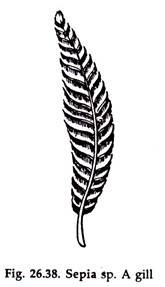
The respiratory organs consist of a pair of gills or ctenidia, one on each side, situated in the mantle cavity. The gills are plume-shaped (Figs. 26.35, 26.38).
1. Each gill consists of a series of paired delicate lamellae along an axis. The lamellae are largest in the middle but become smaller towards the extremities. Each lamella bears a complex system of folding’s by which the surfaces are increased. Internally, the lamellae are not in complete contact, and an axial canal is present.
2. The lamellae of each gill are attached to a long axis known as ctenidial axis. Along the greater part of its length the gill is attached to the wall of the mantle cavity by a thin muscular fold.
Mechanism of respiration:
By sudden contraction of the body, water within the mantle cavity goes out through the opening of the funnel and with the relaxation of the body a fresh current of water enters the mantle cavity through the pallial aperture. The water penetrates freely to all parts of the gills through axial canal.
The blood flows to the gills through afferent branchial vein and reaches gill lamellae through minute branches. After gaseous exchange in the gill lamellae, oxygenated blood is returned to the auricle through the main efferent vein formed by the union of minute vessels in the gill lamellae.
5. Circulatory System of Cuttle Fish:
The circulatory system in Sepia (in all cephalopods) is highly developed and almost the whole of the blood is carried through vessels arteries and veins.
The system consists of a ventricle, two auricles, arteries and veins. The heart is somewhat obliquely placed but the rest of the system is bilateral (Fig 26.39).
Heart and Pericardium of Cuttle Fish:
The heart is enclosed in a pericardium and lies superficially in the middle of the ventral surface.
1. Ventricle:
Median, somewhat asymmetrical in position and divided into two lobes by a median constriction.
2. Auricles:
Two, lateral and symmetrical; they are simple contractile expansions of the efferent branchial veins.
Arteries:
Two aortae arise from the two ends of the ventricle, divide into branches and capillaries in tissues.
1. Oral aorta:
A large vessel, arises from the oral end of the ventricle and sends branches to the organs of the anterior part of the body.
2. Aboral aorta:
A small vessel, arises from the aboral end of the ventricle. It bends over the ink sac, runs aborally and sends branches to the posterior part of the body.
Veins:
The veins are formed in the tissues by the joining of the capillaries and they unite to form a system of veins.
Vena cava:
A large median vein runs from head to near the rectum, in front of which it bifurcates to form the right and left afferent branchial veins.
Afferent branchial vein (two):
Each runs through the renal organ of the side and receives veins from the aboral organs at the base of the gill.
a. At the base of the gill, the afferent branchial vein dilates to form a contractile sac, the branchial heart, to which a round, glandular body, known as appendix is appended.
b. The afferent branchial vein runs through the axis of the gill and sends branches to the gill lamellae.
c. The branchial heart pumps blood to the gills through the afferent branchial vein.
Efferent branchial vein (two):
Each arises from one gill and oxygenated blood is returned to the ventricle through auricles, the dilated contractile base of the efferent branchial veins.
6. Excretory System of Cuttle Fish:
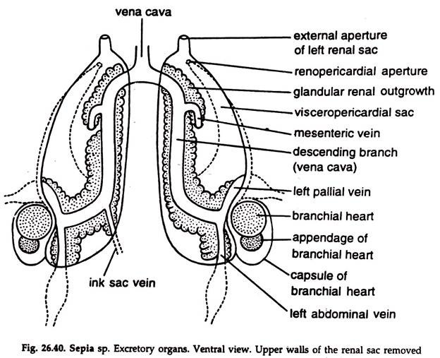
A pair of thin-walled, renal, sacs — right and left—constitute the excretory system. The afferent branchial vein of the side runs through the renal sac. Each sac opens in the mantle cavity through a conspicuous renal aperture on either side of the middle line. Each sac is in communication with the pericardium through a Reno pericardial aperture. The two sacs communicate with one another both orally and aborally.
Masses of glandular tissues (Fig. 26.40) surrounding the renal sacs excrete nitrogenous wastes in the form of guanine and released in the renal sac. The wastes discharged into the mantle cavity through the renal apertures are thrown outside through respiratory water current.
7. Nervous System of Cuttle Fish:
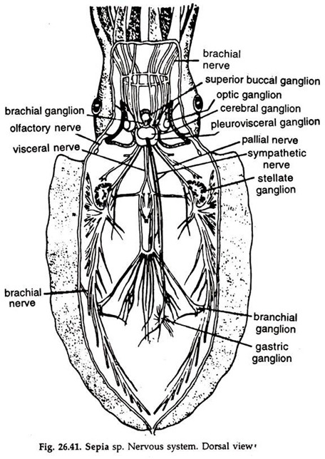
The nervous system is highly developed; the nerve ganglia are large. An extreme condensation of the nervous system has led to the aggregation of all the nerve ganglia in the head region, around the oesophagus and protected by a cartilaginous cranium.
The cerebrals, superior buccals, inferior buccals, pleoroviscerals and pedal ganglia and their nerves constitute the nervous system (Fig. 26.41):
Cerebral ganglia:
Paired, fused to form a large round mass, dorsal to the oesophagus. Anteriorly, they are covered by a tough, fibrous membrane. A pair of very thick band-like nerves around the oesophagus connect the cerebral ganglia with the pleurovisceral ganglia at the sides and posterior border. From each cerebral ganglion arise an optic nerve going to the eye, a statocyst nerve innervating the statocyst and a cerebrosuperior buccal connective.
Superior buccal ganglia:
A small pair, closely united and located dorsal to the oesophagus, close to the buccal mass. They are connected with the cerebral ganglia by cerebrosuperior buccal connectives.
Inferior buccal ganglia:
A pair of small, closely united ganglia on the dorsal surface of the oesophagus, posterior to the superior buccal ganglia, with which they are joined by slender connectives. A loop of sympathetic nerves arising from the ganglia courses along the oesophagus to the stomach and each ends in gastric ganglion.
Pleurovisceral ganglia:
The visceral ganglion is below, and the pleural ganglia on the lateral sides of the oesophagus. The pleurals and the visceral fuse to form a plurovisceral ganglionic mass.
It gives off:
a. Two visceral nerves. Each sends branches to visceral organs and continue as branchial nerve, bearing a branchial ganglion at the base of the gill and runs along the axis to its extremity.
b. Two stout pallial nerves. Each runs through the neck to the inner surface of the mantle cavity and forms a large, flat pallial or stellate ganglion in front of the gill. Nerves emanating from the stellate ganglion innervate different parts of the mantle.
Pedal ganglia:
The two ganglia are fused and located below the oesophagus.
It sends:
a. Ten brachial nerves to the arms. The brachial nerves are connected by a ring commissure.
b. A pair of nerves to the funnel.
Receptors and Sense Organs of Cuttle Fish:
The sense organs are highly developed and consist of a pair of eyes, a pair of statocysts, a pair of ciliary pits and a gustatory organ:
Eyes:
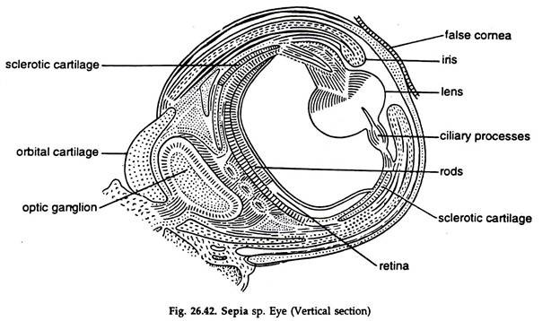
The eyes are simple (Fig. 26.42), located laterally on the head and lodged in some sort of orbit made of curved plates of cartilage, connected with the cranial cartilage.
Structure:
1. False cornea:
A transparent portion of the integument covering the exposed part of the eye.
2. Sclerotic:
It is strengthened by plates of cartilage and forms a firm wall of the eyeball.
3. Pupil:
A large opening of the sclerotic on the outer side towards the head.
4. Iris:
The part of the sclerotic surrounding the pupil. Muscle fibres in the iris can regulate the diameter of the pupil to a limited extent.
5. Lens:
A dense, glassy spherical body just internal to the iris and projecting slightly through the pupil. It consists of two Plano concave lenses in close apposition.
6. Cornea:
A thin layer of cells between two parts of the lens.
7. Ciliary process:
An annular process, projecting inwards from the sclerotic, supporting the lens.
8. Cavities:
The lens with the ciliary process divides the cavity of the eye ball into two portions—a smaller outer cavity containing aqueous humour and a larger inner cavity containing vitreous humour.
9. Retina:
The light sensitive layer lining the wall of the inner chamber.
It is composed of two layers:
a. An inner layer of close set parallel rods.
b. An outer layer of retinal tells in communication with the rods internally and optic nerve fibres externally.
The movement of the eye is very limited and effected by muscles.
Statocysts:
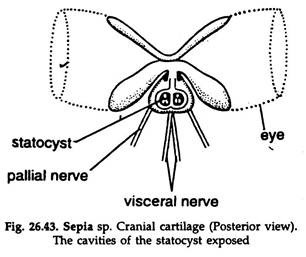
The statocysts are highly developed, closely placed organs (Fig. 26.43), separated by a median cartilaginous septum in the posterior portion of the cranial cartilage, close to the pleurovisceral ganglion.
1. The cavity of a statocyst is about 3 mm in diameter.
2. The inner surface is lined with a flattened epithelium and is thrown into a number of round and pear-shaped elevations.
3. On the posterior surface of the ridge a crista statica and a macula statica are present. These are composed of large cells with hair like processes on the surface and processes of the base continuous with the fibres of the statocyst nerve.
4. A large statolith attached to the macula is present in the cavity. The statocysts are organs of equilibrium.
Olfactory organs:
A pair of ciliated pits opening by slits on the surface behind eyes. The pits are lined by narrow sensory cells connected to the nerve fibres from a small ganglion close to the optic ganglion.
Gustatory organ:
The organ of taste is a small elevation bearing papillae, on the buccal floor, just in front of the odontophore.
8. Reproductive System of Cuttle Fish:
The sexes are separate, sexual dimorphism is pronounced.
Male reproductive system:
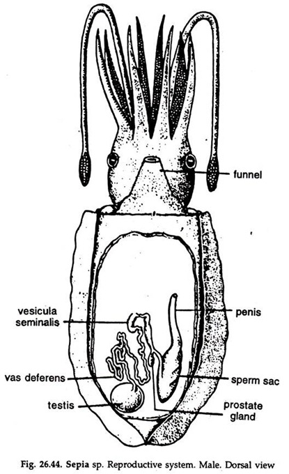
It consists of a testis, a vas deferens, a seminal vesicle, a spermatophoral sac, a penis and the male genital aperture (Fig. 26.44).
Testis:
A compact rounded mass of tubules enclosed in a capsule and situated in the aboral region of the body.
Vas deferens:
A long narrow tube, arises from the capsule of the testis. It is much coiled, moves to the left and ends in the seminal vesicle.
Seminal vesicle:
An elongated tubular structure, the interior of which is provided with grooves and ridges. Appended to it is a glandular body, the prostate. The seminal vesicle expands into a wide sac, the spermatophoral sac or Needham’s sac in which the spermatophores are stored. The spermatophoral sac continues as a stout penis and opens in the mantle cavity by the male genital aperture at its tip.
Sperms produced in the testis are carried to the seminal vesicle through the vas deferens. Here the sperms are rolled up into thin, long bundles by the action of a system of grooves and ridges.
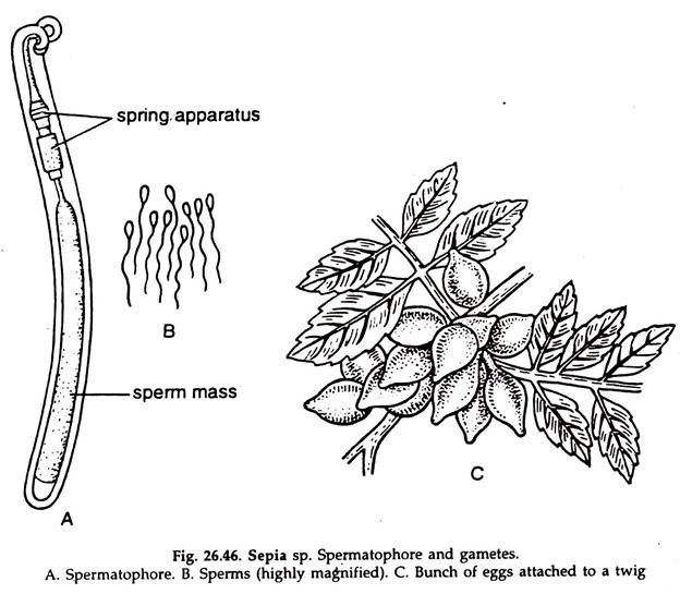
Each bundle is enclosed in a narrow cylindrical chitinoid capsule, the spermatophore. A spring-like structure is present at one end, which causes the rupture of the spermatophore. Spermatophores are stored temporarily in the spermatophoral sac and discharged in the mantle cavity through the genital aperture.
Female reproductive system:
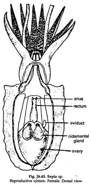
It consists of an ovary, an oviduct, the female genital aperture and a pair of nidamental glands, in association with an accessory nidamental gland (Fig. 26.45):
Ovary:
A rounded body, enclosed in a capsule and lodged in the aboral region of the body. An axial swelling bears numerous follicles, each having a single ovum.
Oviduct:
A short wide tube, arises from the capsule enclosing the ovary and opens in the mantle cavity by the genital aperture at its tip.
Nidamental glands:
A pair of large, flattened pear-shaped glands, situated on the anterior wall of the mantle cavity and to the right and left of the ink duct. A median canal is present in the long axis of each gland, on either side of which are present closely set delicate lamellae.
The median canal opens into the mantle cavity by a slit. The nidamental glands secrete a viscid material by means of which eggs adhere together after deposition. Another glandular mass, accessory nidamental gland, of unknown function is found close to the nidamental glands.
9. Fertilization and Development in Cuttle Fish:
1. Spermatophores (Fig. 26.46A) containing sperms (Fig, 26.46B) are transferred to female during copulation.
2. The hectocotylised arm receives spermatophytes in its specialised tip. The arm is inserted into the mantle cavity of the female and the tip is detached there.
3. Fertilization internal; eggs are large sized, pear-shaped and heavily yoked.
4. The eggs are laid shortly after copulation in masses in a soft, gelatinous substance which becomes attached to some aquatic support. (Fig. 26.46C).
5. Development direct; young resembling the adult hatches out and starts independent life.
