
In this essay we will discuss about:- 1. External Features of Cockroach 2. Body Cavity of Cockroach 3. Digestive System 4. Respiratory System 5. Circulatory System 6. Excretory System 7. Nervous System 8. Endocrine System 9. Reproductive System 10. Fertilization and Development.
Contents:
- Essay on the External Features of Cockroach
- Essay on Body Cavity of Cockroach
- Essay on the Digestive System of Cockroach
- Essay on the Respiratory System of Cockroach
- Essay on the Circulatory System of Cockroach
- Essay on the Excretory System of Cockroach
- Essay on the Nervous System of Cockroach
- Essay on the Endocrine System of Cockroach
- Essay on the Reproductive System of Cockroach
- Essay on the Fertilization and Development in Cockroach
Essay # 1
. External Features of Cockroach:
The entire body surface is invested in a chitinous cuticle of brown colour.
ADVERTISEMENTS:
The body is divisible into four distinct regions—the head, the neck, the thorax and the abdomen (Fig. 25.25):

1. Head:
ADVERTISEMENTS:
The head is triangular. The narrower end is directed downward and the broad end is attached to the body at right angle, through the neck.
i. The mouth is situated in front of the head. It is guarded by the labrum or upper lip in front, labium or lower lip behind and a pair of mandibles on the sides.
ii. A pair of compound eyes, appearing as Reni form black patches, are present on the sides of the head.
iii. A pair of many jointed, long, slender feelers or antennae are present just below the eyes.
ADVERTISEMENTS:
iv. A more flexible pair of jaws, the first maxillae, are situated behind the mandibles.
2. Neck:
The neck is a small narrow region connecting the head with the thorax. It is covered by a thin, flexible cuticle supported by thickened and hardened patches, the cervical sclerites.
3. Thorax:
The thorax consists of three segments, prothorax, mesothorax and metathorax covered by a chitinous dorsal tergum and a ventral sternum.
i. Pronatum is a shield-like hard structure present on the dorsal surface of the prothorax.
ii. A pair of thick, opaque, brown plates, the elytra or anterior wings are attached to the anterior border of the tergum of the mesothorax.
iii. A pair of delicate, membranous posterior wings are attached to the tergum of the metathorax.
iv. A pair of five jointed legs are attached to the sternum of each of the thoracic segments.
ADVERTISEMENTS:
4. Abdomen:
The abdomen gradually narrows down posteriorly. It consists of ten segments. Each segment is covered by a thin tergum and a sternum.
i. A segment is partially covered at the anterior end by its preceding segment.
ii. The eighth and ninth terga are hidden from view by being overlapped by the seventh.
iii. In the female, the sternum of the seventh segment is very much prominent.
iv. In the male, the sternum of the ninth segment bears a pair of short styles.
v. The tergum of the tenth segment projects backward as a flexible plate and bears a deep notch at the middle. The anus is ventral to it and guarded by a pair of many jointed anal cerci at the sides.
vi. The genital aperture is ventral to the anal opening.
vii. Ten pairs of respiratory apertures called stigmata, two in the thorax and eight in the abdomen are present on the sides of the body.
Appendages:
Appendages are seven pairs; four pairs in the head and three pairs in the thorax:
Cephalic Appendages (Figs. 25.25; 25.26):
1. Antennae:
Each antenna is long, slender and many-jointed (Fig. 25.25)
Function:
Sensory.
Mandibles, 1st maxillae, 2nd maxillae and hypo pharynx constitute the mouth parts.
2. Mandibles:
The body is a strong chitinoid structure provided with teeth.
Function:
Cutting of food.
3. 1st maxillae:
Each consists of a protopodite bearing a double endopodite having lacinia and galea and a five-jointed exopodite. The maxillary palp is supposed to represent the endopodite of crustacea.
Functions:
Holding and masticating the food and sensory.
4. 2nd maxillae:
The pair fused to form a labium. The fused base form two sclerites, submentum and mentum. Mentum bears the prementum, which in turn carries an outer, paired and lobed structure, the paraglossae and an inner, paired and lobed glossae; together forming the ligula.
Functions:
Sending the food to the mouth and sensory.

Thoracic Appendages:
7. Three pairs of legs. Each leg consists of five segments, coxa, trochanter, femur, tibia and a six-jointed tarsus ending in a pair of claws (Fig. 25.27).
Function:
Walking.

Abdominal Appendages:
8. The anal cerci are possibly the relics of last abdominal appendages (Fig. 25.25).
Function:
Tactile.
The styles of male and the gonapophyses are possibly relics of appendages.
Muscles:
Muscles concerned with movement of the appendages and feeding, in running and flight and abdominal movement for respiration are striated. Insertion of muscles occur through tendons in many cases.
Essay # 2. Body Cavity of Cockroach:
The embryonic blastocoel and coelom unite to form the adult body cavity, known as haemocoel. Major part of the haemocoel is occupied by fat bodies, digestive, excretory and reproductive organs and these structures are bathed in haemolymph.
Two types of cells—bi-nucleated and with elongated nucleus, are present in the fat bodies. The cells are derived from the walls of the embryonic cavities. Glycogen stored in these cells are used during starvation.
Essay # 3. Digestive System of Cockroach:
The digestive system consists of a long alimentary canal starting at mouth and ending in anus (Fig. 25.28). Salivary glands, hepatic caeca and glands in the lining epithelium of the gut are digestive glands.

Alimentary Canal:
The alimentary canal is divided into three zones:
i. Foregut,
ii. Midgut and
iii. Hindgut.
The fore and hindgut are lined by cells of ectodermal origin, while the lining of the midgut is endodermal.
i. Foregut:
The foregut or stomodaeum starts from mouth, ending in gizzard. It is lined by cuticle:
Mouth:
Ventral in position, located at the base of the buccal cavity.
Buccal cavity:
An ill-defined chamber, bounded anteriorly by epipharynx and labrum, posteriorly by hypo pharynx and labium and laterally by two mandibles.
Pharynx:
A short, vertically oriented tube, opening into the oesophagus. The wall of the opening between the pharynx and the oesophagus is thick, muscular and the opening is guarded by a sphincter.
Oesophagus:
A short, narrow tube, extending up to the prothorax and ending in crop.
Crop:
There is no demarcation between the oesophagus and crop. In fact, the crop is a large, sac-like dilatation of oesophagus. It extends into the abdomen. The crop acts as a temporary reservoir of food, where it can be retained for about 60 days.
Gizzard or proventriculus:
A round, thick- walled, muscular structure, posterior to the crop. It has two parts — the anterior contains six chitinous teeth in the inner wall and the posterior, two circular hairy cushions. The food is crushed by the teeth and the hairy cushions permit only finer food particles pass to the midgut.
ii. Midgut or Mesenteron:
A narrow tube, running from the gizzard to the hindgut. 7 to 8 hepatic caeca are present at the junction of the gizzard and the hindgut. The hepatic caeca presumably secrete digestive enzymes.
iii. Hindgut:
A narrow tube, divisible into three zones — ileum, colon and rectum. The junction of the mid and hindgut is marked by 60 to 70 extremely fine, yellowish Malpighian tubules. (The tubules are excretory in function). The ileum is narrow; colon broad and slightly coiled, with irregular folds of the lining epithelium, covered with a layer of chitin.
The rectum is sac-like, bearing papillae on the inner wall. Rectal glands in the wall absorb water and salt and help in osmoregulation. The rectum opens to the exterior through anus, provided with a sphincter muscle, between two podical plates at the posterior end.
Digestive Glands:
1. Salivary apparatus:
It consists of two salivary glands and two receptacles with their ducts (Fig. 25.29).

The glands and the receptacles (Fig. 25.28) lie on the dorsolateral aspects of the crop. Each gland consists of two leaf-like glands, made of diffused lobes. The receptacles are elongate oval sacs, the anterior ends being narrower.
The ducts of the glands and receptacles run forward by the sides of the crop. The ducts from the two glands unite and those from the receptacles also unite to form two common ducts, which again unite and form an efferent salivary duct opening on the ventral side of the hypo pharynx.
2. Hepatic caeca:
7 to 8 in number, at the junction of the fore and midgut (Fig. 25. 28). The caeca are about one-third of the midgut in diameter and open in it. The cells in the lining epithelium of the caeca are believed to secrete digestive enzymes.
3. Glands of the midgut:
The cells of the lining epithelium of the midgut secrete digestive enzymes.
Feeding and Digestion:
Cockroach is an almost omnivore but it prefers starchly food.
a. The maxillae procure the food, the mandibles cut it into pieces and the food is taken into the buccal cavity.
b. In buccal cavity, the food is mixed with saliva and passes to the crop through oesophagus. Both peristaltic and anti-peristaltic movements occur in the crop.
c. From the crop the food is moved to the gizzard, where it is crushed with cuticular teeth and only the finer particles passing through hairy cushions enter the midgut.
d. Midgut is the site of both digestion and absorption. Carbohydrates, proteinases and lipases are secreted by digestive glands.
e. The undigested part of the food is passed to the hindgut, where water and salts are absorbed, the residue temporarily stored there and finally egested through anus.
Essay # 4. Respiratory System of Cockroach:
The Periplaneta live on land and the respiratory structures, the tracheae, are considered as most, efficient for utilizing atmospheric oxygen.
1. The respiratory organs consist of tracheae or air tubes and their branches or tracheoles which are directly communicated with the exterior (Fig. 25.30).

2. The tracheae open to the exterior by ten pairs of apertures, the spiracles or stigmata. Two are placed on each side of the thorax; one between pro-and mesothorax and the other between meso-and metathorax. Eight occur on each side of the abdomen between terga and sterna of the first eight segments.
The stigmata are guarded by hairs, etc., to prevent the entrance of harmful particles. Each spiracle is controlled by muscular valves. From each spiracle leads inward a small tube which ends in a stout trachea and all of them are connected by longitudinal and transverse tracheae.
3. There are two lateral longitudinal tracheal trunks, one on each side of the body. They are connected by several transverse tracheae, and the tracheae form a system of intercommunicating network. Each trachea divides and subdivides into numerous branches and the ultimate, very fine branches entering the organs are called tracheoles.
The tracheoles end in tissues. The wall of the trachea is strengthened by chitinoid lining in the firm of spiral threads.
Mechanism of Respiration:
The alternate expansion and contraction of the abdomen brings inhalation and exhalation of air in the tracheal system. The opening and closing of the spiracles are also dependent on the CO2 concentration of the inhaled air. The walls of the tracheoles allow gaseous exchange and diffusion takes place between the cell sap and the lumen of the tube. The tissues get direct supply of oxygen from the atmospheric air.
Essay # 5. Circulatory System of Cockroach:
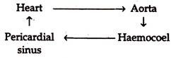
The heart and pericardium:
The heart (Fig 25.31) is an elongated tube, which runs through the whole length of the thorax and abdomen as a dorsomedian contractile vessel. The cavity of the vessel is divided into 13 segmentary arranged chambers, of which 3 are thoracic and 10 are abdominal. Externally, each chamber is marked by constrictions and internally communicates with the adjacent chambers through narrow passages guarded by valves.
The wall of each chamber is perforated by two openings known as Ostia. The whole structure is enclosed in a space known as the pericardial sinus bounded by a thin membranous pericardium. The pericardium also bears a number of perforations and as a result, the pericardial cavity is in communication with the perivisceral cavity or the haemocoel.

Arteries:
The heart is anteriorly continued into the head as a slender tube, the aorta. The aorta runs forward along the dorsal surface of the oesophagus and ends in front of the peripharyngeal nerve ring in the form of a funnel. By the successive contraction of the chambers of the heart, haemolymph is driven forward and poured in the haemocoel through the aorta. Here, the blood bathes all the visceral organs.
Sinuses:
The haemocoel remains filled up with haemolymph coming from the heart. From there it enters the pericardial sinus through apertures in the pericardium.
Haemolymph:
Haemolymph is a watery fluid having no colour. White blood corpuscles, the amoebocytes float in it. The haemolymph bears no respiratory pigment and, as such, it is not concerned with respiration.
Course of Circulation:
Essay # 6. Excretory System of Cockroach:
1. The excretory organs consist mainly of numerous Malpighian tubules and nephrocytes (Fig. 25.28).
2. The Malpighian tubules are thread-like yellow structures of variable number present at the junction of the mid and hindgut. Each tubule is a hollow structure opening into the lumen of the intestine and the wall is made of a single layer of glandular epithelium.
Nitrogenous wastes are absorbed by the cells from the haemolymph and the same are discharged into the lumen of the tubule which, in turn, convey the wastes into the lumen of the intestine.
3. Certain cells, the nephrocytes associated with the fat bodies and the hypodermis also serve as excretory structures.
Essay # 7. Nervous System of Cockroach:
The nervous system comprises of central nervous system, peripheral nervous system and visceral nervous system (Fig 25.32):
A. Central Nervous System:
It consists of a brain or supraoesophageal ganglia, sub-oesophageal ganglia, circumoesophageal connectives and a double ventral nerve cord with ganglia.
1. Brain or supraoesophageal ganglia:
It is a large bilobed mass at the posterior part of the head, below the epicranial plates and above the oesophagus and formed by the fusion of three pairs of ganglia (Fig. 25.32A).

2. Sub-oesophageal ganglia:
Formed by the fusion of three pairs of ganglia associated with the segments that produce mouth appendages and located in the head below the oesophagus.
3. Circumoesophageal connectives:
The brain is connected with the sub-oesophageal ganglia by a pair of short, stout nerves, running around the oesophagus.
4. Ventral nerve cord:
The nerve cord is formed by the fusion of two solid nerve cords and run backwards from the sub-oesophageal ganglia along the mid-ventral line of the thorax and abdomen.
a. The nerve cord bears nine pairs of ganglia, three pairs in the thoracic segments and six pairs in the first six abdominal segments.
b. The thoracic ganglia are large in size. The fifth abdominal ganglia located at the junction of the fifth and sixth segments is largest and occupies the terminal position of the nerve cord.
B. Peripheral Nervous System:
Nerves arising from the ganglia of the central nervous system constitute the peripheral nervous system.
1. Optic and antennary nerves:
Paired, given off from the brain and end in the compound eyes and the antennae.
2. Mandibular, maxillary and labial nerves:
Emanate from sub-oesophageal ganglia and supply mandibles, maxillae and labium.
3. Thoracic nerves:
Several pairs of nerves from each thoracic ganglion innervate the muscles, appendages and other organs in the corresponding segment. The first pair communicate with the ventral nerve cord.
4. Abdominal nerves:
Paired nerves from each abdominal ganglion end in the organs of the corresponding segment.
5. The last abdominal ganglion supplies nerves to the sixth abdominal segment and other segments behind.
C. Visceral or Sympathetic Nervous System:
The system consists of a frontal ganglion, a visceral ganglion, a pair of oesophageal ganglia, a hypocerebial ganglion and their connectives, (Fig. 25.32B).

1. Frontal ganglion:
A median ganglion, just in front of the brain and connected to it by two bilateral connectives and sends a recurrent nerve posteriorly to the visceral ganglion on the dorsal surface of the crop.
2. Oesophageal ganglia:
A pair, on the dorsolateral sides of the oesophagus, just behind the brain. The two are joined by connectives and also connected to a median hypo-cerebral ganglion.
Receptors and Sense Organs in Cockroach:
Photoreceptors, mechanoreceptors, thermo receptors and chemoreceptors constitute sense organs.
Photoreceptors:
Ocelli and eyes are photoreceptors.
1. Ocelli:
An ocellus appears as a white area near the base of each antenna. It consists of an outer transparent corneagen layer of flattened cells; 3 to 5 retinular cells; one rhabdome formed by the inner margins of the retinular cells. The corneal lens in an ocellus is single and a crude image of an object—at a close range—is formed.
2. Eyes:
The compound eyes are Reni form and located on the sides of the head. Structurally, they are similar to those of the prawn except that the pigment between the ommatidia are not retractile.
Mechanoreceptors:
Tactile hairs, campaniform stress receptors and chord atonal organs constitute mechanoreceptors.
1. Tactile hairs:
Present on antennae, mouth palpi, anal cerci and distal leg segments. These are in the form of hairs, bristles, plates, spines, etc.
2. Campaniform stress receptors:
Present on the labial palps and their adjacent areas and ridges on the segments of the leg. They are sensitive to pressure difference on surface.
3. Chordotonal organs:
Aggregates of specialised cells, arranged parallely or in the shape of a fan on the legs. They respond to vibration stimulus.
Thermo receptors:
Sensitive to temperature; located in pads between the first four tarsal segments of the leg.
Chemoreceptors:
Sensitive to chemical stimuli—smell or touch.
1. Olfactory organs:
Distributed on the antennae; represented in two forms—thick- walled bristles and thin-walled hairs.
2. Taste receptors:
Distributed on the tips of the maxillary and labial palps, inner surface of the mouth parts and inner border of the mouth and pharynx.
Essay # 8. Endocrine System of Cockroach:
Endocrine organs, corpora cardiaca, corpora allata and prothoracic glands constitute the endocrine system.
1. Corpora cardiac:
Corpora cardiaca are oblong in shape, 1-2 mm in size and located close to the posterior part of the brain. The neurohormone secreted by the glands controls the contractility of heart, muscles of the gut and Malpighian tubules.
2. Corpora allata:
A pair of small spherical glands, 0.5 mm in diameter, ectodermal in origin, close to the oesophageal glands, behind corpora cardiaca. The secretion initiates moulting, occyte formation and development of secondary sexual organs.
3. Ecdysial gland (Prothoracic gland):
A semitransparent gland in the mid-ventral position of the prothorax. It consists of two bands of tissues that cross in the form of ‘X’: The anterior arms of the cross are relatively long, the overall length being 3.0 ± 2.0 mm. The breadth of the anterior arm is 0.025 ± 0.005 mm.
9. Reproductive System of Cockroach:
The sexes are separate.
Male reproductive system:
1. The male reproductive system consists of a pair of testes, a pair of vasa deferentia, a pair of seminal vesicles, an ejaculatory duct, a conglobate gland and a gonopore (Fig 25. 33).

2. The testes are small bodies made up of a number of vesicles and situated in the 4th and 5th abdominal segments below the terga.
3. Each testis is connected to a narrow duct, the vas deferens, which runs backwards, inwards and downwards.
4. Each vas deferens, before uniting with its fellow of the opposite side, dilates a little forming a seminal vesicle, from which arise a large number of blind pouches and the whole arrangement is known as mushroom gland.
After forming the structure, the two vasa deferentia unite and form a muscular tube, the ejaculatory duct, which opens in the gonopore, below the anus, guarded by gonapophyses. The conglobate gland is associated with the ejaculatory duct.
Female Reproductive System:
1. The female reproductive system consists of a pair of ovaries, a pair of oviducts, a pair of colleterial glands, a pair of spermathecae and a gonopore (vulva) (Fig. 25.34).

2. Ovaries are situated in the abdomen. Each ovary consists of 8 ovarioles connected anteriorly with the body wall by a ligament.
3. From the posterior end of the ovariole runs a narrow tubule and by the union of tubules coming from all the ovarioles of an ovary, a wider tube, the oviduct is formed. The two oviducts unite to form a median chamber, sometimes called the vagina, which opens in the gonopore (vulva) guarded by the gonapophyses on the sternum of the 8th abdominal segment.
A pair of asymmetrical spermathecae of which only one is functional, open together in the genital pouch in the middle of the sternum of the 9th segment. A pair of colleterial glands open behind the spermathecae.
Essay # 10. Fertilization and Development in Cockroach:
1. Spermatophores packed with sperms 0are transferred to female during mating and the sperms are temporarily stored in the spermathecae.
2. Sixteen centrolecithel eggs from each ovary pass to the genital pouch through the oviduct and fertilized there.
3. Sixteen fertilized eggs cure enclosed in a case, the Ootheca, formed by the secretion of the colleterial glands.
4. Development direct and the young come out of the ootheca as nymphs. The pymphs are soft-bodied, white, without gonads and resemble the adult, except being smaller in size and wings lacking.
5. The nymphs undergo several moultings and the stage between two successive moultings is called instar. Wings appear with the completion of last moulting.