In this article we will discuss about:- 1. Description of Scorpion 2. External Features of Scorpion 3. Digestive System 4. Respiratory System 5. Circulatory System 6. Excretory System 7. Nervous System 8. Reproductive System 9. Fertilization and Development.
Contents:
- Description of Scorpion
- External Features of Scorpion
- Digestive System of Scorpion
- Respiratory System of Scorpion
- Circulatory System of Scorpion
- Excretory System of Scorpion
- Nervous System of Scorpion
- Reproductive System of Scorpion
- Fertilization and Development of Scorpion
1. Description of Scorpion:
The Scorpion, Palamnaeus sp. belongs to the class Arachnida, phylum Arthropoda. Scorpions are one of the oldest terrestrial arthropods and the present day species differ very little from their fossil ancestors.
ADVERTISEMENTS:
Scorpions are widely distributed and found in tropical and subtropical regions, from mountains to plains, rain forests to deserts. They live in holes, crevices, under stones, logs of wood, decaying leaves and organic matters, within sand and many other places.
2. External Features of Scorpion:
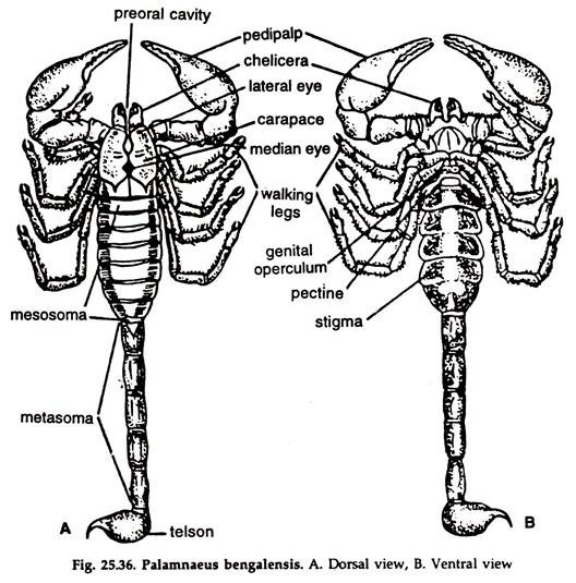
1. The body is narrow, elongated, dorsoventrally flattened and divisible into an anterior Prosoma or Cephalothorax and a posterior Opisthosoma or abdomen (Fig 25.36).
2. The colour of the exoskeletal covering harmonizes to a considerable extent with the surroundings and ranges from yellowish orange to black.
ADVERTISEMENTS:
3. Prosoma:
It is formed by the fusion of five segments:
a. Dorsally, the prosoma is covered by an un-segmented, more or less square Carapace, formed by the fusion of the terga of the segments. The anterior border of the carapace is notched, forming a right and a left frontal lobe.
ADVERTISEMENTS:
b. A single, triangular plate formed by the fusion of sternites, cover the ventral surface of the prosoma.
c. A pair of simple, median eyes at the middle and 2-3 pairs of lateral eyes on the anterolateral margins are present in the carapace.
d. The mouth is a small, transverse, ventrally located opening at the anterior end of the cephalothorax. A small, fleshy labrum overhangs the mouth ventrally.
e. The cephalothorax bears six pairs of appendages:
i. A pair of three-jointed chelicerae anteriorly.
ii. A pair of six-jointed pedipalpi posterior to chelicerae.
iii. Four pairs of seven-jointed walking legs.
4. Opisthosoma:
It is immovably attached to the cephalothorax and divisible into an anterior mesosoma or preabdomen and a posterior metasoma or post abdomen:
ADVERTISEMENTS:
A. Mesosoma:
It is broad, flat and consists of seven segments. Each segment has a dorsal tergum, a ventral sternum, connected by soft integument at the sides.
The mesosoma bears two pairs of modified appendages, and four pairs of stigmata:
a. Genital operculum:
A movable, bifid flap, covering the genital opening, at the middle of the narrow sternum of the first abdominal segment.
b. Pectines:
A pair of comb-like structures attached to the sternum of the second abdominal segment.
The genital operculum and pectines are considered as modified appendages.
c. Stigmata:
Oblique, slit-like openings on the ventrolateral borders of the 3, 4, 5 and 6 pre-abdominal somites.
B. Metosoma:
It is narrow, tubular, five-segmented, enclosed in a complete ring of chitinous material and flexibly jointed. In live specimen, it is turned upward over the mesosoma.
The metosoma bears:
a. Poison bulb:
The terminal metasomal somite is bulb-like at the base and pointed at die apex, forming a poison sting. A pair of poison glands open in the tip of the sting.
b. Anus:
Situated ventrally in the last metasomal somite, immediately in front of the sting.
Appendages:
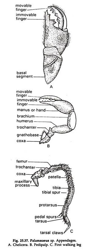
Appendages are six pairs, two pairs in the head and four pairs in the thorax (Fig. 25.37):
Cephalic appendages:
1. Chelicerae:
Paired, preoral in position. The appendage is small, three-jointed, ending in a chela.
Function:
Holding the prey.
2. Pedipalpi:
Paired, post-oral in position. It is a large, six-jointed structure ending in a great chela.
Functions:
Seizure of prey and mastication by the bases.
Thoracic appendages:
Four pairs of legs. Each leg consists of seven segments, the last one ending in a pair of curved, pointed, horny claws. The claws are absent in the 3rd and 4th pairs of legs.
Functions:
Walking but in the first and second pairs, the bases help in mastication of food.
Abdominal appendages:
The operculum bearing the genital pore and the comb-like tactile pectines are probably the remnants of the first and second pairs of abdominal appendages, respectively.
Endoskeleton:
The coxa of the walking legs and the endosternite constitute the endoskeleton:
1. Coxa or apodemes of the walking legs are inserted within the body for attachment of leg muscles.
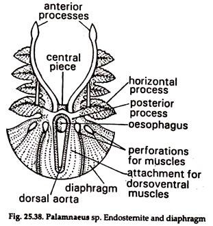
2. Endosternite. A more or less triangular cartilage with perforations for aorta and oesophagus (Fig. 25. 38) is placed obliquely between the prosoma and mesosoma. It has three processes—anterior, posterior and lateral, for attachment of muscles.
3. Digestive System of Scorpion:
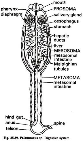
The digestive system consists of a long, alimentary canal, starting from the preoral cavity and ending in anus (Fig 25.39) in the last metasomal somite. Salivary or stomach glands and gastric glands or liver constitute digestive glands.
Alimentary Canal:
The alimentary canal is divided into three zones—foregut, midgut and hindgut. The fore and hindgut are lined by cells of ectodermal origin, while the lining of the midgut is endodermal:
Foregut:
The sclerotised foregut is divisible into a preoral cavity,mouth, a pharynx and an oesophagus:
1. Preoral cavity:
It is located at the anterior end of the cephalothorax and bounded dorsally by two chelicerae, ventrally by gnathobases of the first two pairs of legs and laterally by the coxae of the pedipalpi. Labrum, a club-shaped structure hangs in the cavity.
2. Mouth:
A small transverse opening inside the preoral cavity, near the base of the labrum. It leads to the pharynx.
3. Pharynx:
A well-developed suctorial bulb, leading to the oesophagus. The pharyngeal wall is muscular and a number of radiating bundles of muscle fibres from it run to the walls of the cephalothorax.
4. Oesophagus:
A short tube, opening posteriorly into the midgut.
Midgut:
A long tube with a lining of glandular epithelium and divisible into a stomach and an intestine:
1. Stomach:
A small sac, runs up to the diaphragm and continues posteriorly as intestine.
2. Intestine:
A long, narrow, straight tube runs posteriorly along the middle line of the body and divisible into an anterior mesosomal or pre-abdominal and a posterior metasomal or post-abdominal intestine.
The junction of the two is demarcated by the presence of Malpighian tubules. The mesosomal part is embedded in the liver and receives five to six pairs of narrow hepatic ducts, one after another. The metasomal intestine ends posteriorly in the hindgut.
Hindgut:
It is a small cuticularised tube in the last metasomal segment and opening to the exterior through ventral anus in front of the sting.
Digestive Glands of Scorpion:
Salivary or stomach glands, gastric glands or liver constitute digestive glands. Unicellular glands in the lining epithelium of the midgut secrete digestive juice:
1. Salivary glands:
Paired glands, located on either side of the oesophagus in the prosoma. Saliva, the secretion of the glands, is discharged into the oesophagus through separate ducts from the glands.
2. Liver:
A large, paired glandular mass occupying the dorsal part of the mesosomatic cavity. The secretion of the gland is released in the lumen of the intestine through 5-6 pairs of hepatic ducts. Each duct is formed by the union of a number of smaller ducts arising in the mass of the liver.
Feeding and digestion:
Scorpion is nocturnal and comes out in search of food only at night. It is a predator and depends chiefly on insects, spiders, myriapods, etc. for nourishment.
1. The prey, which comes in contact with the appendages in the dark is grabbed by the chela at the pedipalps and stung.
2. The prey is brought close to the preoral cavity and wounds are made on the body by the chelicerae.
3. The strong suctorial pharynx sucks fluid from the victim’s body and saliva mixes with it in the preoral cavity.
4. The partially digested food is moved to the midgut, where further digestion occurs with the help of enzymes, released by the liver and unicellular glands in the gut wall, and the undigested part of the food is moved to the hindgut.
5. From the midgut food reaches the liver through hepatic ducts, where digestion is completed.
6. Residue collected in the hindgut is egested through the anus.
4. Respiratory System of Scorpion:
The Palamnaeus live on land and the respiratory structures, book-lungs can use atmospheric oxygen. Though physiologically less efficient, the book-lung is a more highly evolved respiratory organ, than those in other arthropods.
Respiratory organs:
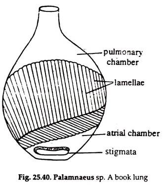
1. The respiratory organs consist of four pairs of book-lungs. The book-lungs open to the exterior by four pairs of apertures, the stigmata (Fig. 25.36B). Sternum of each of the 3rd, 4th, 5th and 6th pre-abdominal segments bears a pair of stigmata on its ventrolateral sides. These are narrow oblique slits. Each stigmata opens directly into a book- lung and they are not interconnected.
2. Each book-lung is a compressed sac lined with a thin cuticle. The lining membrane is folded up into numerous delicate laminae lying parallel to one another like the pages of a book, roughly 130-150 in number (Fig. 25.40).
3. Deoxygenated haemolymph flows through the narrow space in each lamina, separated from the air only by the membranous walls of the lamina.
Mechanism of respiration:
The alternate expansion and contraction of the abdomen brings inhalation and exhalation of air in the book-lungs. Gaseous exchange takes place between the haemolymph in the laminae and the air in between the laminae.
5. Circulatory System of Scorpion:
Heart and Pericardium:
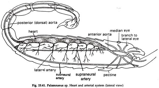
The heart is a dorsal elongated tube lying in a groove in the liver mass, entirely in the pre-abdominal region, extending from the seventh to the thirteenth segment. It is clearly demarcated in its anterior and posterior limits by valves.
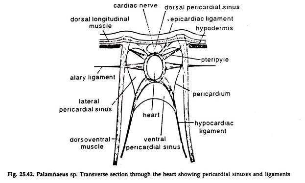
The heart is single-chambered. (Fig, 25.41). Seven pairs of Ostia, guarded by valves, are present on the walls of the heart. The heart is suspended by eight groups of ligaments arranged metamerically (Fig. 25.42). The pericardium is thin, membranous and horizontal in position.
The pericardial cavity is divided into four sinuses, two laterals, one ventral and one dorsal. The cardiac nerve extends from one to the other end of the heart.
Haemolymph:
The freshly shed haemolymph is indigo in colour. White blood corpuscles, the amoebocytes float in it. The haemolymph contains a blue coloured respiratory pigment, proteidhaemocyanin. The oxygenated haemolymph is bright blue, but colourless when deoxygenated.
Arteries (Fig 25.41):
The heart continues anteriorly as the anterior aorta for a considerable distance. Afterwards, it divides into two branches, which embrace the oesophagus on both the sides and unite below in the form of a short arch connecting the aorta with the right and left thoracic sinuses. In the ventral region, from the arch comes out a common vessel that runs over the nerve cord. This vessel is known as the supraneural artery.
On reaching the anterior end of the sub-oesophageal ganglionic mass it turns downward and then runs backward as a sub-neural artery ending behind the ganglionic mass.
The supra and sub-neural arteries are connected by nine vertical arteries.
A pair of large cephalic arteries arise from the aortic arch. Each gives off a number of branches, of which the ophthalmic artery and the cheliceral artery are more important. The ophthalmic supplies haemolymph to the eye and the cheliceral to the chelicera.
Each thoracic sinus sends a small vessel to the supraneural artery and four large vessels to the appendages supplying haemolymph to the pedipalp and legs of the side. The heart continues posteriorly as posterior aorta. It runs straight backward up to the posterior tip and supplies to the intestine as well as various organs by a number Of delicate branches.
Sinuses:
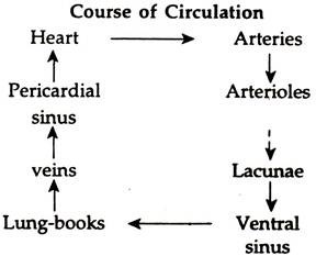
The ventral sinus is a large space where haemolymph from various parts of the body is collected as well as sent to the respiratory organs or lung-books for aeration. Between the roof of the ventral sinus and the floor of the pericardial sinus a special arrangement of muscles is found.
The contraction of these muscles causes the ventral sinus to enlarge and thereby deoxygenated haemolymph is drawn into it. The relaxation of these muscles results in the pumping of haemolymph into the book-lungs. A series of segmental vessels help in the conduction of haemolymph from the lung-books to pericardial sinus.
6. Excretory System of Scorpion:
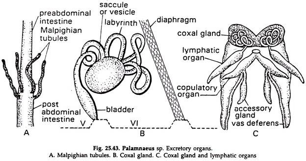
The excretory organs consist of a few Malpighian tubules and a pair of coxal glands (Fig. 25.43):
Malpighian tubules:
The Malpighian tubules (Fig. 25.43A) are delicate tubes and are one or two pairs in number, attached to the intestine. Each tubule is a hollow structure opening into the lumen of the intestine and the wall is made of a single layer of glandular cells.
Nitrogenous wastes are absorbed by the cells from the haemolymph and the same are discharged into the lumen of the tubule which, in turn, convey the wastes into the lumen of the intestine (Fig. 25.43A).
Coxal glands:
A pair of coxal glands are found near the bases of the 3rd walking legs in the 5th segment of the prosoma. These are derived from coelomoducts and function as excretory organs. In the embryo of scorpion, the coelomoducts are found in the 3rd, 4th, 5th, 6th and 8th segments.
All of them disappear in the adult except those in the 5th segment which persist as coxal glands. Each coxal gland consists of an end sac, a colied tube and a bladder opening to the exterior by a duct at the base of the 3rd walking leg (Fig. 25.43B).
Lymphatic organ:
A diverticulum or lymphatic organ, possibly excretory (phagocytic) in function, lies on each side of abdomen connected with the coxal gland of the side (Fig. 25.43C).
Nephrocytes:
The nephrocytes are large cells, located in the walls of the mesosoma and are believed to be of excretory function.
7. Nervous System of Scorpion:
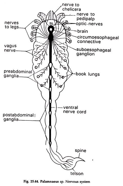
The nervous system consists of a central nervous system, a peripheral nervous system and a visceral nervous system (Fig. 25.44):
Central Nervous System:
The central nervous system includes a brain or supraoesophageal ganglia, sub-oesophageal ganglia, circumoesophageal connectives and a double, ventral ganglionated nerve cord:
1. Brain or Supraoesophageal ganglia:
It is a small bilobed mass, situated in the prosoma, just beneath the median eyes.
2. Sub-oesophageal ganglia:
It is formed by the fusion of ten pairs of ganglia belonging to embryonic 2 to 11 segments and located ventromedially below the oesophagus.
3. Circumoesophageal connectives:
A pair of short and stout nerves arise from the brain, one on each side, encircle the oesophagus and unite ventrally with the sub-oesophageal ganglia.
4. Ventral nerve cord:
A double ventral nerve cord formed by the union of two solid nerve cords runs posteriorly from the sub-oesophageal ganglia up to the fourth segment of the metasoma. The nerve cord bears three pre-abdominal ganglia in the mesosoma and four post-abdominal ganglia in the metasoma, the last one formed by the fusion of two ganglia of the 18 and 19 segments and located in the fourth metasomal segment.
Peripheral nervous system:
Nerves arising from the ganglia of the central nervous system:
1. The brain gives off paired nerves to median and lateral eyes and rostrum and a number of fine nerves to the walls of the preoral cavity, pharynx, oesophagus and adjoining structures.
2. Six pairs of lateral nerves emanating from the sub-oesophageal ganglia innervate the chelicerae, pedipalpi and four pairs of walking legs.
3. Two to four pairs of vagus nerves arising from the sub-oesophageal ganglia run posteriorly to supply the heart and alimentary canal, genital operculum, the pectines and the first two pairs of book-lungs in the mesosoma.
4. Paired nerves emanate from each ganglion of the ventral nerve cord and innervate the structures in the corresponding segments. Nerves from the first two pre-abdominal ganglia supply the last two pairs of book-lungs.
Receptors and Sense Organs of Scorpion:
The sense organs consist of eyes, pectines, sensory setae, trichobothria and slit sense organs:
1. Eyes:
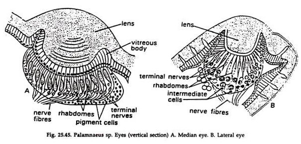
Both the median and lateral eyes are simple eyes and provided with a lens.
a. Median eye:
It is diplostichus type (Fig. 25.45A).
i. In the lower part of the lens the hypo- dermal cells form a vitreous body.
ii. The rhabdome of the retinulae is formed of five rhabdomeres.
b. Lateral eye:
It is monostichus type. The vitreous body is absent; the rhabdome is of irregular shape. (Fig. 25.45B).
Functions:
Not properly understood.
2. Pectines:
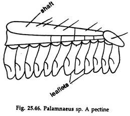
a. Comb-like in appearance and consists of a shaft and movable processes (Fig. 25.46).
b. The shaft is three segmented.
c. Movable processes, also called leaflets are 4 to over 30 and attached in a row along the posterior margin of the shaft.
Functions:
Food procurement and breeding.
3. Sensory setae:
Fine hair-like structures scattered all over the body.
Functions:
Tactile sense organs.
4. Trichobothria:
About 66-68 are present in each chela.
Each organ consists of:
a. A flask-shaped, cuticularised structure, the bothrium.
b. The trichobothrium is a seta, inserted into the bothrium through the middle of a cuticular outer membrane.
c. The seta bends and narrows down within a fluid-filled cylinder supplied by nerve fibres.
Functions:
Response to air current.
5. Slit sense organs:
Scattered all over the body, being crowded on the appendages.
A slit sense organ consists of:
a. A slit on the integument, covered with a membranous epicuticular covering and measures about 0.005-0.16×0.002-0.003 mm.
b. The slit leads to a narrow passage, which is a membrane-lined tube and penetrates in the procuticle.
c. The inner walls of the tube receive rich supply of nerve fibres.
Function:
Concerned with the movement of appendages and also respond to vibration.
8. Reproductive System of Scorpion:
The sexes are separate.
Male reproductive system:
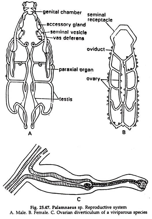
1. The male reproductive system consists of a pair of testes, a pair of vasa deferentia, a pair of genital chambers, a pair of paraxial organs, a pair of flagella, a pair of seminal vesicles, a common genital atrium and a gonopore (Fig. 25.47A).
2. Each testis consists of a pair of longitudinal tubules placed in the pre-abdomen, and connected by transverse tubes.
3. The two longitudinal tubules of each testis unite anteriorly to form a vas deferens, which runs forward and downwards.
4. The terminal portion of each vas deferens opens into the inner side of the genital chamber along with an enlarged seminal vesicle. The genital chambers are muscular tubes and bear the paraxial organ having a chitinoid flagellum. It also receives the accessory gland. The genital chambers unite to form a median genital atrium, which opens to the exterior by the gonopore situated just behind the operculum.
Female reproductive system:
1. The female reproductive system consist of a single ovary, two oviducts, a median genital chamber and a gonopore (Fig. 25.47B).
2. Ovary is situated in the preabdomen. It consists of three longitudinal tubules connected by cross branches. The median tubule is shorter than the other two.
The ovarian tubules and the cross branches bear numerous diverticula (Fig. 25.47C).
3. From the two lateral tubules run two narrow oviducts. Anteriorly each oviduct is slightly swollen and form the seminal receptacle and the two open in the sac-like median genital chamber, the latter opening in the gonopore on the second abdominal segment below the bilobed operculum.
9. Fertilization and Development of Scorpion:
Prior to breeding a nuptial dance is of universal occurrence in scorpion. The dance may continue for 30 to 90 minutes. During the dance, the male scratches a suitable surface with the help of its pectines for depositing the spermatophores.
1. The male deposits the spermatophores on a preselected site.
2. The male drags the female over the site.
3. The lever of the spermatophore being touched by the female genital operculum, the ejecting apparatus shots the sperms within the female genital duct. Fertilization is internal.
4. Palamnaeus is viviparous. Ferried egg develops in the follicle of the ovary, which acts as placenta and provides nourishment to the embryo.
5. Young one resembling the adult is born. Immediately after birth, the young scorpion climbs on mother’s back, remains there for 40 to 63 days and undergoes three moulting, before starting an independent life.
