In this article we will discuss about:- 1. External Features of Prawn 2. Locomotion in Prawn 3. Digestive System 4. Feeding and Digestion 5. Respiratory System 6. Circulatory System 7. Excretory System 8. Nervous System 9. Reproductive System 10. Fertilization and Development.
Contents:
- External Features of Prawn
- Locomotion in Prawn
- Digestive System of Prawn
- Feeding and Digestion in Prawn
- Respiratory System of Prawn
- Circulatory System of Prawn
- Excretory System of Prawn
- Nervous System of Prawn
- Reproductive System of Prawn
- Fertilization and Development in Prawn
1. External Features of Prawn:
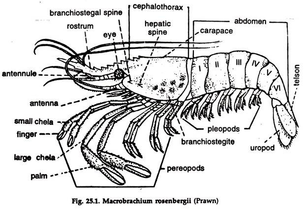
The fresh water prawn Macro brachium (former Palaemon) belongs to subclass Branchiopoda, class Crustacea, phylum Arthropoda. The genus is widely distributed in tropical and temperate countries (Fig. 25.1).
1. The size is variable, the average being 15 to 20 cm.
2. The body is elongated and divisible into an anterior cephalothorax and a posterior abdomen.
Cephalothorax in Prawn:
1. The cephalothorax is formed by the fusion of 5 cephalic and 8 thoracic segments, and covered externally by a hard cephalothoracic shield, the carapace, anteriorly drawn into a serrated and pointed rostrum.
ADVERTISEMENTS:
i. The carapace hangs freely on the sides and encloses, on either side, a narrow gill chamber containing gills or branchiae.
ii. The portion of the carapace covering the gills are called branchiostegite or gill cover.
2. A pair of stalked compound eyes are present near the base of the rostrum.
3. 13 pairs of paired, biramous appendages are present in the cephalothorax.
ADVERTISEMENTS:
The appendages are two pairs of antennae, one pair of jaws, two pairs of maxillae, three pairs of maxillipeds and five pairs of pereopods or walking legs. The first two pairs are chelate.
4. The mouth is a slit-like aperture situated on the ventral surface of the head. It is bounded by the labrum in front, labium behind, and jaws named mandibles on /either side.
5. An excretory pore opens at the base on the inner surface of each of the second antenna.
6. The male genital apertures are present at the base of the last pair of walking legs and the female genital apertures at the base of the third pair of walking legs.
Abdomen of Prawn:
1. The abdomen consists of six distinct, movable segments.
2. Each segment is enclosed in a hard cuticle divisible into a dorsal, convex tergum a pair of lateral pleurons and a ventral sternum.
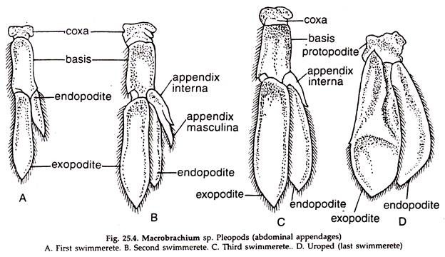
3. A pair of biramous swimming legs called pleopods or swimmerets are present in each segment. The last pair is known as uropod.
4. At the end of the abdomen a median triangular piece called telson is present. It does not carry appendages.
ADVERTISEMENTS:
5. The anus opens on the ventral side near the base of the telson.
Appendages of Prawn:
Appendages are externally projected parts of the body in the formation of which various systems of the body take part. These develop generally at right angles to the long axis of the body and serve as locomotory organs, feelers, food seizers, manipulators and sex organs.
The appendages are jointed in all arthropods (Figs. 25.2-25.4). Biramous appendages are nineteen pairs; five pairs in the head, eight pairs in the thorax and six pairs in the abdomen.
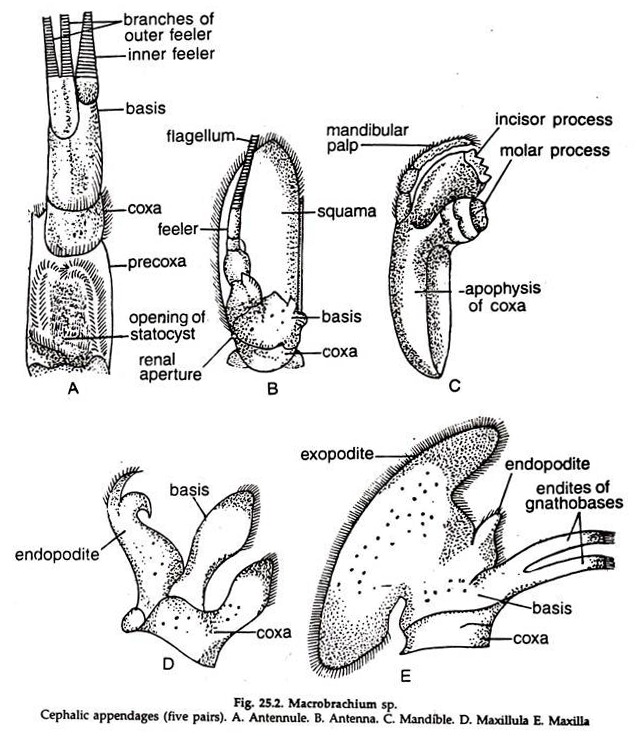
Cephalic Appendages (Fig. 25.2) of Prawn:
1. Antennules:
Each consists of a three- jointed protopodite bearing three many, jointed flagella at the distal end and a statocyst at the base.
Functions:
Sensory and tactile.
2. Antennae:
Each consists of a two- jointed protopodite bearing a flat squama and a many-jointed flagellum.
Functions:
Tactile and balance.
3. Mandibles:
The body is unjoin ted, bears teeth and masticatory lobes known as molar processes and a jointed mandibular palp on the outer surface.
Functions:
Cutting and crushing of the food.
4. 1st maxillae (maxillulae):
Leaf-like, with two inner lobes acting as gnathobases and an outer lobe.
Functions:
Tearing and passing the food to the mouth.
5. 2nd maxillae:
Leaf-like, with a flattened scaphognathite.
Functions:
Masticatory and respiratory.
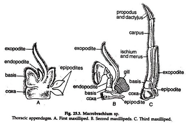
Thoracic appendages (Fig. 25.3):
6. 1st maxillipeds:
Leaf-like protopodite with a whip-like exopodite and a slender endopodite.
Functions:
Respiratory, masticatory and sending the food to the mouth.
7. 2nd maxillipeds:
The protopodite with an epipodite bears a five-jointed endopodite, a whip-like un-jointed exopodite and a gill.
Function:
Respiratory.
8. 3rd maxillipeds:
The protopodite bears a three-jointed leg-like endopodite and a slender un-jointed exopodite. A small epipodite is present.
Functions:
Locomotory and respiratory.
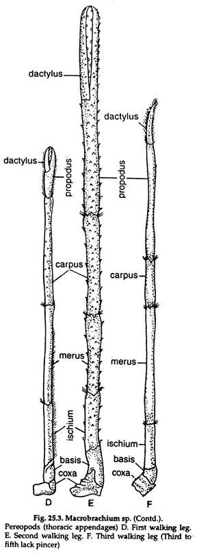
9. Pereopods (walking legs):
Five pairs; each leg consists of seven podomeres or segments.
The first two legs end in chela and the second is the largest. The two basal segments represent the coxopodite and basipodite and the remaining five are ischium, merus, carpus, propodus and dactylus, respectively, in order of succession.
Functions:
Holding the prey and walking in first two, and only walking in the last three.
Abdominal Appendages (Fig. 25.4) of Prawn:
Six pairs; each pleopod consists of a two-jointed protopodite bearing expodite and endopodite. In the second pleopod of male, an appendix masculine, concerned with reproduction is found. In the second to fifth pleopods, appendix internae are present.
Function:
Swimming.
Appendix masculine help in mating. Appendix internae form a basket in female to carry eggs.
Uropod:
Protopodite small, the exo-and endopodite are broad and oval. With the telson it forms the powerful tail fin.
Functions:
Balance and back swimming with a jerk.
2. Locomotion in Prawn:
Change of place in prawn takes place in three ways:
i. Swimming:
The body is kept straight in a horizontal position. The pleopods act like oars. The forward movement of the pleopods is slow, while the back stroke is fast and the animal moves forward. The uropod helps in guiding and the antennae move constantly, presumably feeling the surroundings.
ii. Walking:
The straightened body is supported by all the five pairs of pereopods. The walking legs move in harmony during walking.
iii. Darting:
This is resorted under emergency. With stretched pleopods and uropod the abdomen suddenly moves forward towards the cephalothorax and the animal swiftly moves backward with a jerk due to the sudden thrust.
3. Digestive System of Prawn:
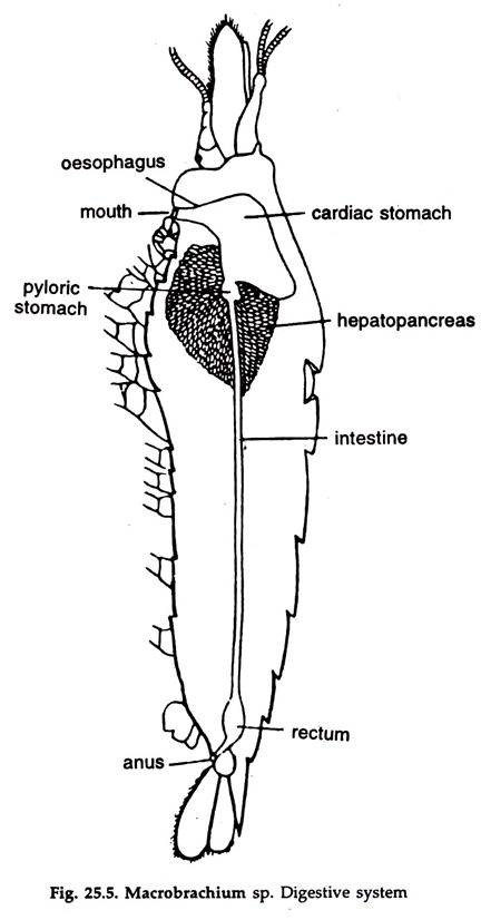
The digestive system consists of an alimentary canal and a hepatopancreas or digestive gland (Fig. 25.5).
Alimentary Canal:
It has three distinct zones—an anterior foregut ending in stomach, a midgut, the constituent of which is intestine and a hind- gut or the rectum. The fore and hindgut are lined by a layer of thick cuticle.
Foregut:
Following structure constitute the foregut.
Mouth:
A slit-like opening situated ventrally in the head region. It is bounded by labrum anteriorly, mandibles laterally and a two- lobed labium posteriorly.
Buccal cavity:
A small, anteroposteriorly compressed chamber, next to mouth, bearing irregular internal folds. Oesophagus. A wide, vertically oriented tube, joining the buccal cavity with the cardiac stomach. The inner lining bears one anterior, one posterior and two lateral folds.
Stomach of Prawn:
A spacious, horizontally oriented sac, divided into two chambers:
A. Cardiac stomach:
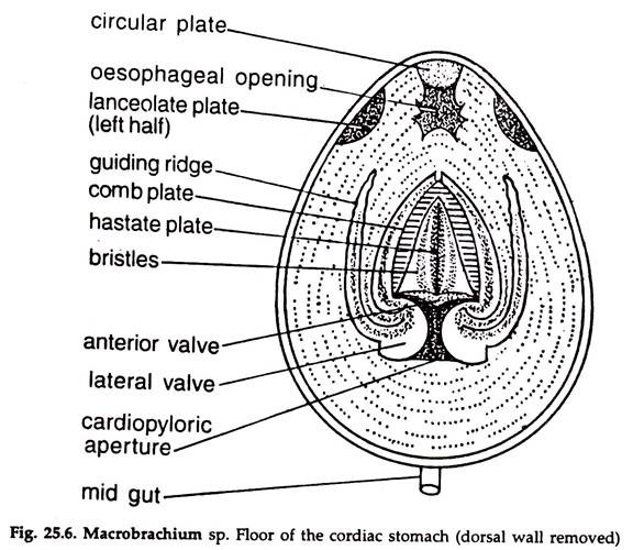
Large, bag-like, constitute the dorsal part, bearing following plates on its walls:
a. A circular plate anteriorly, just behind the oesophagus.
b. A lanceolate plate dorsally in the posterior part.
c. A hastate plate (Fig. 25.6) resembling the head of a spear in the mid-ventral region.
i. The slightly convex upper part of the hastate plate gradually slopes laterally, forming a median ridge in the middle.
ii. Delicate setae are present on both the upper and posterior surfaces of the plate,
iii. An elongated lateral groove is present on either side of the plate.
iv. Each lateral groove is bounded by a supporting rod and a ridged plate, both cuticular, on the inner and outer side, respectively.
v. A Comb plate, bearing rows of comb-like setae is present on the inner side of each ridged plate.
vi. The comb plates join at the anterior end but remain free posteriorly, close to the cardio- pyloric opening.
vii. A longitudinal guiding ridge is formed by the folding of the inner wall of the cardiac stomach, lateral to each comb plate.
viii. The two guiding ridges guide the food to the pyloric stomach through cardio- pyloric opening.
The cardiac stomach opens into the pyloric stomach through a narrow X-shaped cardio-pyloric opening, guarded by an anterior, one posterior and two lateral valves.
B. Pyloric Stomach:
a. Small in size, the lateral walls form prominent folds, imperfectly dividing the cavity into two — a small dorsal and a large ventral chamber.
b. The ventral chamber is subdivided into two lateral compartments and receive the ducts from the hepatopancreas.
c. A rectangular filter plate bearing alternate ridges and grooves is present on the floor of the ventral chamber.
d. The filter plate and the bristles of the lateral walls of the ventral chamber act as a pyloric filtering apparatus and permits only liquefied food to pass.
Midgut or Intestine in Prawn:
It is a long, slender tube. Arising from the posterior end of the pyloric stomach it runs backward, ascending between the two lobes of the hepatopancreas to reach the dorsal groove in the abdomen beyond cephalothorax and runs posteriorly to end in the rectum in the last segment.
Hindgut or Rectum in Prawn:
The last part of the alimentary canal. It is a small chamber, wider anteriorly and narrows down posteriorly to open on the ventral surface, at the base of the telson.
Hepatopancreas of Prawn:
A large, yellow-orange mass, consists of two lobes and occupies major portion of the cephalothorax. The whole of the pyloric stomach, a pail: of the cardiac stomach and the anterior part of the intestine are embedded in it.
A duct arises from each lobe of the hepatopancreas and the two open separately into the ventral chamber of the pyloric stomach, just after the pyloric filter plate. The hepatopancreas plays an important role in digestion and also acts as a storage organ.
4. Feeding and Digestion in Prawn:
Prawn feeds upon small animals, eggs of other animals, algae and decaying plant matters.
Feeding:
1. The food is captured by the chelate legs and brought to the mouth.
2. Maxillipeds, maxillulae and maxillae help in tearing it into pieces.
3. The mandibles crush the food and the crushed food is taken into the buccal cavity and from there to oesophagus.
4. Peristaltic movement of the oesophagus drives the food into the cardiac stomach.
Digestion:
1. The food is churned by the cuticular plates of the cardiac stomach and the fine particles, filtered by the comb plates, reach the lateral grooves and finally guided to the ventral chamber of the pyloric stomach.
2. Enzymes secreted by the hepatopancreas digest proteins, carbohydrates and fats.
3. The filtering apparatus filters the nutrient, which is in a liquid state and passed to the hepatopancreas via the dorsal chamber of the pyloric stomach.
4. The undigested food is moved to the intestine, where certain amount of digestion and absorption take place.
5. The residue reaches the rectum and egested through the anus.
6. The digested food material that is absorbed through the intestinal wall is circulated to different parts of the body through lacunae or sinuses.
5. Respiratory System of Prawn:
Respiration is a mechanism by which gaseous exchange takes place between the organism and the environment, in which oxygen is taken in and carbon dioxide is given out. Macro brachium lives in water and respire by gills, taking up oxygen dissolved in water.
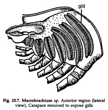
1. The respiratory organs consist of the lining membrane of the branchiostegite, three pairs of epipodites and eight pairs of gills. The gills are lodged in gill chambers, which communicates with the exterior along its anterior, posterior and ventral borders.
2. The gills are crescent-shaped and increase in size anteroposteriorly (Fig. 25.7). The first gill is smallest and the last one the largest. From their point of origin, the first gill is podobranch, being attached to the coxa of the second maxilliped, the 2nd and 3rd are arthrobranchs being attached to the membrane articulating the 3rd maxilliped with the body and the last five are pleurobranchs, being attached to the body above the articulation of the walking legs.
3. Each gill consists of a long, narrow rachis supporting two rows of rhomboidal gill-plates diverging from each other at right angles to the elongated axis. The gill-plates are larger in size in the middle but smaller towards the ends. Each gill-plate is made up of a double layer of cuticle with a single layer of cells in between. The gill is attached to the body about the middle of its length, and is highly vascular.
4. The axis of the gill is roughly triangular in cross-section. Three longitudinal canals, two laterals and one median, run along the axis. The laterals are connected with each other by transverse channels and also with the median canal by marginal channels. Haemolymph enters through the transverse channels and traverses other channels. Gaseous exchange takes place in the gill filaments. The lining membrane of the branchiostegite and the epipodites of the three maxillipeds are highly vascular and aid in the process of respiration.
Mechanism of Respiration:
The movements of scaphognathites maintain a constant backward to forward water current in the gill-chambers. The haemolymph in the respiratory organs gives up CO2 and absorbs O2.
6. Circulatory System of Prawn:
In Arthropoda, a part of the blood vascular system is expanded to surround other organs; as the coelom is reduced, the other space in the mesoderm, the haemocoel, is elaborated and functions as the cavity of the blood vascular system.
The main functions of haemolymph (blood) are to transport food and oxygen and the elimination of respiratory wastes in general, although a number of other functions are complimentary to these.
The blood of arthropods contain 87 to 98 per cent of water, and mainly 3 types of blood corpuscles are found; viz.—amoebocytes, granulocytes and thrombocytes. The respiratory pigment, haemocyanin, is a prosthetic group of copper, dissolved in the haemolymph. Haemocyanin is colourless but oxy-haemocyanin imparts blue colour to the haemolymph.
Heart and Pericardium of Prawn:
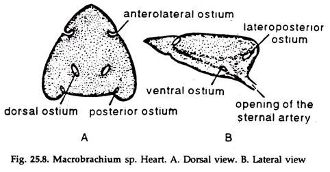
The heart (Fig. 25.8) is a hollow organ, somewhat triangular in outline, and with thick muscular walls. It is situated dorsally at the posterior end of the cephalothorax. It is lodged in a special haemocoel, the pericardial cavity, the walls of which form the pericardium. A horizontal pericardial septum forms the floor of the pericardial sinus. A median cardio pyloric strand and 2 lateral strands support the heart in the pericardium.
Five pairs of valved Ostia are present on the walls of the heart; one pair a little behind the middle on the ventral surface, one on each side; second pair opposite to the first pair on the dorsal surface; third pair on the posterior border; fourth pair behind the apex and the fifth or the last pair, one on each side of the lateral angle of the heart.
The heart is traversed by a large number of interlacing muscle fibres, the interstices of which is the cavity of the heart.
Haemolymph:
Haemolymph of the prawn is a clear fluid having a number of colourless leucocytes. The respiratory pigment is proteidhaemocyanin. The oxygenated haemolymph is shining blue, but colourless when deoxygenated.
Arteries (Fig. 25.9):
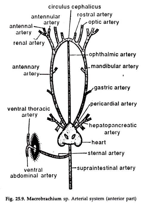
These are thick-walled vessels, through which the heart pumps out its contents—the haemolymph. From the apex of the heart proceeds anteriorly a slender, median ophthalmic artery up to the root of the oesophagus.
Two antennary arteries arise from the inner lateral sides of the heart and run anteriorly, slightly obliquely. Behind the eyes, the arteries of the two sides anastomose and form a loop, the circulas cephalicus, with which the median ophthalmic artery joins. From this loop comes off a rostral artery on each side.
Each antennary artery on its way gives off a pericardial branch to the pericardium, a gastric branch to the cardiac stomach, a mandibular branch to the mandibular muscles, and finally an optic artery supplying the eye of the side. Before giving off the optic artery, the antennary artery sends a common artery, which divides into renal, antennal and antennular branches and supply the respective organs.
A pair of small hepatopancreatic arteries arise from the heart, ventrolateral to the roots of the antennary arteries. They end in branches in the hepatopancreas.
A short and stout dorsomedian artery arises from the posterior and ventral region of the heart. It divides immediately into a supraintestinal and a sternal artery.
The supraintestinal artery runs up to the posterior tip of the abdomen lying dorsal to the alimentary canal. In its course, it gives off a number of small branches to the intestine.
The sternal artery is a large vessel. It runs obliquely to the ventral region of the body either through the right or left side of the midgut. It then pierces through the thoracic ganglionic mass of the ventral nerve cord and divides into two branches. One proceeds anteriorly lying below the nerve cord and is known as ventral thoracic, while the other, the ventral abdominal, runs posteriorly below the nerve cord.
They branch profusely, and the former supplies blood to the thorax, first three pairs of walking legs, the maxillipeds, maxillae and the maxillulae, while the latter supplies blood to the ventral region of the abdomen, fourth and fifth pairs of walking legs, the abdominal appendages and the midgut.
Sinuses:
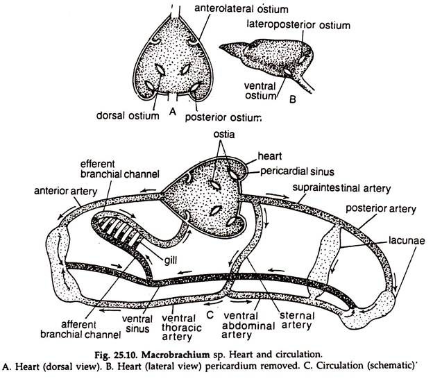
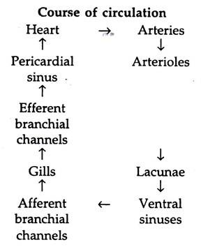
Haemolymph from the arteries is finally received into minute intercommunicating body spaces, the lacunae, which ultimately open into two large spaces, the ventral sinuses, situated lengthwise in the ventral region, beneath the hepatopancreas. The two sinuses are connected with each other at several places.
For aeration, haemolymph from the ventral sinuses is sent through six pairs of afferent branchial channels to the gills. After aeration, haemolymph from the gills is returned to the pericardial sinus through six pairs of efferent branchial channels. (Fig. 25.10).
7. Excretory System of Prawn:
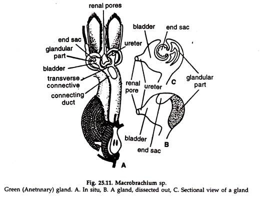
1. The excretory organs consist of a pair of cream-coloured antennary glands with their ducts, a median renal sac and a transverse communicating duct. It also performs the function of osmoregulation (Fig. 25.11).
2. The antennary gland—also called green gland—is placed in the coxa of the second antenna.
It consists of three parts:
a. A small end sac.
b. A glandular mass.
c. A bladder.
The end sac and glandular mass extract excretory products which are carried to the bladder. The bladder occupies the innermost region and is drawn into a narrow tube to open to the exterior through the renal aperture on the inner side of the coxa.
3. The renal sac is a thin-walled median structure lying just above the stomach. It is connected with each antennary gland by a narrow duct anteriorly. The two ducts are again connected by a narrow transverse duck Posteriorly, the renal sac ends blindly at the region of the gonad. The renal sac acts as a temporary reservoir for waste products.
4. No other renal organs.
The thick chitinous layer of the integument is a nitrogenous product secreted by the ectoderm and is cast off in each moult. Moulting is considered as a special mechanism to get rid of nitrogenous wastes.
8. Nervous System of Prawn:
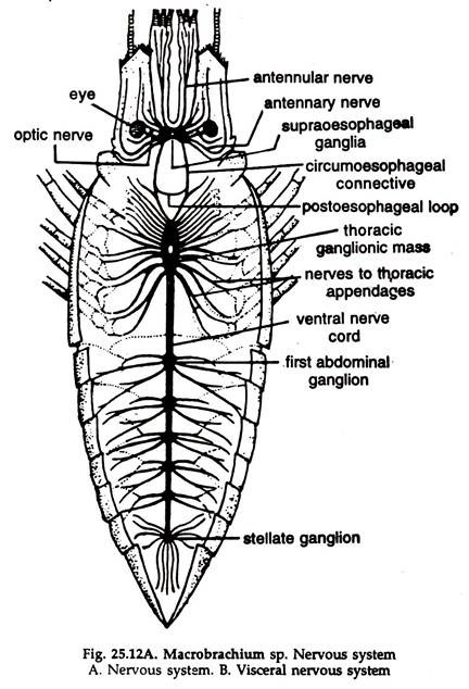
The system which controls and regulates the various activities of an organism is known as nervous system. The nervous system of prawn consists of a central nervous system, a peripheral nervous system and a visceral or sympathetic nervous system.
Central Nervous System:
The central nervous system consists of a pair of supraoesophageal ganglia, a pair of circumoesophageal connectives, a sub-oesophageal or thoracic ganglionic mass and a double ventral ganglionated nerve cord. All these send out nerves which supply the respective organs (Fig. 25.12).
1. Brain or supraoesophageal ganglia:
It is a bilobed structure formed by the fusion of the right and left ganglia and is situated beneath the base of the rostrum just in front of the junction of the oesophagus with the cardiac stomach. The
supraoesophageal ganglia is formed by the fusion of several pairs of ganglia.
2. Circumoesophageal connectives:
Two stout nerve cords, arise from the hinder part of the supraoesophageal ganglia and rim backward and downward round the oesophagus to meet ventrally in the thoracic ganglionic mass.
a. Post-oesophageal or transverse loop:
A loop embracing the oesophagus posteriorly and connects the two circumoesophageal connectives.
3. Thoracic ganglionic mass:
A large ganglion formed by the fusion of several pairs of ganglia and form the anterior-most ganglion of the ventral nerve cord.
4. Ventral nerve cord:
It lies beneath the mass of the abdominal muscles. The ventral nerve cord is formed by the fusion of two nerves and two ganglia unite to form each ganglion of the ventral nerve cord. There are six abdominal ganglia on the nerve cord corresponding to the six abdominal segments. The last ganglion is comparatively large and is known as stellate ganglion; it is possibly formed by the fusion of several ganglia.
Peripheral Nervous System:
The nerves emanating from the central nervous system constitute peripheral nervous system:
1. Optic nerve:
Arising from the outer side of each supraoesophageal ganglion it runs forward and outward and innervate the eye of the side.
2. Antennulary nerve:
Arises from the anterior part of the supraoesophageal ganglion, runs forward and sends branches to the antennule and the statocyst of the side.
3. Antennary nerve:
Arising from the lower portion of the supraoesophageal ganglion and passing downwards and obliquely, curves forward to innervate the antenna.
4. A small nerve:
A small nerve arises along the antennary nerve and innervate the labrum.
5. Cephalothoracic nerves:
Eleven pairs of nerves arise from the thoracic ganglionic mass and innervate all the cepholothoracic appendages except the two pairs of antennae.
6. Abdominal nerves:
Two pairs of nerves arise from each abdominal ganglion and innervate the corresponding muscles and appendages. Two additional pairs of nerves from the stellate ganglion send branches to rectum, telson and adjacent organs.
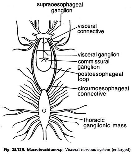
Visceral Nervous System:
1. Two small visceral or oesophageal ganglia are present on the roof of the cardiac stomach, one behind the other.
2. A small nerve arising from the posterior border of the brain connects the two ganglia behind.
3. Two delicate connectives join the anterior visceral ganglion with the two commissural ganglia on the circumaoesophageal connectives.
Receptors and Sense Organs of Prawn:
The sense organs include eyes, statocysts, tactile organs and olfactory setae.
Eyes:
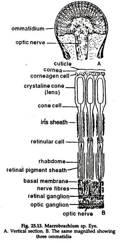
The prawn bears two compound eyes. Each eye is a collection of a large number of visual elements called ommatidia and is borne on a movable stalk.
Structure (Fig. 25.13):
1. The outer convex transparent cuticular covering of the eye is known as cornea. The cornea is divided into a large number of square facets, each corresponding to a single ommatidium. The ommatidia are arranged regularly along the radii of the eye.
2. Each ommatidium is a complete visual unit, made up of cells arranged in end- to-end position along the long axis. Beneath the corneal facet is a pair of flat corneagen cells of epidermal origin which secrete a new cornea when the old one is lost during moulting.
3. Beneath the corneagen cells lie four tall cells—the cone cells—the inner borders of which give rise to a refractive crystalline cone.
4. The basal part of the ommatidium is made of spindle-shaped, transversely striated structure, the rhabdome, which is surrounded by seven elongated cells, the retinular cells. The rhabdome with the retinular cells are known as the retinula bearing a pigment, guanine, the nature of which is said to be of melanin.
5. The optic nerve breaks up into branches and innervate the retinular cells.
6. Each ommatidium is separated from its neighbours by a partition of pigment cells, the chromatophores.
These are arranged in two groups:
a. The distal group surrounding the lens and the cone cells constitute the Irish sheath.
b. The proximal group surrounding the retinula constitute the retinal sheath. The pigment sheaths can extend or retract under the influence of light. In bright light, they are extended and in weak light they are retracted.
Vision in Prawn:
In prawn, two types of visions are found. At daytime, when the intensity of light is high, the vision is of mosaic type. In the night, or dim light, when the intensity of light is less, the vision is of superimposed type.
1. Mosaic vision:
In bright light, the pigment sheath is extended and any jay, of light which falls obliquely on the ommatidium is absorbed by the pigment sheath. Almost parallel rays falling on each ommatidium from an object, reach the rhabdome and an image of a point of the object is formed.
Therefore many images of the many points of the object are formed. Such an image is known as apposition image. By the apposition-of those points of images in a number of ommatidia an erect image of the object is formed. In such a vision, any slight change of the object is quickly detected. Here, the sharpness of the image is dependent upon the number of ommatidia involved and the degree of their separation.
2. Superimposed vision:
In dim light, the pigment sheath is retracted and greater portion of the ommatidium is uncovered. Any ray of light striking obliquely on the sides of the ommatidium passes to the next and, in doing so, becomes refracted to reach the next ommatidium.
In such a case, an overlapping of points of lights occur and a superimposed image is formed, which is not sharp. The crystalline cones, capable of adjusting accordingly, act in unison and behave as a single unit and the whole of the retinal portion act as a single retina.
Statocysts of Prawn:
Statocysts are the balance organs. They are a pair, one in each antennule, located in the cavity of the precoxa or the basal segment.
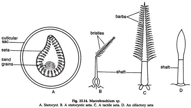
1. A statocyst is a sub-spherical cuticular sac (Fig. 25.14A) attached to the inner surface of the dorsal wall of the precoxa and opens to the exterior through a narrow pore.
2. Sensory setae are arranged in the sac in the form of an oval ring.
3. Each seta has a swollen base and a pointed shaft bearing fine bristles (Fig. 25.14B).
4. Sand grains are present in the space surrounded by the setae.
5. The setae receive fine branches of statocyst nerve, which is a branch of the antennulary nerve.
During movement the sand grains are displaced with the change of position and press against certain setae, which helps the animal to correct its position.
Tactile Sense Organs:
They are present along the margins of the appendages, abundant in antennae and flattened portion of pleopods.
A tactile seta (Fig. 25.14C) consists of:
1. A hollow base or shaft connected to the appendage.
2. A tapering blade bearing double linear rows of tiny barbs is attached to the distal end of the shaft.
The sense organs carry sense of touch.
Olfactory Setae:
A seta is located on the small, middle feeler, between the two long feelers of an antennule.
It has:
1. A two-jointed shaft, proximally attached to the integument by a flexible membrane.
2. The free end of the distal segment is bluntly rounded and covered with a thin membrane (Fig 25. 14D).
3. Innervated by nerve fibres from the olfactory branch of the antennulary nerve.
9. Reproductive System of Prawn:
The sexes are separate.
Male reproductive system:
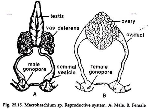
1. The male reproductive system consist of a pair of testes, a pair of vasa deferentia, a pair of seminal vesicles and a pair of gonopores (Fig 25.15A).
2. The testes are soft, white, elongated bodies, fused at both the ends and are situated in the cephalothorax, below the heart and above the hepatopancreas.
3. From each testis arises a narrow tube, the vas deferens, which is much coiled at first and then descends down towards the base of the fifth walking leg of the side.
4. The terminal end of each vas deferens forms a club-shaped swelling, known as seminal vesicle, which opens to the exterior by the male gonopore on the inner side of the coxa of the 5th walking leg.
Female reproductive system:
1. The female reproductive system consist of a pair of ovaries, a pair of oviducts and a pair of female gonopores (Fig 25.15B).
2. Ovaries are small and whitish in off-seasons but large and dark brown in the breeding season. They are fused at both the ends, larger in size than the testes and occupy same position as the testes in the male.
3. From the middle of the outer side of each ovary arises an oviduct, which narrows downwards to open in the gonopore on the inner side of the coxa of the 3rd walking leg of the side.
10. Fertilization and Development of Prawn:
1. Prawns breed in rainy season.
2. The eggs are round and yolk-filled.
3. Fertilization external and the fertilized eggs are carried in the abdominal basket, formed by the appendix internae of the second to fifth pleopods in females. Development direct, the newly hatched young resembling the adult, leave the abdominal basket to lead a free life.
